
Mechanism of regulation of muscle tone
Reading File
Now let me search the medical library for content on muscle tone regulation.
Finding Sources
Finding Sources
Excellent sources found. Let me now read the key sections in parallel - Ganong's physiology, Adams & Victor's neurology, and the Bradley & Daroff sections:
Reading File
Reading File
Reading File
Good start. Now let me search for the deeper mechanistic content - stretch reflex, gamma motor neurons, Golgi tendon organs, and supraspinal control:
Finding Sources
Finding Sources
Now let me read the comprehensive physiology sections from Ganong's and Costanzo in parallel:
Reading File
Reading File
Excellent content. Now let me read the Ganong's section on inverse stretch reflex/Golgi tendon organ and supraspinal control of tone, plus the Costanzo Golgi tendon reflex section:
Reading File
Reading File
Excellent. Now let me get the supraspinal control sections - reticulospinal, vestibulospinal tracts, and alpha-gamma coactivation:
Finding Sources
Finding Sources
Reading File
Reading File
Now let me get the supraspinal control content and the key images:
Finding Sources
Loading Image
Now let me fetch the Golgi tendon organ image and also get the supraspinal control section from Ganong's:
Loading Image
Finding Sources
I have comprehensive content from multiple authoritative textbooks. Let me now compile the complete, well-structured answer.
Mechanism of Regulation of Muscle Tone
Muscle tone is defined as the resistance of a muscle to passive stretch. It is not an active contraction but rather a state of low-level, sustained activation that keeps muscles ready for use and helps maintain posture. Normal tone lies between the extremes of flaccidity (motor nerve severed) and spasticity (hyperactive stretch reflexes). - Ganong's Review of Medical Physiology, 26th ed.
1. What Is Muscle Tone?
- Tone is determined primarily by the rate of gamma (γ) motor neuron discharge.
- Low γ-motor neuron activity → hypotonic muscle.
- High γ-motor neuron activity → hypertonic (spastic) muscle.
- Muscles generally maintain a slight background tension even at rest, mediated through tonic stretch reflex activity.
2. The Peripheral Apparatus: Muscle Spindle
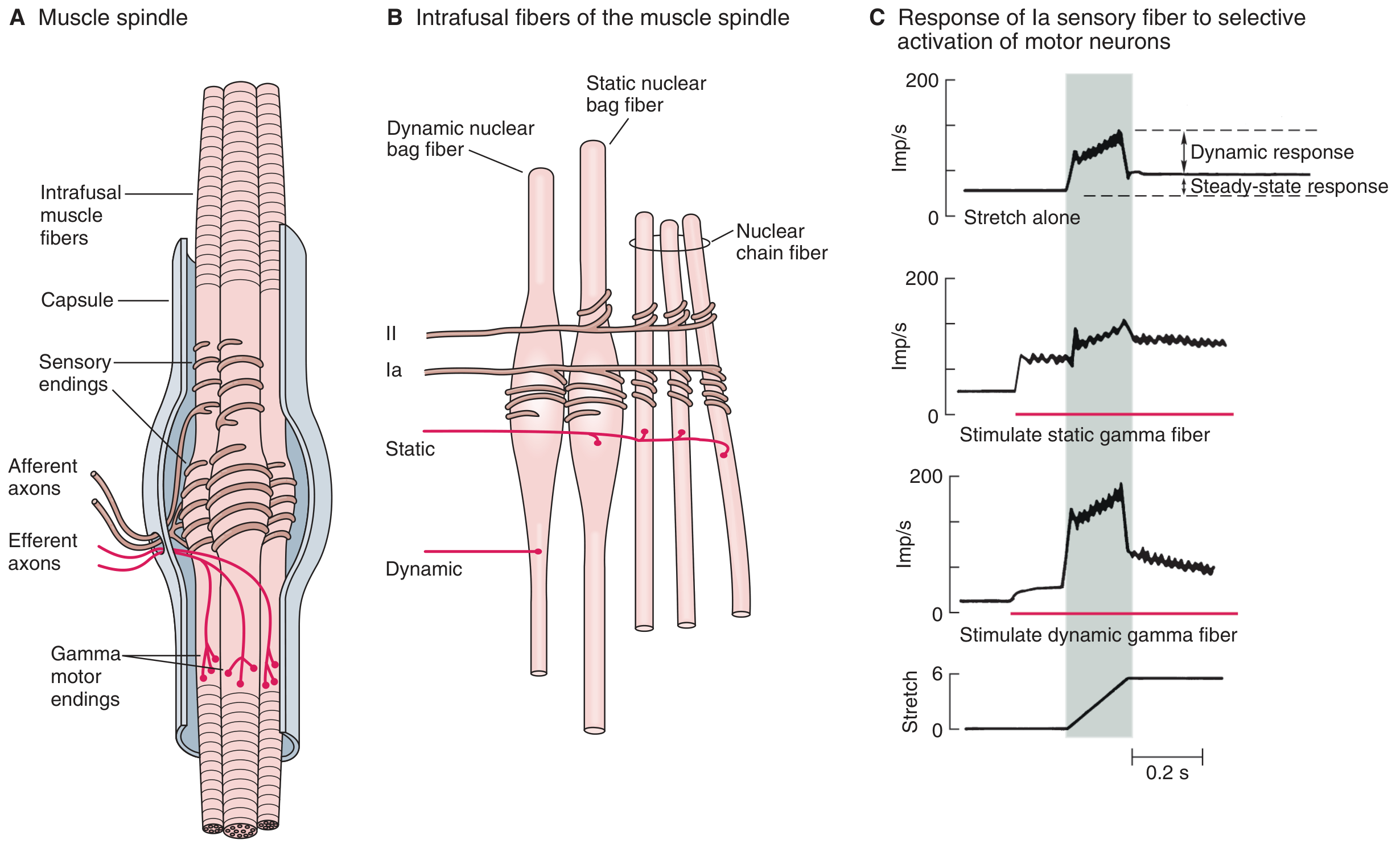
The muscle spindle is the central sensory transducer for tone regulation.
Structure
- Intrafusal fibers (inside the spindle) are of three types:
- Dynamic nuclear bag fibers
- Static nuclear bag fibers
- Nuclear chain fibers
- These are surrounded by the extrafusal (working) muscle fibers.
- The spindle lies in parallel with the extrafusal fibers.
Sensory innervation
| Fiber | Type | What it detects |
|---|---|---|
| Group Ia (primary ending) | Wraps around all intrafusal fiber types | Velocity of length change (dynamic response) |
| Group II (secondary ending) | Innervates static bag + chain fibers | Steady-state length (static/tonic response) |
Motor innervation (the fusimotor system)
- γ-motor neurons (3-6 μm diameter, ~30% of ventral root fibers) innervate only intrafusal fibers.
- Dynamic γ-neurons → innervate dynamic nuclear bag fibers → amplify the velocity-sensitive response of Ia fibers.
- Static γ-neurons → innervate static nuclear bag + chain fibers → increase tonic Ia and II activity; maintain spindle sensitivity during contraction.
- β-axons (collaterals of α-motor neurons) also innervate intrafusal fibers, providing intrinsic alpha-gamma coactivation. - Principles of Neural Science, Kandel, 6th ed.
3. The Stretch Reflex (Myotatic Reflex) - Core Mechanism of Tone
The stretch reflex is the monosynaptic backbone of muscle tone regulation. - Costanzo Physiology, 7th ed.
Sequence of events:
- Muscle is passively stretched → intrafusal fibers lengthen → Ia afferents fire at increased rate.
- Ia afferents enter the spinal cord and synapse directly on α-motor neurons of the same (homonymous) muscle.
- α-motor neurons activate extrafusal fibers → muscle contracts, opposing the stretch.
- Simultaneously: synergistic muscles are excited; antagonistic muscles are inhibited via Ia inhibitory interneurons (reciprocal inhibition).
- γ-motor neurons are coactivated with α-motor neurons (alpha-gamma coactivation), keeping the spindle under tension during contraction so it remains sensitive and does not fall silent. - Kandel's Principles of Neural Science, 6th ed.
This reflex loop continuously adjusts muscle length and provides the tonic background contraction we call "muscle tone."
4. The Inverse Stretch Reflex (Golgi Tendon Reflex) - Protective Modulation
When tension becomes excessive, a counter-regulatory mechanism activates:
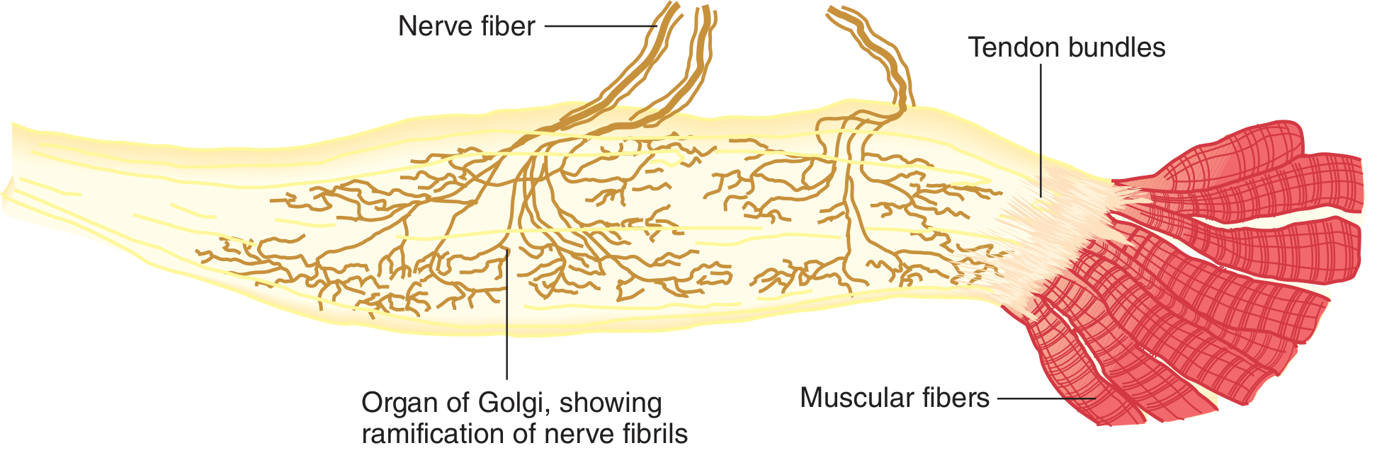
Structure of the Golgi Tendon Organ (GTO):
- Lies in series with extrafusal muscle fibers (unlike spindles, which are parallel).
- Contains knobby nerve endings of Ib myelinated afferent fibers among tendon fascicles.
- Each GTO is served by 3-25 muscle fibers.
Sequence of the Golgi tendon reflex (dysynaptic):
- Strong muscle contraction or stretch activates GTO → Ib afferents fire.
- Ib fibers synapse on inhibitory interneurons in the spinal cord.
- Inhibitory interneurons synapse on α-motor neurons → autogenic inhibition (homonymous muscle relaxes).
- Excitatory connections are made to motor neurons of antagonist muscles.
- Result: muscle tension falls, preventing tendon damage.
The GTO thus functions as a force transducer - a feedback loop regulating muscle force, analogous to how the spindle regulates muscle length. Together, spindle and GTO discharges regulate velocity, length, and force of contraction. - Ganong's Review of Medical Physiology, 26th ed.
Clinical correlate - Clasp-knife phenomenon:
In spasticity, passive flexion of a joint initially meets high resistance (stretch reflex), but as tension builds, sudden relaxation occurs (GTO activation). This is the clasp-knife effect. - Costanzo Physiology, 7th ed.
5. Control of γ-Motor Neuron Discharge (Supraspinal Regulation)
γ-motor neurons are regulated by descending tracts from several brain regions, which modulate spindle sensitivity and thus set the threshold of stretch reflexes across the body. - Ganong's Review of Medical Physiology, 26th ed.
| Supraspinal Center | Effect on Tone | Notes |
|---|---|---|
| Reticular formation (facilitatory area) | Increases tone | Medial reticulospinal tract; facilitates γ-motor neurons |
| Reticular formation (inhibitory area) | Decreases tone | Requires input from cortex and basal ganglia to remain active |
| Vestibular nuclei (vestibulospinal tract) | Increases extensor tone | Particularly in anti-gravity muscles |
| Cerebral cortex (corticospinal tract) | Fine modulation; primarily inhibitory on tone | Damage → spasticity (upper motor neuron lesion) |
| Cerebellum | Coordinates fusimotor activity | Cerebellar lesions → hypotonia |
| Basal ganglia | Modulate cortical output to reticular formation | Lesions → rigidity (Parkinson's) or athetosis |
Decerebrate rigidity: Transection at the midbrain level removes cortical/basal ganglia inhibitory input to the facilitatory reticular formation, causing marked extensor hypertonicity - a classic demonstration of supraspinal control.
Other factors modulating γ-motor neuron activity:
- Anxiety → increased γ discharge → hyperactive tendon jerks.
- Noxious skin stimulation → increased γ activity to ipsilateral flexors, decreased to extensors.
- Jendrassik maneuver (interlocking fingers and pulling) → facilitates knee jerk reflex, likely via increased γ discharge from hand afferents.
- Unexpected movement → greater fusimotor discharge.
- Task difficulty → during precision tasks (e.g., walking a narrow beam), both static and dynamic γ activation increase. - Kandel's Principles of Neural Science, 6th ed.
6. Reciprocal Innervation and Renshaw Cell Inhibition
- When the agonist is excited via the stretch reflex, the antagonist is simultaneously inhibited via Ia inhibitory interneurons → prevents co-contraction and allows smooth movement.
- Renshaw cells (inhibitory interneurons activated by α-motor neuron axon collaterals) inhibit the same motor neuron pool and antagonist's Ia inhibitory interneurons → recurrent inhibition, limiting the duration and spread of motor neuron discharge. - Ganong's Review of Medical Physiology, 26th ed.
7. Summary Diagram of Tone Regulation
Supraspinal centers (cortex, cerebellum, basal ganglia, reticular formation, vestibular nuclei)
            ↓
      γ-motor neurons  ←───── also receive spinal interneuron inputs
            ↓
  Intrafusal muscle fibers → adjust spindle sensitivity
            ↓ (Ia/II afferents)
  α-motor neurons  ←──── Golgi tendon Ib afferents (inhibitory, via interneurons)
            ↓
  Extrafusal muscle fibers → MUSCLE TONE
8. Clinical Disorders of Muscle Tone
| Disorder | Tone Change | Mechanism |
|---|---|---|
| Spasticity (UMN lesion - stroke, spinal cord injury) | Increased (velocity-dependent) | Loss of cortical inhibition → hyperactive stretch reflex; clasp-knife phenomenon |
| Rigidity (Parkinson's, basal ganglia) | Increased (lead-pipe/cogwheel, not velocity-dependent) | Increased γ-motor drive from abnormal basal ganglia output |
| Hypotonia/Flaccidity (LMN lesion, cerebellar lesion) | Decreased | Loss of α-motor neurons (LMN) or reduced γ drive (cerebellum) |
| Clonus | Rhythmic involuntary contraction | Hyperactive stretch reflex + absent Renshaw inhibition; ≥5 beats is pathological |
Sources:
- Ganong's Review of Medical Physiology, 26th ed. - pp. 237-241
- Costanzo Physiology, 7th ed. - pp. 111-112
- Kandel's Principles of Neural Science, 6th ed. - pp. 814-815
- Bradley and Daroff's Neurology in Clinical Practice
Mechanisms of regulation of blood pressure
Reading File
This is a broad topic. Let me run parallel heading and paragraph searches across multiple aspects simultaneously.
Finding Sources
Finding Sources
Finding Sources
Excellent sources found. Now let me read the key sections in parallel - Robbins Pathology (comprehensive BP regulation), Katzung Pharmacology, and Boron Medical Physiology:
Reading File
Reading File
Reading File
Excellent content. Now let me read the continuation of Robbins Pathology (ANP, endothelin, NO) and RAAS from Brenner's Kidney book, plus fetch the key diagrams:
Reading File
Reading File
Loading Image
Now let me fetch the baroreceptor reflex arc diagram and search for more content on endothelin/NO/natriuretic peptides and long-term regulation:
Loading Image
Finding Sources
Finding Sources
Excellent. Let me now read the ANP/BNP section from Robbins (Cotran) and get Guyton's long-term regulation content:
Reading File
Finding Sources
Now let me get Guyton's content on pressure natriuresis and vasopressin/ADH, and also the Harrison's physiology section:
Finding Sources
Finding Sources
Reading File
Reading File
I now have comprehensive content from multiple authoritative sources. Let me compile the full, detailed answer.
Mechanisms of Regulation of Blood Pressure
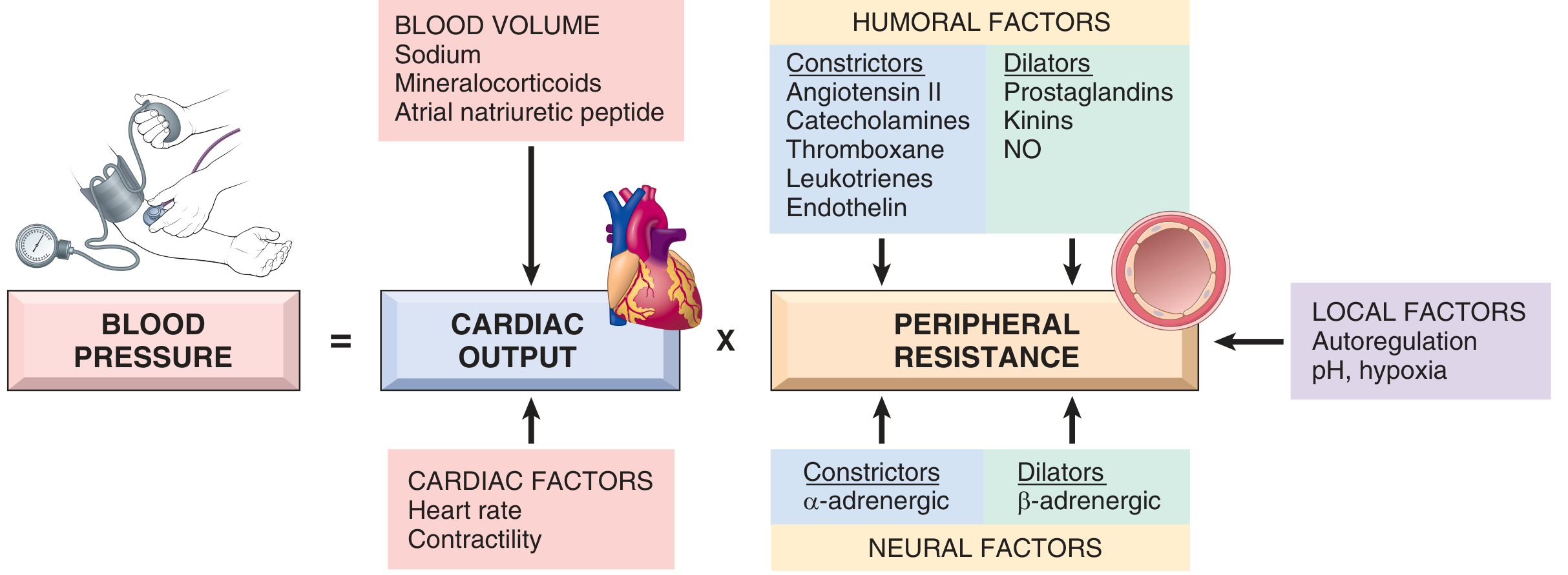
Blood pressure (BP) must be kept within a narrow range. Low BP causes inadequate organ perfusion; high BP damages vessels and end-organs. The fundamental equation that governs BP is:
BP = Cardiac Output (CO) × Peripheral Vascular Resistance (PVR)
Regulation acts at four anatomic control sites: arterioles (PVR), capacitance venules (venous return), heart (CO), and kidney (blood volume). - Katzung's Basic & Clinical Pharmacology, 16th ed.
Master Diagram
I. Determinants of Blood Pressure
A. Cardiac Output
- Stroke volume - the most important determinant is filling pressure, which depends on blood volume and hence sodium homeostasis.
- Heart rate and myocardial contractility - both controlled by α- and β-adrenergic systems.
B. Peripheral Vascular Resistance
- Regulated primarily at the arteriolar level by neural and humoral inputs.
- Vascular tone = balance between vasoconstrictors (angiotensin II, catecholamines, endothelin, thromboxane, leukotrienes) and vasodilators (kinins, prostaglandins, nitric oxide).
- Local autoregulation: increased blood flow induces vasoconstriction to prevent hyperperfusion.
- Fine-tuned by tissue pH and hypoxia to meet local metabolic demands. - Robbins & Kumar Basic Pathology; Robbins, Cotran & Kumar Pathologic Basis of Disease
II. Short-Term (Rapid) Regulation
1. Baroreceptor Reflex (Postural Baroreflex)
The primary moment-to-moment regulator. Acts within seconds.
Mechanism:
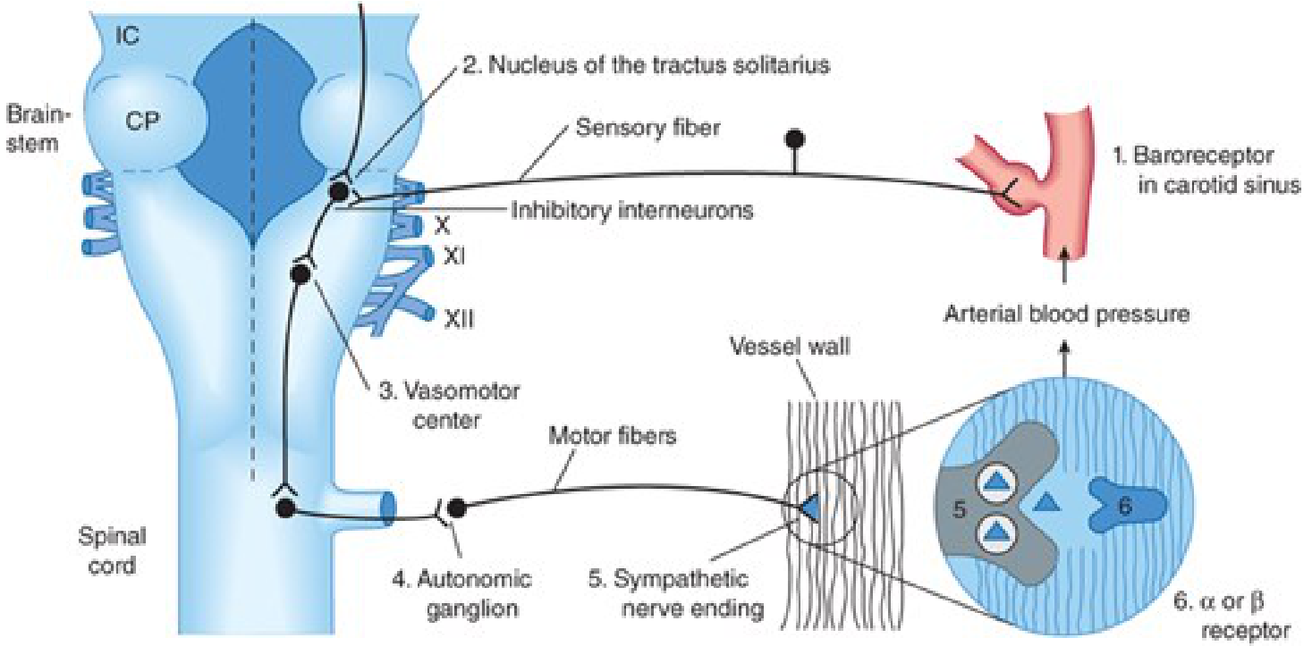
- Baroreceptors in the carotid sinus and aortic arch detect arterial wall stretch (proportional to BP).
- Afferent signals travel via CN IX (carotid) and CN X (aortic) to the nucleus tractus solitarius (NTS) in the medulla.
- NTS projects to the vasomotor (cardiovascular) center in the medulla.
- The vasomotor center tonically drives sympathetic outflow. Baroreceptor activation → inhibits sympathetic discharge (via inhibitory interneurons).
- Efferent sympathetic fibers act on:
- Arterioles (α-adrenergic receptors) → vasoconstriction → ↑ PVR
- Heart (β1-adrenergic receptors) → ↑ heart rate + ↑ contractility → ↑ CO
- Capacitance veins → constriction → ↑ venous return → ↑ stroke volume
Response to a fall in BP (e.g., standing up):
- ↓ baroreceptor stretch → ↓ inhibitory afferent firing → disinhibition of sympathetic center → ↑ sympathetic outflow → ↑ PVR + ↑ CO → BP restored.
Response to a rise in BP:
- ↑ baroreceptor stretch → ↑ inhibitory input → ↓ sympathetic + ↑ parasympathetic → bradycardia, vasodilation → BP falls. - Katzung's Basic & Clinical Pharmacology, 16th ed.
Baroreceptors are reset at higher BP levels in chronic hypertension; they defend the set-point rather than normalizing BP.
2. Chemoreceptor Reflex
- Peripheral chemoreceptors (carotid and aortic bodies) sense hypoxia, hypercapnia, and acidosis.
- When activated: cause peripheral vasoconstriction and stimulate breathing (secondary rise in BP).
- Central chemoreceptors in the medulla respond to CO₂/H⁺ and influence the vasomotor center. - Medical Physiology (Boron)
3. CNS Ischemic Response (Cushing Reflex)
- When cerebral perfusion pressure falls critically, massive sympathetic discharge ensues.
- Produces severe systemic vasoconstriction and hypertension - a last-resort protective mechanism to maintain cerebral flow.
III. Intermediate Regulation (Minutes to Hours)
4. Renin-Angiotensin-Aldosterone System (RAAS)
The central humoral regulator of both vasoconstriction and volume.
Triggers for renin release from juxtaglomerular cells:
- ↓ Blood pressure in afferent arterioles (stretch-sensitive mechanism)
- ↓ Sodium delivery to the macula densa in the distal tubule (tubuloglomerular feedback)
- ↑ Circulating catecholamines (β1-adrenergic stimulation of JG cells)
- Reduced renal perfusion / ↓ GFR
Cascade:
Angiotensinogen (liver)
        ↓  [Renin - from JG cells]
Angiotensin I
        ↓  [ACE - mainly pulmonary/vascular endothelium]
Angiotensin II
Actions of Angiotensin II (raises BP by 3 mechanisms):
| Mechanism | Site | Effect |
|---|---|---|
| Direct vasoconstriction | Vascular smooth muscle | ↑ PVR |
| Stimulates aldosterone release | Adrenal cortex (zona glomerulosa) | ↑ Na⁺ + H₂O reabsorption → ↑ blood volume |
| Direct sodium reabsorption | Renal proximal tubule | ↑ blood volume |
Aldosterone:
- Increases ENaC (epithelial sodium channel) activity in the distal convoluted tubule and collecting duct.
- Na⁺ (+ water) retention → ↑ blood volume → ↑ filling pressure → ↑ CO → ↑ BP.
- Causes potassium excretion. - Robbins, Cotran & Kumar Pathologic Basis of Disease; Katzung's Basic & Clinical Pharmacology
Renal counterbalancers: The kidney also produces prostaglandins and NO to counterbalance the vasopressor effects of angiotensin II. - Robbins, Cotran & Kumar Pathologic Basis of Disease
5. Vasopressin (ADH - Antidiuretic Hormone)
- Released from the posterior pituitary in response to:
- ↑ plasma osmolality (detected by hypothalamic osmoreceptors) - primary trigger
- ↓ blood pressure / ↓ blood volume (detected by high-pressure arterial baroreceptors and low-pressure atrial mechanoreceptors)
- Actions:
- Inserts aquaporin-2 channels in collecting duct → water reabsorption → ↑ blood volume
- V1 receptors on vascular smooth muscle → vasoconstriction
- Plays a role in maintenance of BP especially during hemorrhage and hypovolemia. - Katzung's Basic & Clinical Pharmacology; Tietz Laboratory Medicine
IV. Long-Term Regulation
6. Renal Pressure Natriuresis and Diuresis
The ultimate long-term controller of blood pressure. The kidney is the only organ that can provide infinite-gain regulation over extended periods. - Guyton & Hall Textbook of Medical Physiology
Mechanism:
- ↑ Arterial BP → ↑ renal perfusion pressure → kidney excretes more Na⁺ and water (pressure natriuresis/diuresis) → ↓ blood volume → ↓ CO → BP returns to normal.
- This is amplified by indirect effects: ↑ BP → ↓ sympathetic activity → ↓ RAAS → further ↓ antinatriuretic hormones → more Na⁺ excretion.
- Conversely: ↓ BP → ↓ natriuresis → Na⁺ retention → ↑ volume → BP restored.
In healthy individuals with normal kidneys, this mechanism can accommodate Na⁺ intake varying 6-fold with minimal change in BP. Salt sensitivity occurs when this renal output curve is "flattened" by kidney disease, RAAS excess, or reduced nephron number. - Guyton & Hall
7. Natriuretic Peptides (ANP and BNP) - Counter-Regulatory
Released in response to volume expansion / atrial stretch - opposite to RAAS.
| Peptide | Source | Release Trigger |
|---|---|---|
| ANP (Atrial Natriuretic Peptide) | Atrial myocytes | Atrial stretch (normal levels constitutive) |
| BNP (B-type / Brain Natriuretic Peptide) | Ventricular myocytes | Volume overload, wall stress |
Actions (both lower BP):
- Systemic vasodilation
- Inhibit sodium reabsorption in the distal renal tubules → natriuresis and diuresis → ↓ blood volume
- Inhibit renin and aldosterone secretion
- Inhibit ADH release
Elevated ANP/BNP signals hypervolemia - most commonly in congestive heart failure. - Robbins, Cotran & Kumar Pathologic Basis of Disease
V. Local / Endothelial Regulation
8. Endothelial Vasoactive Substances
The vascular endothelium is an active paracrine regulator of underlying smooth muscle tone:
| Substance | Type | Mechanism |
|---|---|---|
| Nitric oxide (NO) | Vasodilator | Synthesized by eNOS (shear stress, bradykinin) → activates guanylyl cyclase → ↑ cGMP → smooth muscle relaxation |
| Prostacyclin (PGI₂) | Vasodilator | Arachidonic acid pathway → ↑ cAMP → relaxation + anti-platelet |
| Endothelin-1 | Vasoconstrictor | Most potent vasoconstrictor known; released by endothelial injury/stress; acts on ETA receptors |
| Thromboxane A₂ | Vasoconstrictor | Pro-aggregant and vasoconstrictive |
9. Local Metabolic Autoregulation
- Tissues adjust their own vascular tone based on local metabolic needs.
- Vasodilators produced locally: CO₂, H⁺ (acidosis), adenosine, K⁺, lactate, hypoxia.
- When metabolic demand rises → local vasodilation → ↑ blood flow to active tissue.
- Myogenic autoregulation: vascular smooth muscle contracts in response to increased intraluminal pressure (Bayliss effect) - protects capillary beds from hyperperfusion. - Robbins & Kumar Basic Pathology
VI. Summary - Timescale of Regulation
| Mechanism | Speed | Primary Effect |
|---|---|---|
| Baroreceptor reflex | Seconds | ↑ or ↓ sympathetic output → HR, contractility, PVR |
| Chemoreceptor reflex | Seconds-minutes | Vasoconstriction + ventilation |
| CNS ischemic (Cushing) | Seconds | Emergency vasoconstriction |
| Catecholamines (adrenal medulla) | Minutes | α1 vasoconstriction, β1 cardiac stimulation |
| RAAS | Minutes-hours | Vasoconstriction + Na⁺ retention |
| ADH/Vasopressin | Minutes-hours | Water retention + vasoconstriction |
| ANP/BNP | Hours | Natriuresis + vasodilation (counter-regulatory) |
| Renal pressure natriuresis | Days-weeks | Ultimate long-term volume and BP control |
VII. Clinical Implications
| Condition | Mechanism Disrupted |
|---|---|
| Primary (essential) hypertension | Multifactorial: RAAS upregulation, reduced pressure natriuresis, increased sympathetic tone, endothelial dysfunction |
| Renovascular hypertension | ↓ Renal perfusion → uncontrolled RAAS activation |
| Primary hyperaldosteronism | Autonomous aldosterone → Na⁺ retention + hypertension |
| Pheochromocytoma | Catecholamine excess → sustained vasoconstriction |
| SIADH | ↑ ADH → water retention → dilutional hyponatremia + volume expansion |
| Orthostatic hypotension | Failure of baroreceptor reflex (autonomic neuropathy, volume depletion) |
Sources:
- Robbins & Kumar Basic Pathology - Fig. 8.2 and Blood Pressure Regulation, pp. 309-310
- Robbins, Cotran & Kumar Pathologic Basis of Disease - pp. 462-464
- Katzung's Basic & Clinical Pharmacology, 16th ed. - pp. 269-271
- Guyton & Hall Textbook of Medical Physiology - pp. 235-236 (pressure natriuresis)
This is a shared conversation. Sign in to Orris to start your own chat.