
histology of lungs
Reading File
Now let me search for lung histology content in the textbook library and relevant images in parallel.
Finding Sources
Searching Images
lung histology microscopy alveoli bronchioles
Reading File
Finding Sources
Now let me fetch the textbook histology image and search for more lung structural images in parallel.
Loading Image
Finding Sources
Searching Images
bronchus bronchiole wall layers smooth muscle cartilage histology cross section
Junqueira's Basic Histology has a dedicated section. Let me read it in full.
Reading File
Reading File
Reading File
Loading Image
Loading Image
Reading File
Loading Image
Loading Image
Reading File
I now have comprehensive material from Junqueira's Basic Histology and Schwartz's Surgery. Let me compile the full, well-illustrated answer.
Histology of the Lungs
The lung has two linked functional components: the conducting airway system (tracheobronchial tree) and the gas exchange system (alveolar spaces). Below is a systematic account from the largest airways down to the alveoli.
1. Tracheobronchial Tree (Conducting Airways)
Bronchi
The bronchial wall is organized into four layers:
| Layer | Contents |
|---|---|
| Mucosa | Pseudostratified ciliated columnar epithelium + goblet cells; rests on a basement membrane |
| Lamina propria | Loose connective tissue; contains a distinct smooth muscle layer encircling the entire bronchus |
| Submucosa | Hyaline cartilage (as irregular plates); mixed submucosal glands (mucous + serous + neuroendocrine Kulchitsky cells) |
| Adventitia | Fibrous connective tissue with blood vessels and nerves; merges with surrounding lung tissue |
The submucosal glands are mixed salivary-type structures that can give rise to mucoepidermoid and adenoid cystic carcinomas. Goblet cells (mucous cells) significantly increase after injury such as cigarette smoke exposure.
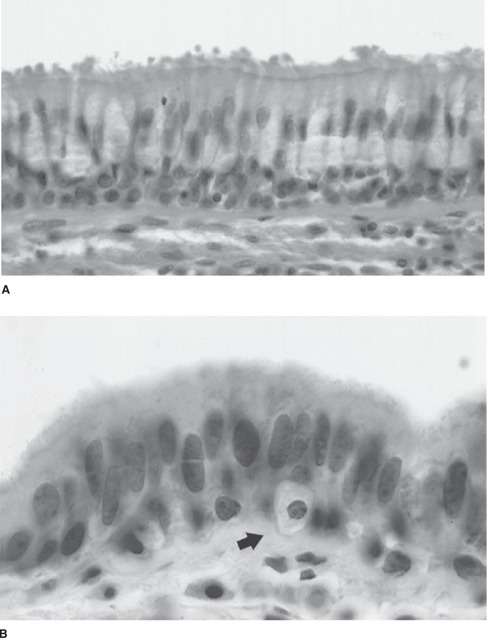
Figure 19-10 (Schwartz's Surgery): Normal bronchial mucosa showing pseudostratified ciliated columnar cells (panel A) and a Kulchitsky neuroendocrine cell indicated by arrow (panel B).
Bronchioles
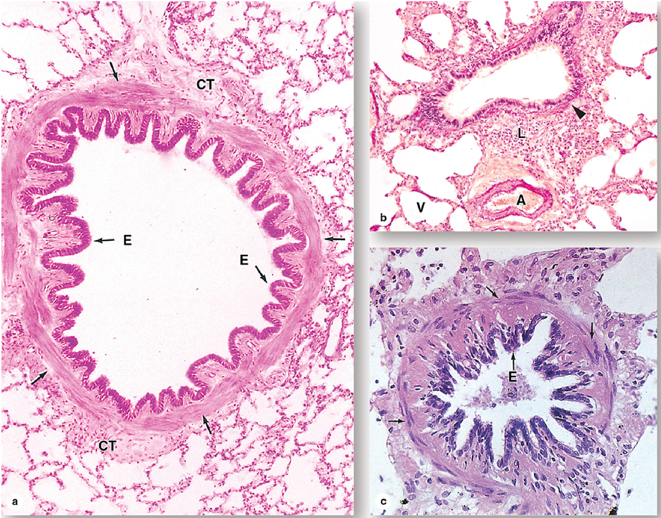
Bronchioles are intralobular airways with diameters of 1 mm or less (formed after ~10 generations of branching). Key distinguishing features:
- No cartilage, no submucosal glands
- Epithelium transitions progressively:
- Large bronchioles: still ciliated pseudostratified columnar
- Smaller bronchioles: simple ciliated columnar
- Terminal bronchioles (last conducting segment): simple cuboidal with cilia
- Smooth muscle becomes proportionally more prominent, with prominent elastic fibers
- Diffuse mucosal lymphoid tissue (MALT) increases as cartilage decreases
- Club cells (formerly Clara cells) are non-ciliated secretory cells that increase toward terminal bronchioles - they produce surfactant components, enzymes, and bacteriostatic agents
Figure 17-9 (Junqueira's Histology): Bronchioles at three levels - note the folded epithelium (E), prominent smooth muscle (arrows), and surrounding fibrous connective tissue (CT) in the absence of cartilage.
2. Respiratory Zone
Respiratory Bronchioles
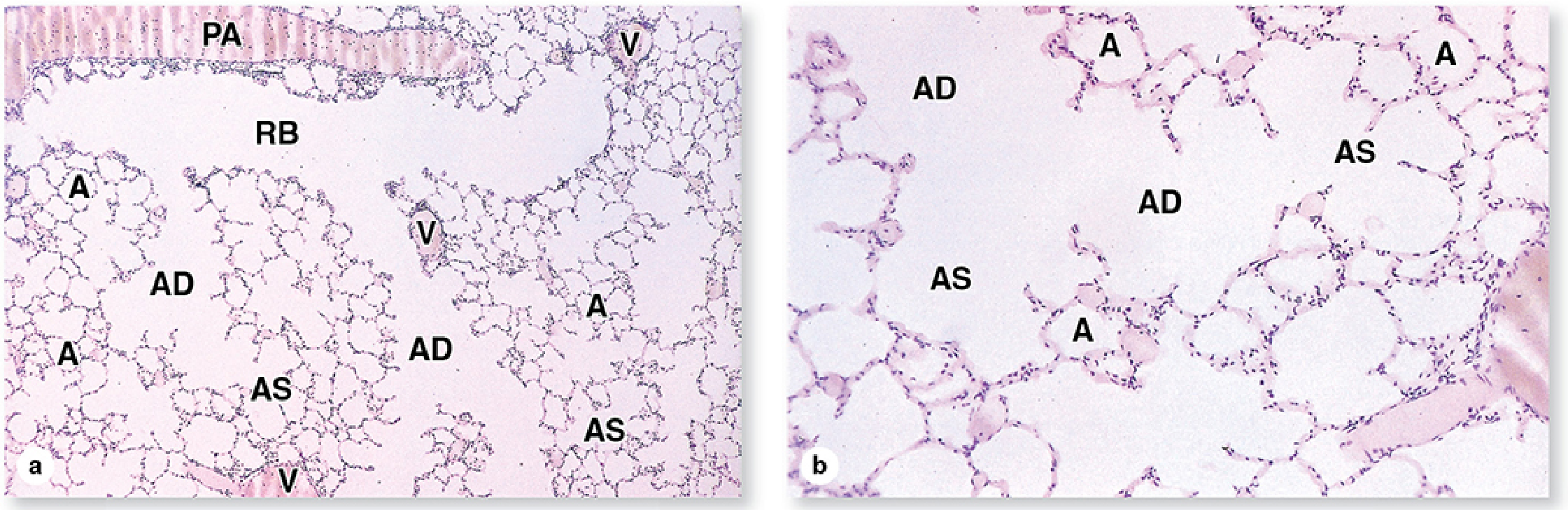
Terminal bronchioles subdivide into 2-3 respiratory bronchioles, which are the transition zone. They are lined by simple cuboidal epithelium interrupted by scattered alveolar outpouchings - the first site of gas exchange. They still contain a smooth muscle layer.
Alveolar Ducts
Alveolar ducts are completely lined by the openings of alveoli - they have no wall of their own other than smooth muscle "knobs" (eosinophilic nodules visible on H&E) around each alveolar opening, and a sparse lamina propria of elastic and reticular fibers plus capillaries.
Alveolar Sacs
Clusters of alveoli opening into a common chamber at the terminal end of alveolar ducts. The lamina propria here is essentially a delicate web of elastic and reticular fibers.
Figure 17-12 (Junqueira's Histology): Low-power view of lung parenchyma showing respiratory bronchioles (RB), alveolar ducts (AD), alveolar sacs (AS), individual alveoli (A), pulmonary artery (PA), and veins (V).
3. Alveoli
Each adult lung contains approximately 200 million alveoli, each ~200 µm in diameter, giving a total surface area of ~75 m².
Alveolar Wall (Interalveolar Septa)
The septa contain:
- The richest capillary network in the body
- Sparse connective tissue: elastic fibers (allow expansion/contraction) + reticular fibers (prevent collapse and overdistension)
- Fibroblasts
- Alveolar macrophages (dust cells)
Cell Types of the Alveolar Epithelium
| Cell | Proportion | Surface coverage | Key features |
|---|---|---|---|
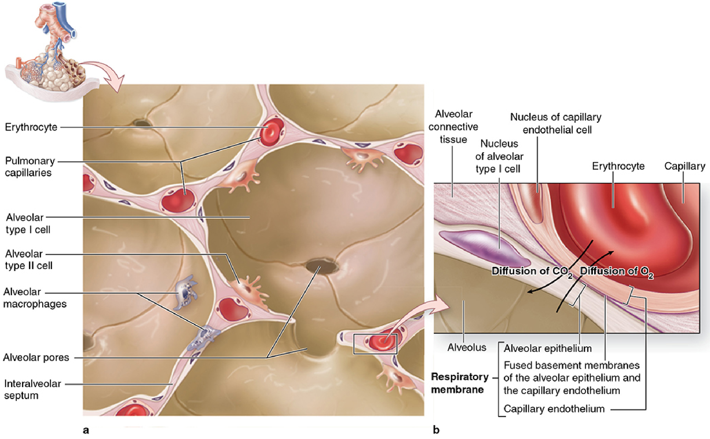
| Type I pneumocyte | 40% of cells | 95% of surface area | Extremely flattened (attenuated); non-mitotic; forms the gas exchange surface; continuous (non-fenestrated) |
| Type II pneumocyte | 60% of cells | ~3-5% of surface area | Cuboidal, bulge into lumen; contain lamellar bodies (surfactant precursors); produce surfactant (DPPC + SP-A, SP-B, SP-C, SP-D); regenerate after injury |
| Alveolar macrophages | Variable | Luminal/septal | Derived from monocytes; phagocytose surfactant, particulate matter, erythrocytes; contain carbon/hemosiderin; "dust cells" |
Figure 17-13 (Junqueira's Histology): Diagram of alveolar structure showing Type I cells, Type II cells, alveolar macrophages, pulmonary capillaries, alveolar pores of Kohn, and the interalveolar septum:
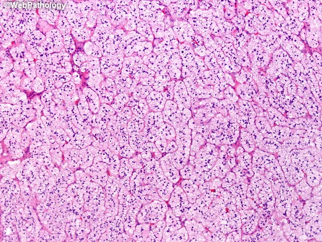
H&E photomicrograph of normal lung parenchyma showing multiple thin-walled alveoli with delicate interalveolar septa and capillary networks:
4. Blood-Air Barrier (Respiratory Membrane)
The barrier between alveolar air and capillary blood consists of three layers:
- Type I alveolar cell cytoplasm (extremely thin, 0.1-0.2 µm)
- Fused basement membranes of the alveolar epithelium and capillary endothelium
- Capillary endothelial cell (continuous, non-fenestrated; thin with perinuclear organelle clustering)
Total thickness: 0.1 to 1.5 µm - designed for maximum diffusion efficiency.
Alveolar pores of Kohn (10-15 µm diameter) penetrate interalveolar septa and equalize air pressure between adjacent alveoli and allow collateral air circulation if a bronchiole is blocked.
5. Surfactant
Produced by type II pneumocytes, surfactant contains:
- DPPC (dipalmitoylphosphatidylcholine) - the main phospholipid; reduces surface tension
- Cholesterol
- SP-A and SP-D - hydrophilic glycoproteins; innate immune protection
- SP-B and SP-C - hydrophobic proteins; orient DPPC in the surfactant film
Surfactant appears in the last weeks of fetal gestation. Inadequate surfactant causes neonatal respiratory distress syndrome in premature infants. Type II pneumocytes are the primary target of SARS-CoV-2 (COVID-19) via ACE2 receptor docking, leading to reduced surfactant production and alveolar collapse.
Summary Table
| Structure | Epithelium | Key Feature |
|---|---|---|
| Bronchi (large) | Pseudostratified ciliated columnar + goblet cells | Cartilage plates + submucosal glands |
| Bronchi (small) | Columnar ciliated | Less cartilage, more smooth muscle |
| Bronchioles | Ciliated columnar → simple cuboidal | No cartilage, no glands; Club cells present |
| Terminal bronchioles | Simple cuboidal, ciliated | Last conducting segment |
| Respiratory bronchioles | Simple cuboidal + alveolar outpouchings | First gas exchange site |
| Alveolar ducts | Alveolar lining only | Smooth muscle "knobs" at openings |
| Alveoli | Type I + Type II pneumocytes | Blood-air barrier; surfactant |
Sources: Junqueira's Basic Histology, 17e; Schwartz's Principles of Surgery, 11e
This is a shared conversation. Sign in to Orris to start your own chat.