
What is horners syndrome
Horner's syndrome ptosis miosis clinical photo
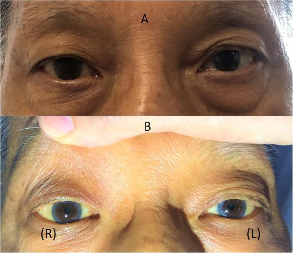
Two-panel clinical photograph demonstrating Horner’s syndrome in a patient. Panel A shows the patient's eyes at rest, highlighting a mild right-sided ptosis (drooping of the upper eyelid) and a subtle 'upside-down ptosis' (elevation of the right lower eyelid). Panel B provides a close-up view of the eyes with the upper eyelids manually retracted to reveal anisocoria; the right pupil (labeled R) is significantly smaller (miotic) compared to the left pupil (labeled L). The clinical findings illustrate the classic triad of sympathetic denervation: ptosis, miosis, and enophthalmos appearance due to narrowing of the palpebral fissure. This visual evidence supports a diagnosis of Horner’s syndrome, often associated with neurological or vascular pathology affecting the oculosympathetic pathway. The image serves as an educational tool for identifying autonomic ocular dysfunction in neurology and ophthalmology.
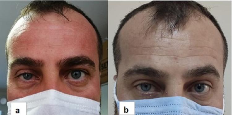
Clinical photograph comparing the ocular features of a patient with right-sided Horner's syndrome at two time points: one month (a) and six months (b) post-surgery. Figure (a) clearly demonstrates the classic triad of miosis (constricted right pupil) and partial ptosis (drooping of the right upper eyelid) when compared to the left eye. Figure (b) shows the progression after six months of recovery, where there is a subtle improvement in the right upper eyelid position (reduction in ptosis) and a decrease in the degree of anisocoria (pupil size discrepancy). The image serves as a diagnostic visual for neurological and ophthalmological assessment, illustrating the clinical manifestation of sympathetic pathway disruption and its subsequent partial resolution over time. The anatomical focus is on the periocular region and pupillary symmetry, which are key indicators for evaluating brachial plexus or cervical sympathetic chain pathologies.
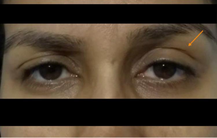
This clinical photograph displays a frontal view of a patient's eyes, highlighting diagnostic signs of Horner's syndrome. The image demonstrates a clear asymmetry between the left and right ocular regions. The left eye exhibits visible ptosis (drooping of the upper eyelid), indicated by an orange arrow, where the eyelid margin sits lower on the iris compared to the contralateral side. Furthermore, there is evidence of miosis (pupillary constriction) in the left eye, making the pupil appear significantly smaller than the right. The right eye appears normal with a baseline pupil size and expected eyelid position. Clinically, this presentation is characteristic of sympathetic nerve pathway disruption, which in this context followed the administration of neuraxial analgesia. The image serves as a high-quality educational example for identifying the ocular components of a partial Horner's syndrome in a clinical setting.
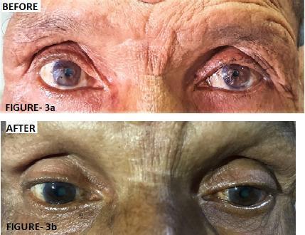
Two-panel clinical photograph displaying the 'Before' and 'After' ophthalmic findings in a patient with left-sided Horner's syndrome. The 'Before' image (Figure 3a) demonstrates classic clinical signs including left upper eyelid ptosis (drooping) and left-sided miosis (constricted pupil) compared to the right eye. The 'After' image (Figure 3b) illustrates the clinical response following three cycles of chemotherapy for an underlying superior sulcus tumor (Pancoast tumor). There is visible improvement in the left ocular presentation, characterized by the elevation of the left upper eyelid, reduction in ptosis severity, and a comparative increase in left pupil diameter (resolution of miosis). The image highlights the ocular manifestations of sympathetic nerve pathway disruption and its subsequent recovery during systemic treatment of the primary thoracic malignancy. Educational focus includes the recognition of Horner's syndrome triad (ptosis, miosis, and anhidrosis) and its clinical association with apical lung lesions.
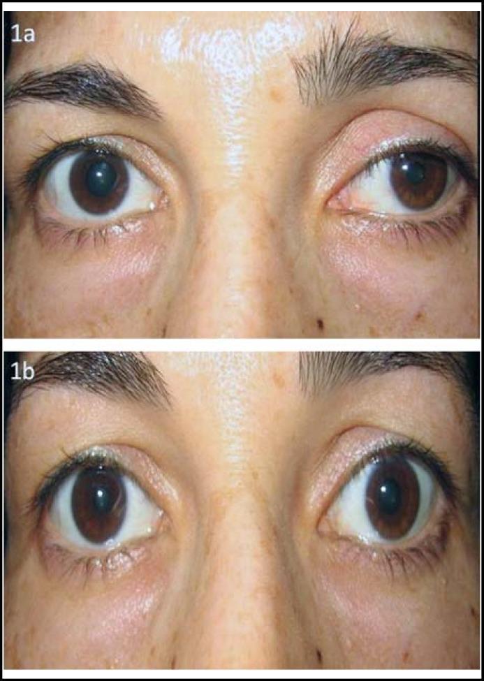
Clinical photograph showing a comparative view of a 37-year-old female patient's eyes (Figure 1a and 1b) demonstrating findings characteristic of Horner syndrome and its pharmacological testing. In Figure 1a (baseline), the patient exhibits left-sided blepharoptosis (drooping of the upper eyelid) and miosis (a smaller, constricted pupil) compared to the right eye, resulting in significant anisocoria. In Figure 1b, following the topical instillation of apraclonidine (0.5%), there is a visible reversal of these signs: the left eyelid ptosis has resolved, and the left pupil has dilated while the right pupil has slightly constricted (reversal of anisocoria). This 'apraclonidine test' result is diagnostic for Horner syndrome, reflecting the hypersensitivity of the denervated dilator muscle to alpha-adrenergic stimulation. This image is medically significant for illustrating the clinical presentation of sympathetic nerve pathway disruption and the objective results of diagnostic pupil testing in neuro-ophthalmology.
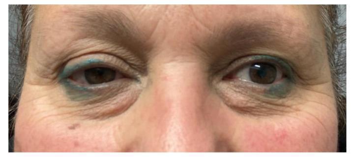
This clinical photograph shows a close-up, frontal view of a patient's periorbital region, demonstrating the classic triad of Horner syndrome on the left side. Key diagnostic features include visible ptosis (drooping) of the left upper eyelid and mild elevation of the left lower eyelid (inverse ptosis), resulting in a narrowed palpebral fissure. Comparison of the pupils reveals miosis (constriction) of the left pupil compared to the right, indicating an anisocoria that is typically more pronounced in dim light. The surrounding skin exhibits signs of aging, including rhytids (wrinkles) and mild dermatochalasis. The patient has blue cosmetic tattooing or permanent eyeliner on both the upper and lower eyelids. This presentation is characteristic of a disruption in the oculosympathetic pathway, which in this clinical context followed the surgical resection of a parapharyngeal space schwannoma involving the cervical sympathetic chain.
Horner syndrome sympathetic pathway anatomy diagram oculosympathetic
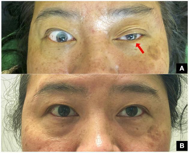
This clinical comparison photograph shows a 47-year-old woman presenting with classic oculofacial features of Horner’s syndrome following a clavicle fracture. Image A demonstrates the acute presentation of the oculosympathetic defect in the left eye, characterized by significant blepharoptosis (drooping of the upper eyelid) and ipsilateral miosis (constriction of the pupil) compared to the normal right eye. A red arrow points to the affected left eye to emphasize these findings. Image B shows the same patient at a three-month follow-up after surgical fixation of the fracture. It illustrates complete resolution of the syndrome, with symmetric eyelid positions and equal pupillary diameters (isocoria) in both eyes. This visual progression highlights the classic clinical triad (ptosis and miosis, with anhidrosis typically associated) and the potential for recovery following the management of the underlying traumatic compression of the preganglionic sympathetic pathway. The image is a valuable educational resource for neurology, ophthalmology, and trauma surgery.
Two-panel clinical photograph demonstrating Horner’s syndrome in a patient. Panel A shows the patient's eyes at rest, highlighting a mild right-sided ptosis (drooping of the upper eyelid) and a subtle 'upside-down ptosis' (elevation of the right lower eyelid). Panel B provides a close-up view of the eyes with the upper eyelids manually retracted to reveal anisocoria; the right pupil (labeled R) is significantly smaller (miotic) compared to the left pupil (labeled L). The clinical findings illustrate the classic triad of sympathetic denervation: ptosis, miosis, and enophthalmos appearance due to narrowing of the palpebral fissure. This visual evidence supports a diagnosis of Horner’s syndrome, often associated with neurological or vascular pathology affecting the oculosympathetic pathway. The image serves as an educational tool for identifying autonomic ocular dysfunction in neurology and ophthalmology.
This clinical photograph shows a close-up, frontal view of a patient's periorbital region, demonstrating the classic triad of Horner syndrome on the left side. Key diagnostic features include visible ptosis (drooping) of the left upper eyelid and mild elevation of the left lower eyelid (inverse ptosis), resulting in a narrowed palpebral fissure. Comparison of the pupils reveals miosis (constriction) of the left pupil compared to the right, indicating an anisocoria that is typically more pronounced in dim light. The surrounding skin exhibits signs of aging, including rhytids (wrinkles) and mild dermatochalasis. The patient has blue cosmetic tattooing or permanent eyeliner on both the upper and lower eyelids. This presentation is characteristic of a disruption in the oculosympathetic pathway, which in this clinical context followed the surgical resection of a parapharyngeal space schwannoma involving the cervical sympathetic chain.
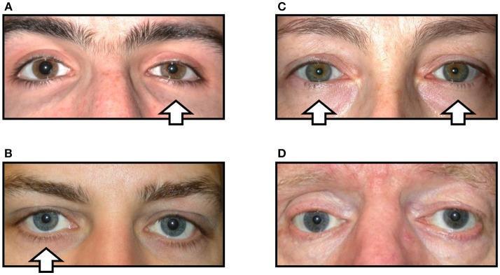
A clinical comparison chart consisting of four color photographs (A-D) demonstrating variations in oculosympathetic paresis (Horner syndrome) signs. (A) Displays classic unilateral Horner syndrome of the right eye, characterized by mild upper eyelid ptosis, elevation of the lower eyelid (reverse ptosis), miosis (pupillary constriction), and mild conjunctival injection. (B) Shows 'incomplete' Horner syndrome with right-sided miosis but absence of visible ptosis. (C) Illustrates bilateral Horner syndrome, where symmetry in pupil size and lid position masks the pathology due to lack of a normal comparative side. (D) Demonstrates 'Pseudo-Horner syndrome' in the right eye; lid asymmetry, enophthalmos, and hypoglobus are present due to a prior orbital floor fracture rather than sympathetic denervation, accompanied by physiological anisocoria. White arrows indicate the affected side(s) in each case. This comparison teaches the importance of distinguishing true sympathetic paresis from structural orbital pathology and identifying partial or bilateral presentations.
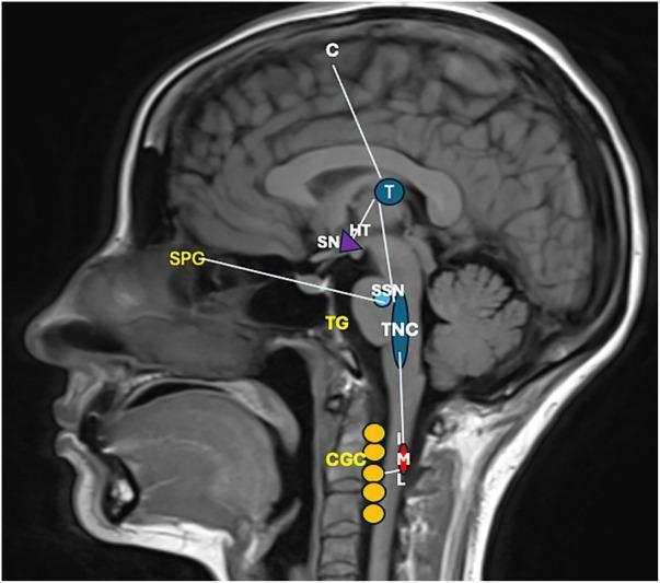
This diagnostic image consists of a midsagittal T1-weighted MRI scan of the human head and neck, overlaid with a schematic pathophysiology diagram illustrating the neuroanatomical pathways of cluster headache (CH). The diagram maps three primary systems: the trigeminovascular system, the autonomic reflex, and the hypothalamic system. Key labeled structures include the Primary Sensory Cortex (C), Thalamus (T), Hypothalamus (HT), and Suprachiasmatic Nucleus (SN). In the brainstem and peripheral regions, the diagram highlights the Superior Salivatory Nucleus (SSN), Trigeminal Caudal Nucleus (TNC), Trigeminal Ganglion (TG), and Sphenopalatine Ganglion (SPG). Descending pathways are shown reaching the Intermediolateral Nucleus (IML) of the spinal cord and the Cervical Ganglionic Complex (CGC). Functional connections illustrate nociceptive afferents from the TG to the TNC, ascending projections to the thalamus and cortex for pain perception, and hypothalamic activation of autonomic centers. This educational visual explains the clinical manifestations of CH, including trigeminal distribution pain, cranial autonomic symptoms like lacrimation via the SPG, and sympathetic dysfunction leading to Horner's syndrome via the IML-CGC pathway.
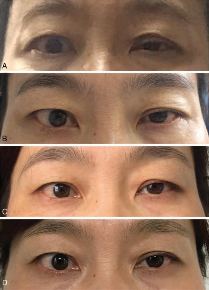
A series of four clinical photographs (A-D) demonstrating the longitudinal progression and recovery of left-sided Horner syndrome in a female patient over a one-year postoperative period. Image A (2 days postoperative) shows the classic triad of ptosis (drooping of the upper eyelid), miosis (constricted pupil), and apparent enophthalmos (sunken eye) on the left side compared to the normal right eye. Image B (one month) and Image C (six months) illustrate a gradual recovery of eyelid elevation and pupillary dilation. Image D (one year) displays a near-complete resolution of symptoms with restoration of ocular symmetry, though a slight residual ptosis may remain. The images serve as an educational timeline for the spontaneous or medically managed recovery of iatrogenic Horner syndrome following neck surgery, such as thyroidectomy. The progression highlights the clinical features of sympathetic pathway disruption and the objective markers used to track neurologic recovery of the oculosympathetic fibers.
Horner's Syndrome (Oculosympathetic Palsy)
Definition
| Sign | Mechanism |
|---|---|
| Ptosis (upper lid drooping) | Loss of innervation to Müller's smooth muscle in the upper eyelid |
| Miosis (small pupil) | Loss of sympathetic innervation to the pupillary dilator muscle |
| Anhidrosis (decreased sweating) | Loss of sympathetic innervation to facial sweat glands |
- Inverse ptosis - slight elevation of the lower eyelid
- Apparent enophthalmos - sunken-eye appearance due to narrowing of the palpebral fissure (not true enophthalmos)
- Redness of the conjunctiva (conjunctival injection)
- Heterochromia iridis in congenital cases (the affected iris is lighter)
- Dilation lag - the miotic pupil dilates more slowly than the normal side when lights are dimmed
The Three-Neuron Sympathetic Pathway
1st-order (Central) Neuron
- Runs from the hypothalamus down through the brainstem and lateral spinal cord to the ciliospinal center of Budge (C8-T2)
- Causes: lateral medullary (Wallenberg) infarct, brainstem hemorrhage, cervical cord trauma
2nd-order (Preganglionic) Neuron
- Exits the cord, loops over the apex of the lung and around the subclavian artery, and synapses at the superior cervical ganglion
- Causes: Pancoast tumor (apical lung), cervical rib, thyroid mass, neck trauma, aortic aneurysm
3rd-order (Postganglionic) Neuron
- Runs along the internal carotid artery, through the cavernous sinus, into the orbit
- Causes: carotid artery dissection, cavernous sinus thrombosis/tumor, cluster headache, orbital disease
Causes Summary
- Lateral hypothalamus or brainstem (e.g., infarct or hemorrhage)
- Spinal cord trauma
- First/second thoracic roots - apical lung tumor (Pancoast syndrome), disc herniation
- Sympathetic chain - tumor or trauma
- Carotid plexus - carotid dissection (important, do not miss)
- Cavernous sinus - thrombosis, aneurysm, neoplasm
- Orbit - infection or neoplasm
- Congenital - perinatal injury to the sympathetic chain
Sweating Patterns for Localization
- Lesion at common carotid level - loss of sweating over the entire ipsilateral face
- Lesion distal to carotid bifurcation - anhidrosis absent, or confined only to the medial forehead and side of nose
Diagnosis & Pharmacological Testing
- Apraclonidine is a weak alpha-1 agonist; normally it causes minimal dilation
- In Horner's, denervation hypersensitivity causes the miotic pupil to dilate (and the normal side may constrict)
- A reversal of anisocoria is diagnostic - the affected pupil becomes larger than the unaffected one
- One drop (0.5%) in each eye; check at 30-45 minutes
- Cocaine prevents norepinephrine reuptake, dilating a normal pupil
- In Horner's, the affected pupil does NOT dilate
- A difference of ≥0.8 mm is diagnostic
- Releases stored norepinephrine from intact nerve terminals
- Pupil dilates if 1st or 2nd order lesion (3rd-order neuron intact)
- No dilation if 3rd-order neuron is damaged
Imaging
- MRI of brain, neck, and chest to localize the lesion
- MRA/CTA of the carotid arteries if dissection is suspected (a medical emergency)
- Chest imaging to rule out Pancoast tumor
Clinical Photographs
Key Clinical Pearls
- Carotid dissection must be ruled out in any new-onset Horner's - it can present with only ipsilateral neck pain + Horner's, and carries risk of stroke
- Horner's in an infant should prompt urgent evaluation for neuroblastoma
- Bilateral Horner's is rare - seen in autonomic neuropathies and high cervical cord transection
- The ciliospinal reflex (painful pinch to neck causes pupil dilation on the normal side but not the Horner side) can be useful at bedside