
Open fracture management
Reading File
Searching sources...
Finding Sources
Finding Sources
Searching PubMed
"open fracture" management
Good results. Now let me read the most authoritative sources in parallel - Rockwood & Green (the gold standard orthopaedic trauma text), Campbell's Operative Orthopaedics, and Miller's Review.
Reading File
Reading File
Reading File
Good. Let me now read the critical sections on wound management, debridement, timing of fixation, and soft tissue coverage from Rockwood & Green and Campbell's:
Reading File
Reading File
Reading File
Excellent content from Rockwood & Green. Let me read more of the wound excision section and Campbell's open fractures section:
Reading File
Reading File
Now let me get the critical section from Rockwood & Green on wound closure, fixation, and irrigation:
Reading File
Reading File
I now have comprehensive material. Let me fetch the degloving image and read a bit more from Rockwood & Green on wound closure and fixation:
Reading File
I now have a comprehensive dataset. Let me compile the full answer.
Open Fracture Management
An open fracture (compound fracture) is one in which the fracture communicates with the external environment through a skin wound. The priorities of management are infection prevention, skeletal stabilization, and soft tissue reconstruction.
1. Classification
Gustilo-Anderson Classification (most widely used clinically)
| Type | Description |
|---|---|
| I | Clean wound <1 cm; minimal soft tissue damage; no periosteal stripping |
| II | Wound 1-10 cm; moderate muscle damage; little periosteal stripping |
| IIIA | Wound >10 cm OR high-energy mechanism (gunshot, farm injury, shotgun); extensive periosteal stripping but adequate soft tissue coverage |
| IIIB | As IIIA but requires flap coverage; extensive periosteal stripping |
| IIIC | As IIIA/IIIB but with vascular injury requiring repair |
Important: There is considerable interobserver variability; the type may change after further debridement. Energy of injury matters more than absolute wound length.
OTA/AO Open Fracture Classification (OTA-OFC) - newer, more predictive
Developed to address shortcomings of Gustilo-Anderson (which was designed only for open tibia fractures and uses treatment to determine classification). Assesses five categories, each graded 1-3:
| Category | Grade 1 | Grade 2 | Grade 3 |
|---|---|---|---|
| Skin (S) | Can be approximated | Cannot be approximated | Extensive degloving |
| Muscle (M) | No necrosis, intact function | Functional muscle loss, intact muscle-tendon unit | Dead muscle, loss of function, compartment excision |
| Arterial (A) | No injury | Vessel injury, no ischemia | Vessel injury with distal ischemia |
| Contamination (C) | None/minimal | Surface contamination | Embedded in bone/deep tissue or high-risk environment (barnyard, fecal) |
| Bone loss (B) | None | Missing/devascularized but some contact | Segmental bone loss |
A cumulative OTA-OFC score ≥10 correlates with the need for amputation. Studies show the OTA-OFC is superior to Gustilo-Anderson in predicting infection, need for soft tissue coverage, amputation, and number of debridements.
- Rockwood and Green's Fractures in Adults, 10th ed, p.154
- Campbell's Operative Orthopaedics 15th Ed, p.8063
- Miller's Review of Orthopaedics 9th Ed, p.813
2. Initial Emergency Management
Field/ED Care
- Wound irrigation and sterile dressing
- Splinting/immobilization to reduce ongoing soft tissue damage
- Neurovascular assessment (document distal pulses, sensation, motor function)
- Photography before dressing application
Antibiotics (single most effective infection-prevention measure)
Administer within 3 hours of injury - this is the most effective way to decrease infection risk.
| Fracture Type | Antibiotic |
|---|---|
| Types I & II | First-generation cephalosporin (cefazolin 1 g IV q8h) |
| Type III | Cephalosporin + aminoglycoside (gentamicin weight-adjusted or levofloxacin 500 mg q24h) |
| Farm/heavily contaminated | Cephalosporin + aminoglycoside + high-dose penicillin |
| Freshwater wounds | Fluoroquinolone or 3rd/4th gen cephalosporin (ceftazidime) |
| Saltwater wounds | Doxycycline + ceftazidime or fluoroquinolone |
Duration:
- Continue 24 hours if wound is primarily closed
- Continue 24 hours after final closure if not closed at initial debridement
2022 Surgical Infection Society Guidelines: For Type I/II, recommend gram-positive coverage only (avoid broad-spectrum). For Type III, also recommend against extended coverage beyond gram-positive. For Type III with bone loss, use local + systemic antibiotics.
Note: Fluoroquinolones should NOT be used for prophylaxis due to adverse effects on bone healing.
- Miller's Review of Orthopaedics 9th Ed, p.813
- Campbell's Operative Orthopaedics 15th Ed, Box 58.6
Tetanus Prophylaxis
- Tetanus-prone wounds: >6 hours old, >1 cm deep, devitalized tissue, or grossly contaminated
- Unknown immunization status or <3 prior immunizations + tetanus-prone wound: toxoid + human tetanus immunoglobulin (at different sites)
- Fully immunized: tetanus toxoid if wound is severe, >24 hours old, or no booster in 5 years
3. Surgical Wound Management
Timing of Debridement - the "6-hour rule" revisited
The historic "6-hour rule" originated from an 1898 guinea pig study by Friedrich and has been widely challenged. Current evidence shows no significant difference in infection rate between debridement <6 hours vs. 6-24 hours, as long as systemic antibiotics are given promptly.
Serial wound debridement should be avoided. Definitive soft tissue coverage should not be delayed beyond 72 hours if possible, and early definitive coverage is preferred.
- Rockwood and Green's Fractures in Adults, 10th ed, p.632
Wound Excision (Debridement) Principles
- "Debridement" = wound extension to expose injured tissue; "excision" = removal of devitalized material
- An orthoplastic approach (orthopaedic + plastic surgery together from the start) is recommended for all significant open fractures
- Key goals:
- Reduce infective burden by removing contaminated debris
- Excise devitalized tissue that acts as a nidus for biofilm formation
- Minimize dead space and hematoma
Systematic excision by tissue layer:
- Skin: Excise only nonviable edges (confirmed by absence of bleeding). Retain viable skin flaps. Fascia that is detached, shredded, or nonviable must be excised.
- Subcutaneous fat: Excise areas with thrombosis or loss of integrity.
- Muscle: Assessed by the 4 Cs - Color, Contractility, Consistency, Capacity to bleed
- Normal muscle: bright red, contracts on stimulation, firm, bleeds when cut
- Nonviable muscle: dark/gray, no contraction, mushy, does not bleed
- Bone: Devascularized cortical fragments (those without soft tissue attachments) should be removed; however, large segments attached to periosteum may be retained if soft tissue reconstruction is planned
Wound Irrigation
- Lavage with normal saline reduces bacterial load
- Studies (FLOW trial) found low-pressure irrigation with saline is as effective as high-pressure with additives
- Volume: typically 3-6 L for Type I-II, 6+ L for Type III
Wound Closure Options
- Primary closure: Only if wound is clean, <6 hours, Type I with minimal contamination
- Delayed primary closure: Most Type II/IIIA wounds - close at 48-72 hours
- Split skin graft: For clean granulating wounds without exposed bone
- Flap coverage: Required when bone, tendon, or hardware is exposed
Coverage timeline by tibial location (the "rule of thirds"):
| Level | Flap of choice |
|---|---|
| Proximal third | Gastrocnemius flap |
| Middle third | Soleus flap |
| Distal third | Fasciocutaneous flap or free tissue transfer |
Definitive coverage goal: <7 days. After 7 days, infection risk increases significantly.
Negative-pressure wound therapy (NPWT/VAC) is useful as a bridge but is not a substitute for definitive coverage.
- Miller's Review of Orthopaedics 9th Ed, p.895
4. Skeletal Stabilization
The choice of fixation depends on the ability to achieve wound closure:
| Scenario | Fixation |
|---|---|
| Wound can be closed, minimal contamination | Proceed to definitive internal fixation + simultaneous soft tissue closure |
| Wound cannot be closed | Temporizing spanning external fixator |
| Polytrauma/damage control | External fixation as "bridge" before conversion to IM nail |
Key principle: If definitive internal fixation is used, definitive soft tissue coverage must be achieved at the same time. Internal fixation + delayed soft tissue coverage = higher infection rates.
External Fixator Tips
- Place pins through intact skin, not through the wound
- Predrill pins to avoid thermal necrosis
- Ensure pin placement does not compromise later soft tissue reconstruction or internal fixation conversion
- Plan the frame to allow access for subsequent flap coverage
Common Definitive Fixation Methods
-
IM nail: Gold standard for diaphyseal tibial fractures (including open)
-
Plate fixation: Periarticular fractures; anatomic reduction required
-
External fixation: Definitive in some pelvic/foot injuries, or as bridge
-
Rockwood and Green's Fractures in Adults, 10th ed, p.638+
5. Vascular Injury (Type IIIC)
- Warm ischemia time is critical - revascularization should occur within 6 hours
- Temporary vascular shunting can maintain perfusion while fracture stabilization proceeds
- Sequence: skeletal stabilization first, then vascular repair (to avoid disrupting anastomosis during manipulation) - OR vascular shunt, then skeletal fixation, then definitive vascular repair
- Fasciotomy is mandatory after revascularization (prophylactic or therapeutic for compartment syndrome)
6. Special Considerations
Compartment Syndrome
- May complicate any high-energy open fracture
- An open wound does NOT exclude compartment syndrome (only decompresses the compartment directly under the wound)
- Four compartments of the leg must be released: anterior, lateral, superficial posterior, deep posterior
Local Antibiotic Delivery
-
PMMA antibiotic beads (tobramycin/vancomycin) can augment systemic antibiotics in contaminated wounds
-
Meta-analysis (2,738 patients): local antibiotic prophylaxis reduced fracture-related infection risk by 11.9%
-
Topical vancomycin powder at closure reduces gram-positive SSI rates in tibial plateau/pilon fractures (METRC trial)
-
Rockwood and Green's Fractures in Adults, 10th ed, p.1752
Amputation vs. Limb Salvage
- IIIC injuries with combined bone, nerve, and vascular injury have high amputation rates
- MESS (Mangled Extremity Severity Score), NISSSA, HFS-97 scoring systems help guide decision making
- OTA-OFC cumulative score ≥10 best correlates with need for amputation
7. Summary Algorithm
Open Fracture
    |
    ├── ABCs, polytrauma assessment
    ├── IV antibiotics within 3 hours
    ├── Tetanus prophylaxis
    ├── Wound dressing + splint in ED
    |
    └── SURGERY (ideally same day, not necessarily <6h)
            |
            ├── Wound extension + systematic excision (orthoplastic)
            ├── Irrigation (saline lavage)
            ├── Skeletal stabilization
            |       ├── Closure possible → definitive fixation + closure
            |       └── Closure not possible → external fixator + planned return
            |
            └── Soft tissue coverage
                    ├── Primary / delayed primary
                    ├── SSG (no exposed bone)
                    └── Flap (exposed bone/tendon/hardware)
                    Goal: <7 days definitive coverage
Key References:
- Rockwood and Green's Fractures in Adults, 10th ed (2025) - Classification, debridement principles, skeletal stabilization
- Campbell's Operative Orthopaedics 15th Ed (2026) - Antibiotic protocols, OTA classification
- Miller's Review of Orthopaedics 9th Ed - Summary tables, coverage options
Neck of femur and intertrochanteric fracture
Searching sources...
Finding Sources
Finding Sources
Searching PubMed
"hip fracture" management
Let me search more specifically in the orthopaedic textbooks:
Finding Sources
Finding Sources
Finding Sources
Excellent - found the key sources. Now let me read the main sections in parallel:
Reading File
Reading File
Reading File
Excellent content. Now let me read the management/treatment sections from Campbell's and Rockwood & Green, plus intertrochanteric fracture content:
Reading File
Reading File
Reading File
Now let me read the intertrochanteric fracture content and the treatment decision-making section:
Reading File
Reading File
Reading File
Now let me get the intertrochanteric fracture details and Campbell's treatment algorithm:
Reading File
Reading File
Let me get the intertrochanteric fracture classification and management details:
Finding Sources
Finding Sources
Now let me read the intertrochanteric fracture classification and treatment section from Campbell's block 32:
Reading File
Reading File
Let me get the intertrochanteric classification section from Campbell's:
Reading File
Reading File
Reading File
Let me now also fetch a key image and read the complications + avascular necrosis section:
Reading File
Loading Image
I now have comprehensive material across all key areas. Let me compile the full detailed answer.
Neck of Femur (NOF) & Intertrochanteric Fractures
These are the two major categories of proximal femur fractures, distinguished by their anatomical location relative to the hip capsule. This distinction is clinically fundamental because it determines blood supply risk, surgical strategy, and prognosis.
Anatomical Overview & Key Distinction
| Feature | Neck of Femur (NOF) | Intertrochanteric |
|---|---|---|
| Location | Intracapsular | Extracapsular |
| Blood supply risk | HIGH - AVN is a major complication | Low - blood supply preserved |
| Typical treatment | Arthroplasty (displaced) or fixation | Internal fixation (DHS or IM nail) |
| Fracture healing | Prone to non-union | Generally heals well |
| Age group | Elderly (low energy) or young (high energy) | Predominantly elderly |
PART 1: Neck of Femur Fracture (Intracapsular)
Blood Supply - The Core Problem
The femoral head receives its blood supply primarily from the medial circumflex femoral artery (MCFA), with contributions from the inferior retinacular artery, the lateral circumflex femoral artery, and the artery of the ligamentum teres (minimal contribution in adults). These vessels run along the femoral neck within the capsule - making them vulnerable to disruption at the time of fracture.
Consequence: Displaced NOF fractures interrupt this blood supply, causing avascular necrosis (AVN) of the femoral head in 15-30% of cases and non-union in up to 30%.
- Campbell's Operative Orthopaedics 15th Ed, p.3432
Classification Systems
1. By Anatomical Location (AO/OTA)
- Subcapital - just below the femoral head (most common; highest AVN risk)
- Transcervical - through the mid-neck
- Basicervical - at the base of the neck (functionally behaves more like intertrochanteric)
2. Garden Classification (most widely used clinically)
Based on the degree of displacement on AP radiograph, using trabecular alignment:
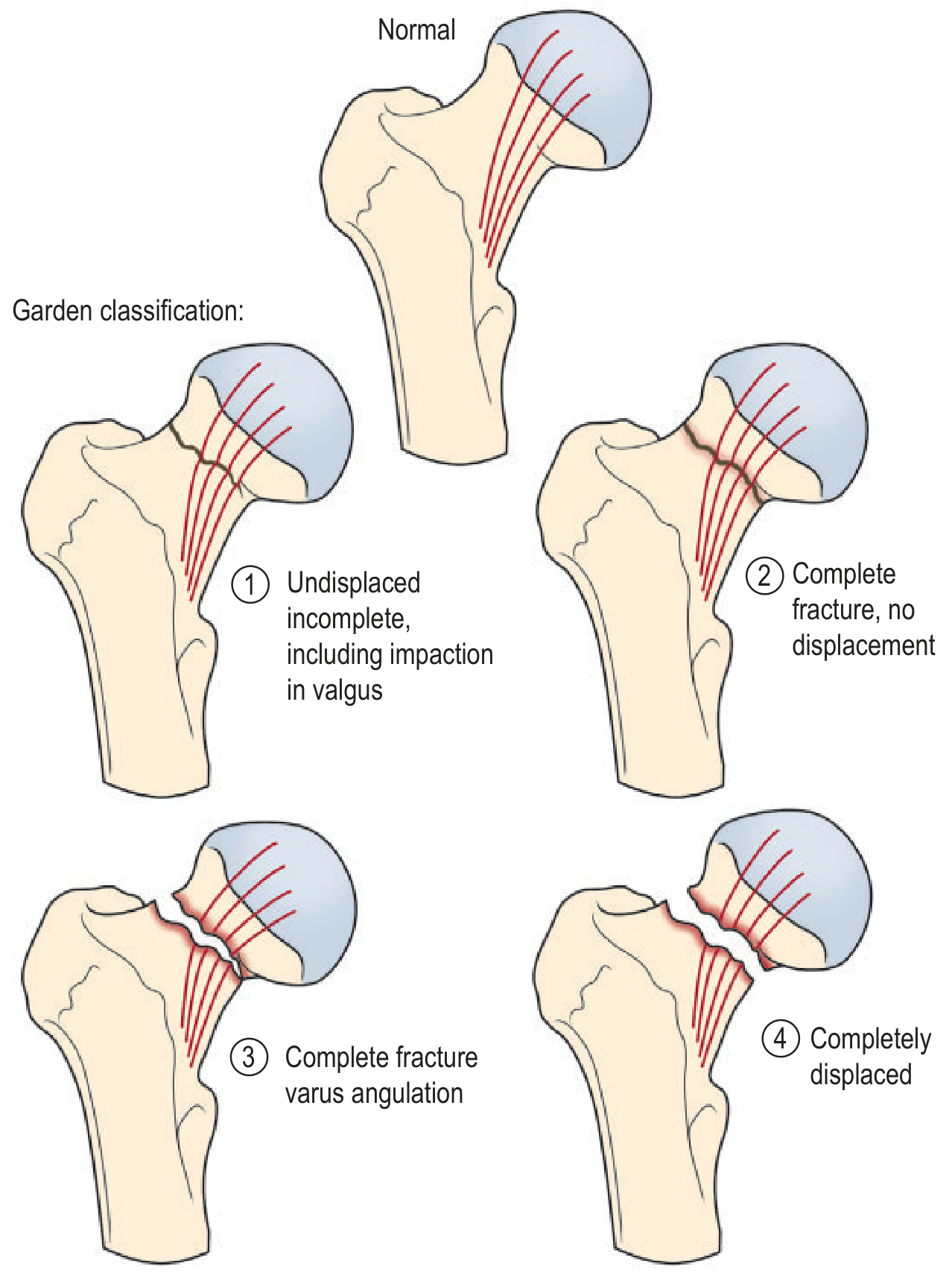
| Stage | Description | Union rate |
|---|---|---|
| Garden I | Incomplete fracture - valgus impacted; trabeculae angulated but continuous | ~100% |
| Garden II | Complete fracture; non-displaced; trabeculae still aligned | ~100% |
| Garden III | Complete fracture; partially displaced; trabeculae misaligned but head-neck contact maintained | ~93% |
| Garden IV | Complete fracture; fully displaced; trabeculae between head and acetabulum re-align | ~57% |
Practical simplification: Clinically, most surgeons reliably differentiate only undisplaced (I+II) vs displaced (III+IV) - interobserver reliability for all 4 stages is poor (complete agreement in only 22% of cases). Garden I/II with ≥20 degrees of posterior (sagittal) tilt should NOT be treated as undisplaced.
- Rockwood & Green's Fractures in Adults, 10th ed, p.2638
- Campbell's Operative Orthopaedics 15th Ed, p.3432
3. Pauwels Classification
Based on the angle of the fracture line relative to horizontal - describes shear forces at the fracture site:
| Type | Angle to horizontal | Dominant force | Stability |
|---|---|---|---|
| Type I | 0-30° | Compressive | Stable |
| Type II | 30-50° | Mixed | Moderately unstable |
| Type III | >50° | Shear | Unstable - high fixation failure risk |
Type III Pauwels fractures have high shear forces and require augmented fixation strategies.
- Campbell's Operative Orthopaedics 15th Ed, p.3433
4. Posterior Tilt Classification
More recently, posterior tilt angle on lateral radiograph ≥20 degrees has been identified as a major risk factor for treatment failure:
-
Posterior tilt ≥20°: 24.4% treatment failure rate
-
Posterior tilt <20°: 10.9% treatment failure rate
-
Risk ratio 2.73 (95% CI 1.77-4.21)
-
Rockwood & Green's Fractures in Adults, 10th ed, p.2640
Diagnosis
- Clinical presentation: Shortened, externally rotated leg; inability to weight-bear
- Radiographs: AP pelvis + cross-table lateral; traction internal rotation view is often helpful
- If plain X-ray negative but clinical suspicion high: MRI is the investigation of choice (most sensitive and specific); CT if MRI contraindicated
- ~1% of fractures are initially occult on X-ray; if patient cannot weight-bear at 24h, further imaging is mandatory
Preoperative Traction
No longer routinely recommended - arteriographic studies show traction + internal rotation reduces MCFA perfusion and venous drainage. Nine RCTs showed no benefit in pain control or quality of reduction.
Management
Timing of Surgery
Surgery should not be delayed. Studies show:
- 44.9% 30-day mortality for NOF treated nonoperatively vs. 2.0% operative
- 1-year mortality: 65.3% nonoperative vs. 17.6% operative
- Even nondisplaced fractures treated nonoperatively: 38.7% 30-day mortality
Operative treatment is effectively mandatory except in extreme circumstances.
The Core Treatment Decision Tree
NOF Fracture
    |
    ├── UNDISPLACED (Garden I/II, posterior tilt <20°)
    │       └── Internal fixation
    │               ├── Cannulated screws (3 screws, inverted triangle)
    │               └── Sliding Hip Screw (DHS) - for basicervical/valgus impacted
    │
    └── DISPLACED (Garden III/IV)
            ├── YOUNG PATIENT (<65 or physiologically fit)
            │       └── Urgent reduction + internal fixation
            │               (emergent within 6-12h to preserve head)
            │
            └── ELDERLY PATIENT (physiologically older)
                    ├── Not independently ambulant / cognitively impaired
                    │       └── HEMIARTHROPLASTY (cemented)
                    │
                    └── Independently ambulant, cognitively intact
                            └── TOTAL HIP ARTHROPLASTY (THA)
                                    (NICE guidelines)
Internal Fixation (for undisplaced and young displaced)
Cannulated Screws (most common for NOF)
- 3 partially threaded screws (6.5, 7.0, or 7.3 mm) in inverted triangle configuration
- Inferior screw along calcar (most important for stability)
- Aim for center-center screw position in femoral head
- Garden Alignment Index: trabeculae should measure 160-180° on AP; deviation >20° on lateral indicates malreduction
Sliding Hip Screw (DHS)
- Better for basicervical fractures and valgus-impacted fractures
- Basicervical fractures are NOT well treated with cannulated screws (high failure rate)
- Provides dynamic compression along the femoral neck axis
Arthroplasty (for displaced NOF in elderly)
Hemiarthroplasty (HA)
- Standard treatment for most elderly patients with displaced NOF
- Shorter operative time, less blood loss than THA
- Implant types: Unipolar vs Bipolar
- Unipolar: 20% wear rate at 12 months vs. 5% bipolar; however no significant long-term difference
- Acetabular erosion rate: 5.3% conversion to THA for patients <75 vs. 1.4% for patients >75
- Cemented vs. uncemented: Cemented preferred - periprosthetic fracture rate 0.5% cemented vs. 2.1% uncemented; less pain and better mobility
- Risk: Bone Cement Implantation Syndrome (fat embolism) - use second-generation cementing technique
Total Hip Arthroplasty (THA)
-
NICE guidelines: recommended for patients who are cognitively intact AND independently ambulant (walking with at most one stick) AND medically fit
-
Better functional outcomes and quality of life than HA in selected patients
-
Higher dislocation risk than HA (RR 2.02; 95% CI 1.26-3.25) - use anterior/anterolateral approach to minimize this
-
THA with Dual Mobility Components (THA-DMC): dislocation rate 1.5% (lower than standard THA)
-
Head size: increasing from 28 mm to 40 mm reduces dislocation from 2.0% to 0.1%
-
Osteosynthesis/Hemiarthroplasty/THA textbook, p.3-8
-
Campbell's Operative Orthopaedics 15th Ed, p.3434-3448
Complications of NOF Fracture
| Complication | Frequency | Comment |
|---|---|---|
| Avascular necrosis (AVN) | 15-30% displaced | Presents 6-24 months post-injury |
| Non-union | 10-30% displaced | Higher with varus malreduction |
| Implant cut-out | 5-10% | TAD >25 mm exponentially increases risk |
| Periprosthetic fracture | 0.5-2.1% | Higher with uncemented stems |
| Dislocation (post-THA) | ~2-4% | Higher posterior approach |
| DVT/PE | Common | Thromboprophylaxis mandatory |
| Mortality (1-year) | 20-30% | Mostly due to pre-existing comorbidities |
PART 2: Intertrochanteric Fractures (Extracapsular)
Key Features
- Fracture line runs between the greater and lesser trochanters, outside the hip capsule
- Blood supply to femoral head is NOT at risk - AVN is extremely rare
- Prognosis for healing is generally good
- Treatment is always internal fixation (arthroplasty reserved for failed fixation or highly comminuted in frail patients)
Classification
AO/OTA Classification (31-A) - most commonly used
| Group | Description | Stability |
|---|---|---|
| A1 | Simple two-part fracture along intertrochanteric line | Stable |
| A2 | Comminuted; fracture extends over two or more levels of the medial cortex | Unstable |
| A3 | Reverse oblique pattern; fracture line extends through the lateral cortex distal to the vastus ridge | Very unstable |
Boyd-Griffin Classification (historical)
- Type 1: Fracture along intertrochanteric line (stable)
- Type 2: Comminuted; main fracture along intertrochanteric line
- Type 3: Subtrochanteric extension
- Type 4: Fracture in trochanteric and subtrochanteric regions with at least two fracture planes
Evans Classification
Classifies intertrochanteric fractures as stable vs. unstable based on whether the posteromedial cortex (lesser trochanter) is intact:
- Stable: Posteromedial cortex intact; can be reduced to restore medial cortical support
- Unstable: Posteromedial comminution; medial cortex cannot be restored; higher failure rates
Management
General Principle
All intertrochanteric fractures should be treated surgically (internal fixation) - nonoperative management in the elderly carries prohibitive mortality.
Fixation Choice: DHS vs. IM Nail
| Feature | Dynamic Hip Screw (DHS) | Cephalomedullary IM Nail |
|---|---|---|
| Best for | Stable (A1, many A2) | Unstable (A3, A2 with lateral wall compromise) |
| Biomechanics | Longer lever arm, more stress on bone | Shorter lever arm, load-sharing |
| Blood loss | More | Less |
| Incision | Larger | Smaller |
| Cost | Lower | Higher |
| Evidence | Cochrane meta-analysis: similar functional outcomes to IM nail in stable fractures | Preferred for unstable/reverse oblique patterns |
Tip-Apex Distance (TAD) - The most important predictor of fixation failure (cut-out):
- TAD = distance from lag screw tip to femoral head apex on AP + same on lateral view
- Target TAD: <25 mm (ideally <20 mm)
- Risk of cut-out increases exponentially when TAD >25 mm
Lateral Wall Integrity:
- Lateral wall thickness <21 mm predicts lateral wall fracture with 95% sensitivity
- Compromised lateral wall → DHS may cause iatrogenic fracture → use IM nail instead
- Preoperative CT may be indicated to assess lateral wall in borderline A2 fractures
Special Pattern: Reverse Oblique & Transverse (A3)
- DHS is contraindicated - the fracture line runs parallel/perpendicular to the screw, allowing medialization of the shaft
- Must be treated with IM nail (95° blade plate or 95° condylar screw are alternatives)
DHS Technique Summary
- Patient supine on fracture table, closed reduction
- Guide wire placed in center of femoral head (center-center position on AP and lateral)
- Lag screw at 130-135° along femoral neck axis
- Sliding compression mechanism allows dynamic collapse and union
- 2-hole vs. 4-hole side plate: 4-hole preferred for most cases
- Weight-bearing as tolerated postoperatively (for stable fractures)
IM Nail Technique Summary
- Fracture table, closed reduction
- Entry point at piriformis fossa or greater trochanter tip (trochanteric entry preferred with modern nails)
- 10 mm diameter nail typically used; 130° nail angle
- Lag screw placed center-center in femoral head; TAD confirmed fluoroscopically
- Nail should not perforate anterior cortex (respect the anterior bow)
- Campbell's Operative Orthopaedics 15th Ed, p.3448-3456
Special Situations
Atypical Femoral Fractures (Bisphosphonate-associated)
- Long-term bisphosphonate use causes subtrochanteric atypical fractures
- Typically transverse, originating at the lateral cortex (tension side)
- Associated with lateral cortical beaking/thickening; often bilateral
- Management: IM nail; stop bisphosphonates; teriparatide may aid healing
Occult Fractures
- ~1% of hip fractures are occult on plain radiograph
- If patient cannot weight-bear at 24h: MRI (investigation of choice)
- A significant proportion of isolated "greater trochanter fractures" on X-ray have intertrochanteric extension on MRI
Lesser Trochanter Avulsion in Adults
- Isolated lesser trochanter fracture in an adult without significant trauma = assume pathological until proven otherwise (metastatic disease, myeloma)
Perioperative Care
| Element | Recommendation |
|---|---|
| Timing | Within 48 hours of admission (ideal: within 24h) |
| Anaesthesia | Regional (spinal) preferred; reduces blood loss and DVT |
| Thromboprophylaxis | LMWH + TED stockings; extend to 28-35 days post-discharge |
| Osteoporosis workup | DEXA + anti-osteoporotic treatment (bisphosphonates) for patients <75 |
| Orthogeriatric co-management | Reduces mortality and length of stay |
| Rehabilitation | Full weight-bearing postoperatively encouraged; early physiotherapy |
Summary Comparison
PROXIMAL FEMUR FRACTURES
        |
        ├── INTRACAPSULAR (Neck of Femur)
        │       ├── Undisplaced (Garden I/II)
        │       │       └── Internal fixation
        │       │               (cannulated screws / DHS)
        │       └── Displaced (Garden III/IV)
        │               ├── Young patient → urgent ORIF
        │               └── Elderly
        │                       ├── Active, fit → THA
        │                       └── Less active → Hemiarthroplasty
        │
        └── EXTRACAPSULAR (Intertrochanteric, AO 31-A)
                ├── Stable (A1) → DHS
                ├── Unstable (A2) → DHS or IM nail (depends on lateral wall)
                └── Very unstable/reverse oblique (A3) → IM nail
Key References:
- Campbell's Operative Orthopaedics 15th Ed 2026 - Classification, fixation techniques, arthroplasty
- Rockwood and Green's Fractures in Adults, 10th ed 2025 - Classification reliability, posterior tilt, outcome data
- Grainger & Allison's Diagnostic Radiology - Radiological appearances, occult fractures
- Osteosynthesis, Hemiarthroplasty, Total Hip Arthroplasty (NOF textbook) - HA vs. THA evidence, NICE guidelines
This is a shared conversation. Sign in to Orris to start your own chat.