Here are comprehensive answers to all questions across all the papers:
KATHMANDU UNIVERSITY - BPT Semester I
Paper II: Musculoskeletal System (Upper & Lower Limb)
Complete Answers - All Papers
SECTION B - SHORT ANSWER QUESTIONS
ANATOMY
1. Intrinsic Muscles of the Hand
Thenar muscles (3):
- Abductor pollicis brevis
- Flexor pollicis brevis
- Opponens pollicis
- (Adductor pollicis - sometimes listed separately)
Hypothenar muscles (3):
- Abductor digiti minimi
- Flexor digiti minimi brevis
- Opponens digiti minimi
Lumbricals (4): Arise from flexor digitorum profundus tendons; flex MCP joints and extend IP joints
Interossei:
- 4 Dorsal interossei (DAB - Dorsal ABduct fingers)
- 3 Palmar interossei (PAD - Palmar ADduct fingers)
2. Boundaries and Contents of the Axilla
Boundaries:
- Apex: Bounded by the clavicle, 1st rib, and superior border of scapula
- Base (floor): Axillary fascia and skin
- Anterior wall: Pectoralis major, pectoralis minor, subclavius, clavipectoral fascia
- Posterior wall: Subscapularis, teres major, latissimus dorsi
- Medial wall: Serratus anterior (covering upper ribs)
- Lateral wall: Intertubercular sulcus of humerus
Contents:
- Axillary artery (1st, 2nd, 3rd parts) with its branches
- Axillary vein
- Brachial plexus (cords and branches)
- Axillary lymph nodes (anterior, posterior, lateral, central, apical)
- Long thoracic nerve (nerve of Bell)
- Intercostobrachial nerve
- Subscapular vessels
- Areolar/fatty tissue
3. Adductor Magnus - Attachment, Nerve Supply, and Actions
Origin:
- Adductor part: inferior ramus of pubis and ramus of ischium
- Hamstring part: ischial tuberosity
Insertion:
- Adductor part: medial lip of linea aspera and medial supracondylar line
- Hamstring part: adductor tubercle of medial femoral condyle
Nerve supply:
- Adductor part: Obturator nerve (L2, L3, L4)
- Hamstring part: Sciatic nerve (tibial part) (L4)
Actions:
- Adduction of thigh (primary action)
- Medial rotation of thigh (adductor part)
- Extension of thigh (hamstring part)
4. Formation of Medial Longitudinal Arch of Foot
The medial longitudinal arch is the highest and most important arch of the foot.
Bones forming it: Calcaneus → Talus → Navicular → 3 Cuneiforms → 1st, 2nd, 3rd Metatarsals
Keystone: Talus (head of talus)
Factors maintaining it:
- Bony: Shape of the bones (especially talus)
- Ligaments:
- Spring ligament (plantar calcaneonavicular) - most important, supports head of talus
- Long plantar ligament
- Short plantar (plantar calcaneocuboid) ligament
- Plantar aponeurosis (most important passive support - "tie-beam")
- Muscles:
- Tibialis posterior (main muscle support)
- Flexor digitorum longus
- Flexor hallucis longus
- Intrinsic foot muscles (short plantar muscles)
- Peroneus longus (from the lateral side)
5. Abduction of Shoulder Joint
Normal range: 0-180°
Phases:
- 0-90°: Supraspinatus initiates first 15°; deltoid (middle) is the prime mover for 15-90°
- 90-150°: Scapular rotation (serratus anterior + trapezius = force couple)
- 150-180°: Lateral flexion of spine
Muscles involved:
- Supraspinatus (initiates, 0-15°)
- Middle deltoid (15-90°)
- Serratus anterior + Trapezius (scapular rotation, 90-150°)
Important points:
- At 90°, the greater tuberosity impinges against the acromion - prevented by lateral rotation of humerus
- Supraspinatus is more of a stabilizer (prevents superior subluxation)
6. Boundaries and Contents of Popliteal Fossa
Boundaries:
- Roof (superficial): Skin, superficial fascia, popliteal fascia, small saphenous vein, sural nerve, posterior femoral cutaneous nerve
- Floor (deep): Posterior femur (popliteal surface), oblique popliteal ligament, posterior capsule, popliteus muscle
- Superolateral: Biceps femoris
- Superomedial: Semitendinosus and semimembranosus
- Inferolateral: Lateral head of gastrocnemius + plantaris
- Inferomedial: Medial head of gastrocnemius
Contents (from medial to lateral - "V.A.N" = Vein, Artery, Nerve):
- Popliteal artery (deepest)
- Popliteal vein
- Tibial nerve (most superficial)
- Common fibular (peroneal) nerve
- Small saphenous vein (entering the fossa)
- Posterior cutaneous nerve of thigh
- Lymph nodes and fat
7. Ligaments of Knee Joint (8 Ligaments)
Extracapsular:
- Patellar ligament (ligamentum patellae)
- Medial collateral ligament (tibial)
- Lateral collateral ligament (fibular)
- Oblique popliteal ligament
- Arcuate popliteal ligament
Intracapsular (but extrasynovial):
6. Anterior cruciate ligament (ACL) - prevents anterior tibial displacement
7. Posterior cruciate ligament (PCL) - prevents posterior tibial displacement
Meniscal:
8. Transverse ligament (connecting menisci anteriorly)
(Also: coronary ligaments; medial and lateral menisci are fibrocartilaginous)
8. Contents of Cubital Fossa
The cubital fossa is a triangular depression anterior to the elbow.
Boundaries:
- Medial: Pronator teres
- Lateral: Brachioradialis
- Base: Imaginary line between medial and lateral epicondyles
- Floor: Brachialis + supinator
- Roof: Deep fascia + bicipital aponeurosis + skin
Contents (medial to lateral: "M.B.R.N" = acronym aid):
- Median nerve
- Brachial artery (with its bifurcation into radial and ulnar arteries)
- Biceps tendon (brachii)
- Radial nerve (and its deep branch)
(Plus: recurrent radial artery, fat, lymphatics)
9. Adductor Compartment of Thigh - Muscles with Nerve Supply
| Muscle | Nerve Supply |
|---|
| Gracilis | Obturator nerve (L2, L3) |
| Adductor longus | Obturator nerve (L2, L3, L4) |
| Adductor brevis | Obturator nerve (L2, L3) |
| Adductor magnus | Obturator nerve (adductor part) + Sciatic nerve (hamstring part) |
| Obturator externus | Obturator nerve (L3, L4) |
| Pectineus | Femoral nerve (L2, L3); sometimes obturator nerve |
10. Movements and Muscles at Ankle Joint
Ankle (Talocrural) Joint Movements:
| Movement | Range | Muscles |
|---|
| Dorsiflexion | 0-20° | Tibialis anterior, Extensor hallucis longus, Extensor digitorum longus, Peroneus tertius |
| Plantarflexion | 0-50° | Gastrocnemius, Soleus (prime movers), Tibialis posterior, Flexor digitorum longus, Flexor hallucis longus, Peronei |
(Subtalar/intertarsal joints):
| Inversion | - | Tibialis anterior, Tibialis posterior |
| Eversion | - | Peroneus longus, Peroneus brevis, Peroneus tertius |
11. Branches of Axillary Artery
1st Part (medial to pectoralis minor):
- Superior thoracic artery
2nd Part (behind pectoralis minor):
2. Thoracoacromial artery (acromial, clavicular, deltoid, pectoral branches)
3. Lateral thoracic artery
3rd Part (lateral to pectoralis minor):
4. Anterior circumflex humeral artery
5. Posterior circumflex humeral artery
6. Subscapular artery (→ thoracodorsal + circumflex scapular)
Mnemonic: "She Loves Anatomy - Anterior Posterior Sub" (Superior, Lateral, Thoracoacromial, Anterior circumflex, Posterior circumflex, Subscapular)
12. Plantar Aponeurosis
A thick, strong band of fibrous tissue on the sole of the foot.
Attachment:
- Proximally: Medial process of calcaneal tuberosity
- Distally: Splits into 5 bands → base of proximal phalanges and deep transverse metatarsal ligament
Functions:
- Maintains medial longitudinal arch (acts as a "tie beam")
- Protects plantar structures
- Acts like a windlass mechanism during push-off (dorsiflexion of toes tightens aponeurosis, raising arch)
13. Peculiarities/Characteristics of Clavicle
- First bone to ossify in fetal life (6th week of intrauterine life, intramembranous)
- Last bone to complete ossification (25 years - medial end epiphysis)
- Only long bone that ossifies by intramembranous ossification (the sternal end has a secondary epiphysis)
- No medullary cavity (trabecular bone throughout)
- Only bone that articulates with the axial skeleton via the sternoclavicular joint
- Double curve (S-shaped): Medial 2/3 convex forward; lateral 1/3 concave forward
- Most commonly fractured bone (at junction of middle and outer 1/3)
- Subclavian vein runs in a groove on its inferior surface
14. Medial Longitudinal Arch of Foot (formation - see Q4 above for full detail)
15. Erb's Point and Clinical Relations
Erb's Point: Junction of C5 and C6 nerve roots, 2.5 cm above clavicle at the lateral border of sternocleidomastoid.
Clinical significance:
- Upper brachial plexus injuries (Erb's palsy) affect C5, C6
- Mechanism: Forcible depression of shoulder with lateral neck flexion (difficult deliveries, motorcycle accidents)
- "Waiter's tip position": Arm adducted, internally rotated, forearm pronated, wrist flexed
- Muscles paralyzed: Deltoid, supraspinatus, infraspinatus, biceps, brachioradialis, supinator
- Loss of: shoulder abduction, external rotation, elbow flexion, forearm supination
16. Branches of Brachial Plexus (Diagram)
Roots: C5, C6, C7, C8, T1
↓
Trunks: Upper (C5,C6), Middle (C7), Lower (C8,T1)
↓ (each trunk divides into anterior and posterior division)
Cords: Lateral (ant. div. upper + middle), Posterior (post. div. all 3), Medial (ant. div. lower)
↓
Terminal branches:
- Lateral cord: Musculocutaneous nerve + lateral root of median nerve
- Posterior cord: Radial nerve + Axillary nerve
- Medial cord: Ulnar nerve + medial root of median nerve
Terminal 5 branches (MARMU):
- Musculocutaneous nerve (C5,C6,C7)
- Axillary nerve (C5,C6)
- Radial nerve (C5-T1)
- Median nerve (C5-T1)
- Ulnar nerve (C8,T1)
17. Brachial Plexus Well-Labeled Diagram (Feb 2025, Q2)
(As above - the diagram should show roots → trunks → divisions → cords → branches)
18. Short Note on Axilla (KUSMS Internal 2024)
(See Q2 above for full boundaries and contents)
19. Median Nerve - Short Note
Root value: C6, C7, C8, T1 (from both lateral and medial cords)
Course in arm: Runs lateral to brachial artery, crosses to medial side at midarm; enters forearm between heads of pronator teres
Branches in hand (enters via carpal tunnel):
- Recurrent branch (motor to thenar muscles - APB, FPB, opponens pollicis)
- Palmar digital nerves to lateral 3.5 fingers (sensory)
- Motor to lateral 2 lumbricals
LOAF muscles (median nerve in hand):
- L - Lateral 2 lumbricals
- O - Opponens pollicis
- A - Abductor pollicis brevis
- F - Flexor pollicis brevis
Clinical: Carpal tunnel syndrome → thenar wasting, ape hand deformity, loss of sensation on lateral 3.5 fingers
20. Shoulder Joint - Abduction (see Q5 above)
21. Lymphatic Drainage of Mammary Gland
Groups of axillary lymph nodes (main drainage ~75%):
- Anterior (pectoral) group: Drains lateral breast
- Central group
- Apical group (final common pathway → subclavian trunk)
Medial drainage (~25%): Internal mammary (parasternal) nodes → thoracic duct or bronchomediastinal trunk
Other pathways:
- Interpectoral (Rotter's) nodes
- Posterior intercostal lymphatics
- Subdiaphragmatic: to liver (Gerota's pathway)
- Cross drainage to contralateral breast
Clinical significance: Axillary lymph node dissection in breast cancer surgery
22. 1st Carpometacarpal (CMC) Joint
- Type: Saddle joint (bicondylar/sellar)
- Bones: Trapezium + base of 1st metacarpal
- Movements: Flexion/extension, abduction/adduction, circumduction, opposition (most mobile CMC joint)
- Ligaments: Anterior oblique, posterior oblique, radial, ulnar ligaments
- Clinical: Osteoarthritis here = "Bennett's fracture" area; joint laxity leads to CMC OA (common in post-menopausal women)
23. Radial Nerve - Formation, Course, Distribution
Root value: C5, C6, C7, C8 (from posterior cord)
Course:
- Axilla: Behind axillary artery, in front of subscapularis
- Arm: Spiral groove of humerus (with profunda brachii artery)
- Lateral side of arm: Pierces lateral intermuscular septum
- Anterior to lateral epicondyle: Divides into superficial (sensory) and deep (posterior interosseous nerve)
Muscles supplied:
- In arm: Triceps, anconeus, brachioradialis, ECRL
- In forearm: ECRB, supinator, finger extensors (via PIN)
Sensory: Posterior arm, posterior forearm, anatomical snuffbox and dorsal surface of lateral 3.5 fingers (proximal to nails)
Clinical: Wrist drop (radial nerve palsy) - Saturday night palsy (compression in spiral groove)
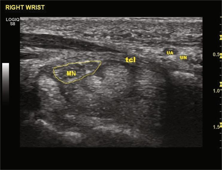
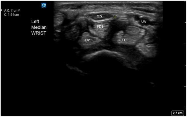
24. Formation of Carpal Tunnel
Floor and sides: Concave arch formed by carpal bones (scaphoid, trapezium laterally; triquetrum, pisiform, hamate medially)
Roof: Flexor retinaculum (transverse carpal ligament)
- Medial attachment: Pisiform and hook of hamate
- Lateral attachment: Scaphoid tubercle and trapezium
Contents (9 tendons + 1 nerve):
- Flexor digitorum superficialis tendons (×4) - superficial and middle
- Flexor digitorum profundus tendons (×4) - deep
- Flexor pollicis longus tendon (×1)
- Median nerve (most superficially placed and most vulnerable)
(Note: Flexor carpi radialis runs in its own tunnel in the flexor retinaculum; palmaris longus is outside)
25. Inguinal Ligament - Attachments, Triangle, Contents
Attachments: Anterior superior iliac spine (ASIS) → Pubic tubercle (rolled-under lower border of external oblique aponeurosis)
Femoral triangle (formed beneath inguinal ligament):
- Base: Inguinal ligament
- Medial: Medial border of adductor longus
- Lateral: Medial border of sartorius
- Floor: Iliopsoas, pectineus, adductor longus
- Roof: Fascia lata + cribriform fascia
Contents (lateral to medial: "NAVEL" from lateral to medial):
- Nerve (femoral) - lateral
- Artery (femoral)
- Vein (femoral)
- Empty space (femoral canal - medial compartment - site of femoral hernia)
- Lymphatics (in femoral canal)
26. Knee Joint - Articulating Bones, Ligaments, Movements
Articulating bones: Femur, tibia, patella (fibula does NOT articulate)
Extracapsular ligaments: See Q7 above
Intracapsular: ACL, PCL
Movements:
- Flexion (0-130°): Hamstrings, gracilis, sartorius, popliteus
- Extension (0-10° hyperextension): Quadriceps femoris
- Medial rotation (unlocking): Popliteus
- Lateral rotation (locking in full extension): ITB, biceps femoris
27. Supracondylar Ridges of Humerus - Muscular Attachments
Medial supracondylar ridge:
- Brachialis (partially)
- Coracobrachialis (partial)
- Medial head of triceps
Lateral supracondylar ridge:
- Brachioradialis
- Extensor carpi radialis longus (ECRL)
- Lateral head of triceps proximally
Volkmann's Ischaemic Contracture:
- Caused by compartment syndrome after supracondylar fracture of humerus
- Ischaemia → fibrosis of forearm flexor muscles (flexor digitorum superficialis, profundus, flexor pollicis longus)
- Results in: Forearm pronated, wrist and fingers flexed (characteristic posture)
- Brachial artery branches: Profunda brachii, superior ulnar collateral, inferior ulnar collateral, anterior ulnar recurrent, posterior ulnar recurrent, radial artery, ulnar artery
BIOCHEMISTRY
28. Uric Acid and Related Disease
Uric acid:
- End product of purine (adenine and guanine) metabolism in humans
- Formed by xanthine oxidase action on xanthine and hypoxanthine
- Normal serum level: 3.5-7.0 mg/dL (men), 2.5-6.0 mg/dL (women)
- Mostly excreted by kidneys (70%) and gut (30%)
Related disease - Gout (Hyperuricaemia):
- Deposition of monosodium urate (MSU) crystals in joints and soft tissues
- Types: Primary gout (overproduction or underexcretion) vs Secondary gout
- Acute gout: Intense inflammatory arthritis, commonly 1st MTP joint (podagra)
- Chronic tophaceous gout: Tophi in ear helix, tendons, skin
- Crystals: Needle-shaped, negatively birefringent under polarized light
- Pathogenesis: Hyperuricaemia → crystal deposition → neutrophil phagocytosis → IL-1β release → inflammation
29. Cori Cycle (Importance)
Definition: The metabolic cycle between muscle/RBC and liver involving lactate.
Steps:
- Muscle undergoes anaerobic glycolysis → Pyruvate → Lactate
- Lactate released into blood → transported to liver
- In liver: Lactate → Pyruvate (lactate dehydrogenase) → Gluconeogenesis → Glucose
- Glucose released back into blood → taken up by muscle
Importance:
- Removes lactate from muscles (prevents acidosis and fatigue)
- Recycles carbon for new glucose production
- Maintains blood glucose during exercise
- Glucose-sparing effect for the brain
- Links anaerobic (muscle) and aerobic (liver) metabolism
30. Creatine Phosphate (Phosphocreatine)
Definition: A high-energy phosphate compound in muscle and brain
Synthesis:
- Arginine + Glycine → Guanidinoacetate + Ornithine (enzyme: AGAT, in kidney)
- Guanidinoacetate + SAM → Creatine + SAH (enzyme: GAMT, in liver)
- Creatine → transported to muscle → Creatine kinase adds phosphate → Phosphocreatine
Function:
- Immediate ATP reserve (Lohmann reaction): Phosphocreatine + ADP ⇌ Creatine + ATP
- First energy source used during sudden intense exercise (first 5-10 seconds)
- Creatinine is spontaneous degradation product of creatine → excreted in urine (marker of GFR)
31. Role of PTH and Calcitriol in Calcium Homeostasis
PTH (Parathyroid Hormone):
- Secreted in response to low serum Ca²⁺
- Actions:
- Bone: Stimulates osteoclast activity → Ca²⁺ and PO₄³⁻ release
- Kidney: Increases Ca²⁺ reabsorption in DCT; decreases phosphate reabsorption; activates 1-alpha hydroxylase
- Gut: Indirect (via calcitriol synthesis)
- Net effect: ↑ serum Ca²⁺, ↓ serum PO₄³⁻
Calcitriol (1,25-dihydroxycholecalciferol / Active Vitamin D):
- Synthesized: Skin (UV) → Cholecalciferol → 25-OH in liver → 1,25(OH)₂D₃ by 1-alpha hydroxylase in kidney (PTH stimulates this step)
- Actions:
- Gut: Increases Ca²⁺ and PO₄³⁻ absorption (main action)
- Bone: Works with PTH on osteoclasts
- Kidney: Mild increase in Ca²⁺ reabsorption
- Net effect: ↑ serum Ca²⁺ and ↑ serum PO₄³⁻
32. ATP Sources for Skeletal Muscle Contraction
3 Systems:
| System | Duration | ATP Source |
|---|
| Phosphagen (immediate) | 0-10 sec | Phosphocreatine + ADP → Creatine + ATP |
| Anaerobic glycolysis (short-term) | 10 sec - 2 min | Glucose/glycogen → Lactate + 2 ATP |
| Aerobic oxidative (long-term) | >2 min | Glucose, fatty acids, amino acids via TCA + ETC → up to 38 ATP/glucose |
33. Hormones in Calcium Homeostasis (Short Answer)
(See Q31 above - PTH and Calcitriol) Additional hormones:
- Calcitonin (from thyroid C cells): Opposes PTH; inhibits osteoclasts; lowers Ca²⁺
- Estrogen/Androgens: Inhibit bone resorption
- Glucocorticoids: Decrease intestinal Ca²⁺ absorption; increase renal excretion (→ osteoporosis)
MICROBIOLOGY
34. Pathogenesis of Osteomyelitis
Definition: Infection of bone (and bone marrow) most commonly by bacteria.
Common causative agents:
- Most common overall: Staphylococcus aureus
- Neonates: Group B Streptococcus, S. aureus
- Sickle cell: Salmonella (classically) + S. aureus
- Post-surgical/trauma: S. aureus, gram-negative rods
- Immunocompromised: Pseudomonas, fungi
Pathogenesis - Hematogenous (most common in children):
- Bacteremia → seeding of metaphysis (rich blood supply, slow sinusoidal flow, lack of phagocytes)
- Bacteria multiply → acute inflammation → pus formation
- Pus spreads under periosteum → subperiosteal abscess → strips periosteum → avascular necrosis
- Dead bone = sequestrum (surrounded by pus and granulation tissue)
- Periosteum lays down new bone = involucrum (shell of new bone around sequestrum)
- Channels in involucrum = cloacae (drain pus)
- Sinus tract formation to skin
- Chronic osteomyelitis if untreated
35. Types of Leprosy (Ridley-Jopling Classification)
Causative agent: Mycobacterium leprae (acid-fast bacillus; cannot be cultured in vitro)
Classification based on host immune response:
| Type | Immunity | Bacilli | Skin Lesions | Nerves |
|---|
| Tuberculoid (TT) | High | 0 (paucibacillary) | 1-3, well-defined, anesthetic, dry | Thickened, early nerve damage |
| Borderline tuberculoid (BT) | - | Few | Similar to TT | - |
| Borderline (BB) | - | Moderate | Multiple, variable | - |
| Borderline lepromatous (BL) | - | Many | - | - |
| Lepromatous (LL) | Low | Many (multibacillary) | Multiple, bilateral, symmetric, nodular; leonine facies | Late, global |
WHO Classification (treatment-based):
- Paucibacillary (PB): ≤5 lesions → Rifampicin + Dapsone (6 months)
- Multibacillary (MB): >5 lesions → Rifampicin + Clofazimine + Dapsone (12 months)
PATHOLOGY
36. Morphology of Osteosarcoma
Definition: Most common primary malignant bone tumor (excluding myeloma).
Gross:
- Large, destructive tumor, crosses epiphyseal plate (but rare in epiphysis)
- Grey-white, hemorrhagic, necrotic
- Codman's triangle: Periosteal elevation (reactive bone)
- Sunburst pattern on X-ray: Perpendicular spicules of reactive bone
Microscopy:
- Malignant pleomorphic spindle cells (osteoblasts) producing osteoid (woven bone) - HALLMARK
- Bizarre mitoses, nuclear atypia
- May have chondroblastic or fibroblastic areas
- Necrosis common
Location: Metaphysis of long bones (distal femur > proximal tibia > proximal humerus)
Age: Bimodal: 10-20 years (primary) and >60 years (secondary to Paget's disease/radiation)
37. Etiopathogenesis of Pott's Spine (Spinal Tuberculosis)
Definition: Tuberculosis of the spine (most common site of skeletal TB)
Most common level: Thoracolumbar junction (T10-L2)
Pathogenesis:
- Primary pulmonary TB → hematogenous spread to vertebral body (via Batson's plexus or systemic arterial spread)
- Bacilli settle in anterior part of vertebral body (near disc, blood-rich)
- Infection spreads to adjacent disc → disc destruction (TB destroys discs, unlike pyogenic which also does but later; distinguishing: in pyogenic, disc preserved early)
- Caseous necrosis of vertebral body → vertebral collapse → kyphosis/gibbus
- Cold abscess: Pus collects → does NOT produce heat/redness (hence "cold")
- Thoracic: Paraspinal abscess → tracks along posterior mediastinum
- Lumbar: Psoas abscess → tracks to groin/iliac fossa → presenting in femoral triangle
- Cord compression → paraplegia (Pott's paraplegia)
38. Clinical Features and Complications of Rheumatoid Arthritis
Clinical Features:
Joints (symmetrical, small joints first):
- Morning stiffness >1 hour
- Symmetrical polyarthritis (MCP, PIP joints - spares DIP)
- Swelling, warmth, tenderness
- Deformities: Swan neck, boutonniere, ulnar deviation, Z-thumb
Systemic (extra-articular):
- Fever, fatigue, anorexia, weight loss
- Rheumatoid nodules (subcutaneous, at pressure points)
- Anemia of chronic disease
- Felty's syndrome (RA + splenomegaly + leukopenia)
Complications:
- Articular: Joint destruction, atlantoaxial subluxation (C1-C2 → cervical myelopathy)
- Cardiac: Pericarditis, myocarditis, valvular lesions
- Pulmonary: Pleuritis, rheumatoid nodules, fibrosing alveolitis
- Ocular: Episcleritis, scleritis, keratoconjunctivitis sicca (secondary Sjogren's)
- Neurological: Mononeuritis multiplex, carpal tunnel syndrome
- Renal: Secondary amyloidosis (AA amyloid)
- Vascular: Vasculitis
- Hematological: Anemia, thrombocytosis
39. Fracture - Definition, Types
Definition: Breach in the continuity of bone.
Classification:
By cause:
- Traumatic (most common)
- Stress/fatigue fracture (repeated loading)
- Pathological (diseased bone - osteoporosis, tumor)
By skin integrity:
- Closed (simple): Skin intact
- Open (compound): Communicates with external environment (infection risk)
By pattern:
- Transverse: Perpendicular to long axis
- Oblique: Angled
- Spiral: Twisting force
- Comminuted: Fragmented into >2 pieces
- Greenstick: Incomplete (children; cortex on one side intact)
- Impacted: Ends driven into each other
By displacement:
40. Fracture Healing
Primary healing: Rigid fixation, no callus; direct bone remodeling
Secondary healing (most common):
- Hematoma formation (0-48 hrs): Bleeding, clot formation; inflammatory mediators release
- Fibrocartilaginous callus (Days 3-7): Fibroblasts and chondroblasts invade; cartilaginous bridge
- Bony (hard) callus (Weeks 3-12): Endochondral ossification; woven bone formed
- Remodeling (Months to years): Woven bone → lamellar bone; medullary canal restored
41. Pott's Disease - see Q37 above
42. Duchene Muscular Dystrophy
Definition: X-linked recessive muscle disease; mutation in dystrophin gene (Xp21)
Pathology: Absent dystrophin → membrane instability → Ca²⁺ influx → fiber necrosis → fibrofatty replacement
Features:
- Onset: 2-5 years; boys
- Proximal muscle weakness (Gower's sign - uses hands to climb up own legs to stand)
- Pseudohypertrophy of calves (fibrofatty replacement)
- Waddling gait, lumbar lordosis
- Cardiac involvement (dilated cardiomyopathy)
- Progressive: wheelchair by age 12; death by 20-25 (respiratory failure)
Lab: Massively elevated serum CK (creatine kinase)
43. Differences between Osteoporosis and Osteoarthritis
| Feature | Osteoporosis | Osteoarthritis |
|---|
| Definition | Decreased bone mass (normal mineralization) | Degenerative joint disease |
| Bone quality | Quantitatively reduced; histology normal | Normal bone, cartilage lost |
| Affected area | Entire skeleton | Joints (cartilage + subchondral bone) |
| Symptoms | Often silent until fracture | Joint pain, stiffness, crepitus |
| X-ray | Decreased bone density | Osteophytes, joint space narrowing, sclerosis |
| Age/sex | Post-menopausal women, elderly | Elderly, obese, post-trauma |
| Lab | Ca, PO₄ normal; ↑ bone resorption markers | No specific labs |
44. Characteristic Morphology of Osteoclastoma (Giant Cell Tumor)
Gross: Expansile, lytic lesion; epiphyseal/epiphysio-metaphyseal; soap-bubble appearance on X-ray; reddish-brown, soft, cystic
Microscopy:
- Uniformly distributed multinucleate giant cells (osteoclast-like) in a background of spindle-shaped mononuclear stromal cells
- Nuclei of giant cells identical to stromal cells (key feature)
- Hemorrhagic areas common
Location: Epiphysis of long bones (distal femur, proximal tibia, distal radius)
Age: 20-40 years; more common in women
Behavior: Locally aggressive; 1-2% malignant transformation
PHARMACOLOGY
45. Ibuprofen - Therapeutic Uses and Adverse Effects
Mechanism: Reversibly inhibits COX-1 and COX-2 → reduces prostaglandin synthesis
Therapeutic Uses:
- Mild-moderate pain (headache, dental pain, dysmenorrhoea)
- Fever (antipyretic)
- Rheumatoid arthritis, osteoarthritis (anti-inflammatory)
- Patent ductus arteriosus closure in neonates
- Ankylosing spondylitis
Adverse Effects:
- GI: Nausea, dyspepsia, peptic ulcer, GI bleeding (inhibits PGE₂/PGI₂ that protect gastric mucosa)
- Renal: Decreased GFR, fluid retention, acute kidney injury (especially in volume-depleted patients)
- Cardiovascular: Increased risk of MI/stroke (especially COX-2 selective NSAIDs)
- Platelet: Reversibly inhibits platelet aggregation (temporary prolonged bleeding time)
- Hypersensitivity: Aspirin-sensitive asthma (can trigger bronchospasm)
- CNS: Headache, dizziness (rare)
46. DMARDs (Disease-Modifying Anti-Rheumatic Drugs)
Definition: Drugs that slow/halt joint damage progression in RA (unlike NSAIDs which only relieve symptoms)
List:
Conventional (csDMARDs):
- Methotrexate (gold standard, most used first-line)
- Hydroxychloroquine
- Sulfasalazine
- Leflunomide
- Gold salts (historical)
- D-penicillamine (historical)
- Azathioprine
Biologic (bDMARDs):
8. TNF-α inhibitors: Etanercept, Infliximab, Adalimumab
9. IL-6 inhibitors: Tocilizumab
10. IL-1 inhibitor: Anakinra
11. B-cell depleter: Rituximab
12. T-cell costimulation blocker: Abatacept
Targeted synthetic (tsDMARDs):
13. JAK inhibitors: Tofacitinib, Baricitinib
47. Aspirin - Mechanism and Important Adverse Effects
Mechanism of action:
- Irreversible inhibition of COX-1 and COX-2 (acetylates serine residue)
- Reduces prostaglandins (PGE₂, PGF₂α, PGI₂) and thromboxane (TXA₂)
- At low doses: selectively inhibits platelet COX-1 → inhibits TXA₂ → antiplatelet effect (irreversible for 7-10 days - platelet lifespan)
- At high doses: Also inhibits PGI₂ in vessel walls; antipyretic, anti-inflammatory
Important Adverse Effects:
- GI irritation, ulcers, bleeding
- Reye's syndrome (children with viral illness - encephalopathy + liver failure - CONTRAINDICATED in children <16)
- Aspirin-sensitive asthma (bronchoconstriction via LTs)
- Salicylism (tinnitus, dizziness, hearing loss at high doses)
- Respiratory alkalosis → metabolic acidosis (high dose toxicity)
- Hypoglycemia (high doses)
- Antiplatelet (beneficial for cardiovascular; bleeding risk for surgery)
48. Allopurinol - Mechanism and Adverse Effects
Mechanism:
- Structural analog of hypoxanthine
- Inhibits xanthine oxidase (enzyme that converts hypoxanthine → xanthine → uric acid)
- Its active metabolite oxypurinol is the main inhibitor
- Reduces uric acid synthesis → prevents gout, urate nephropathy, tumor lysis syndrome
Adverse Effects:
- Hypersensitivity reactions: Maculopapular rash (most common); severe: DRESS syndrome (Drug Reaction with Eosinophilia and Systemic Symptoms), Stevens-Johnson syndrome (especially with HLA-B*5801 in Asian populations)
- GI upset, nausea
- Drug interactions: Azathioprine/6-MP toxicity (allopurinol inhibits their metabolism by xanthine oxidase → dose must be reduced by 75%)
- Acute gout flare at initiation (mobilization of urate deposits)
- Hepatotoxicity (rare)
- Xanthine and oxypurinol urolithiasis (rare)
49. Drugs Used in Leprosy (MDT)
WHO Multidrug Therapy (MDT):
Paucibacillary (PB - ≤5 lesions), 6 months:
- Rifampicin 600 mg once monthly (supervised)
- Dapsone 100 mg daily
Multibacillary (MB - >5 lesions), 12 months:
- Rifampicin 600 mg once monthly (supervised)
- Clofazimine 300 mg once monthly + 50 mg daily
- Dapsone 100 mg daily
Individual drug notes:
- Rifampicin: Bactericidal; inhibits bacterial RNA polymerase; red-orange urine/secretions
- Dapsone: Bacteriostatic; inhibits folate synthesis; adverse: hemolytic anemia, methemoglobinemia
- Clofazimine: Bactericidal + anti-inflammatory; adverse: skin hyperpigmentation (red-brown), GI effects
50. Drugs in Chronic Gout
- Allopurinol (xanthine oxidase inhibitor - uric acid synthesis inhibitor)
- Febuxostat (selective xanthine oxidase inhibitor; preferred in renal impairment)
- Probenecid (uricosuric - inhibits renal tubular reabsorption of urate)
- Rasburicase (recombinant uricase; converts uric acid to allantoin; used in tumor lysis)
- Colchicine (acute gout attack prevention; also for prophylaxis)
- NSAIDs (for acute attacks; not for chronic)
PHYSIOLOGY
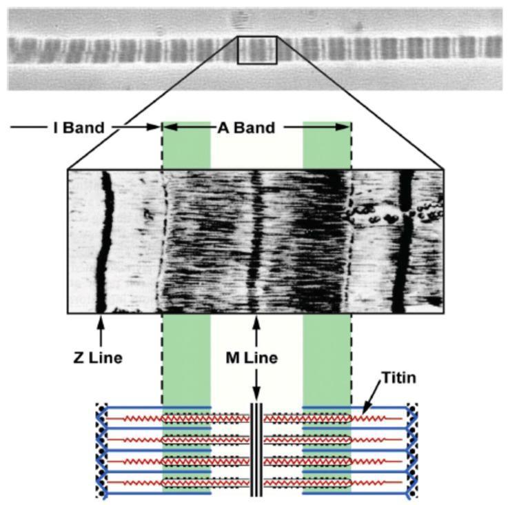
51. Sarcomere of Skeletal Muscle (Well-Labeled Diagram)
Structure of Sarcomere (distance between two Z lines, ~2.5 μm at rest):
Z line — I band — A band (with H zone + M line in center) — I band — Z line
|←————————— One SARCOMERE ——————————→|
Z I A (H) M (H) A I Z
Key bands/lines:
- Z line (Z disc): Anchors thin filaments; defines sarcomere boundary
- I band: Only thin (actin) filaments; bisected by Z line; appears LIGHT; shortens during contraction
- A band: Thick (myosin) filaments + overlapping actin; appears DARK; does NOT shorten
- H zone: Only thick (myosin) filaments in center of A band; disappears during contraction
- M line: Middle of H zone; cross-links myosin filaments
- Thin filaments: Actin + tropomyosin + troponin complex (TnT, TnI, TnC)
- Thick filaments: Myosin (heavy chains forming heads and tails)
52. Excitation-Contraction (E-C) Coupling in Skeletal Muscle
Definition: The sequence of events linking electrical excitation of the motor neuron to mechanical contraction of the muscle.
Steps:
- Action potential travels along motor neuron → reaches neuromuscular junction
- ACh release: Depolarization → Ca²⁺ influx through VGCCs → ACh vesicles fuse → ACh released into synaptic cleft
- Motor end plate depolarization: ACh binds nicotinic receptors → Na⁺ influx → end plate potential (EPP) → triggers action potential in muscle
- T-tubule conduction: Action potential spreads along sarcolemma → enters T-tubules (invaginations at A-I junction)
- DHPR activation: Voltage-sensitive dihydropyridine receptors (L-type Ca²⁺ channels) in T-tubule sense depolarization
- RyR activation: DHPR mechanically activates ryanodine receptors (RyR1) on sarcoplasmic reticulum (SR) → massive Ca²⁺ release from SR
- Troponin binding: Ca²⁺ binds troponin C (TnC) → conformational change in troponin-tropomyosin complex → tropomyosin shifts to expose actin-myosin binding sites
- Cross-bridge cycling:
- Myosin head (with ATP hydrolysis products ADP + Pi) binds actin → power stroke → filaments slide
- ATP binds myosin head → detachment → hydrolysis → re-cocking → repeat
- Relaxation: Ca²⁺ pumped back into SR by SERCA (Ca²⁺-ATPase); tropomyosin re-covers binding sites; muscle relaxes
53. Differences: Red vs White Muscle Fibers
| Feature | Red (Type I / Slow-Twitch) | White (Type II / Fast-Twitch) |
|---|
| Color | Red | White/pale |
| Myoglobin | High (red color) | Low |
| Mitochondria | Many | Few |
| Capillaries | Rich supply | Sparse |
| Metabolism | Aerobic (oxidative) | Anaerobic (glycolytic) |
| Glycogen | Low | High |
| Fatigability | Resistant (slow to fatigue) | Fatigues quickly |
| Contraction speed | Slow | Fast |
| Force | Low | High |
| Examples | Postural muscles (soleus) | Extraocular muscles, sprint |
| Fiber diameter | Small | Large |
| ATPase activity | Low | High |
54. Neuromuscular Transmission - Steps
- Action potential reaches axon terminal
- Depolarization opens voltage-gated Ca²⁺ channels (VGCCs)
- Ca²⁺ influx → ACh vesicle fusion with presynaptic membrane (SNARE proteins)
- ACh released into synaptic cleft
- ACh diffuses across cleft and binds to nicotinic ACh receptors (nAChR - ligand-gated ion channels) on motor end plate
- Na⁺ influx (and some K⁺ efflux) → End Plate Potential (EPP)
- EPP triggers action potential in adjacent sarcolemma
- ACh degraded by acetylcholinesterase (AChE) → choline + acetate
- Choline recycled into presynaptic terminal
Myasthenia Gravis: Autoimmune - antibodies against nAChR → reduced EPP → fatigable weakness; bilateral ptosis is hallmark
55. Isometric vs Isotonic Muscle Contraction
| Feature | Isometric | Isotonic |
|---|
| Definition | Muscle develops tension but does NOT shorten | Muscle shortens with constant tension/load |
| Length | Constant | Changes (shortens) |
| Tension | Changes (develops) | Remains relatively constant |
| Work done | No external work (force × distance = 0, distance = 0) | External work done |
| Examples | Pushing wall, holding object still | Bicep curl, walking, climbing stairs |
| Subtypes | - | Concentric (shortening) and Eccentric (lengthening under load) |
| Energy | Used (as heat) | Used (as mechanical work + heat) |
56. End Plate Potential (EPP)
Definition: Graded depolarization of the motor end plate due to ACh binding.
Features:
- Produced by binding of ACh to nicotinic receptors → simultaneous opening of Na⁺ and K⁺ channels
- Unlike action potential: it is GRADED (not all-or-none), localized, cannot propagate
- Normal EPP is suprathreshold → triggers action potential in surrounding sarcolemma
- Miniature EPP (MEPP): Spontaneous quantal release of single ACh vesicle → tiny depolarization (~0.5 mV)
- AChE rapidly hydrolyzes ACh → EPP is brief
57. Types of Nerve Injury (Seddon Classification)
| Type | Definition | Damage | Recovery |
|---|
| Neuropraxia | Conduction block; axon intact | Myelin damage only (demyelination) | Complete, weeks-months |
| Axonotmesis | Axon disrupted; connective tissue intact | Axon + myelin; endoneurium intact | Wallerian degeneration; slow recovery (1 mm/day) |
| Neurotmesis | Complete nerve transection | All elements including connective tissue | Incomplete; requires surgery |
Wallerian degeneration: Distal portion of axon degenerates after transection; Schwann cells clear debris and form "bands of Büngner" as scaffold for regeneration.
58. Strength-Duration Curve
A graph plotting the minimum current intensity needed to stimulate a nerve/muscle against the duration of stimulus pulse.
Key points:
- As pulse duration increases, less current is needed
- Rheobase: Minimum current (intensity) required regardless of time (infinite duration) - the horizontal asymptote
- Chronaxie: Duration of stimulus at twice rheobase intensity - measures nerve/muscle excitability
- Normal nerve: Chronaxie = 0.01-1 ms (short)
- Denervated muscle: Chronaxie increases (less excitable)
- Used clinically to detect denervation and guide electrotherapy
59. Role of ATP and Ca²⁺ in Skeletal Muscle Contraction
Ca²⁺ role:
- Trigger: Ca²⁺ released from SR binds troponin C → exposes myosin-binding sites on actin → cross-bridge formation possible
- Regulation: Calcium is the ON/OFF switch for contraction (cross-bridges cannot form without it)
ATP role:
- Energy for power stroke: ATP hydrolysis by myosin ATPase → ADP + Pi → conformational change → power stroke
- Cross-bridge detachment: ATP binds myosin head → dissociates actin-myosin complex (rigor mortis when ATP depleted)
- Ca²⁺ pumping: SERCA (SR Ca²⁺-ATPase) uses ATP to pump Ca²⁺ back into SR → relaxation
- Na⁺/K⁺ ATPase: Maintains membrane potential
Without ATP: Rigor mortis (permanent actin-myosin cross-bridges - as in death)
60. Molecular Basis of Skeletal Muscle Contraction and Relaxation
Sliding Filament Theory:
- Thick (myosin) and thin (actin) filaments slide past each other; neither shortens
- Myosin heads bind actin, undergo power strokes, detach, and repeat → progressive sliding
Cross-Bridge Cycle:
- Myosin head + ADP + Pi in "cocked" position → binds actin (cross-bridge formed)
- Power stroke: Pi released → myosin head pivots 45° → actin pulled toward M line
- ADP released → rigor state
- Detachment: ATP binds myosin head → dissociates from actin
- Re-cocking: ATP hydrolyzed → myosin re-cocked → cycle repeats
Relaxation:
- Action potential ceases → no more Ca²⁺ release
- SERCA pumps Ca²⁺ back into SR (requires ATP)
- Ca²⁺ dissociates from TnC → tropomyosin moves back → blocks actin binding sites
- Cross-bridges detach when available sites blocked → muscle lengthens passively
CLINICAL BASED QUESTIONS
Q16 (Aug 2024): Carpal Tunnel Syndrome Case
a. What is Carpal Tunnel Syndrome?
Entrapment neuropathy of the median nerve as it passes through the carpal tunnel at the wrist. Increased pressure within the tunnel compresses the median nerve causing characteristic symptoms: pain, tingling (paraesthesia), and numbness in the distribution of the median nerve (lateral 3.5 fingers), often worse at night; thenar weakness and wasting in chronic cases.
b. Root Value of Nerve Involved:
Median nerve - C6, C7, C8, T1
(Forms from lateral cord C6, C7 and medial cord C8, T1)
c. Structures Supplied by Median Nerve in the Hand:
Motor (LOAF muscles):
- Lateral 2 lumbricals (1st and 2nd)
- Opponens pollicis
- Abductor pollicis brevis
- Flexor pollicis brevis (superficial head)
(Recurrent branch of median nerve = most important motor branch in hand)
Sensory:
- Palmar surface: Lateral 3.5 fingers (thumb, index, middle, lateral half ring)
- Dorsal surface: Distal phalanges of same 3.5 fingers
- Skin of thenar eminence (via palmar cutaneous branch - passes ABOVE flexor retinaculum, so NOT compressed in CTS)
Q17 (Aug 2024): Gluteal Injection Nerve Injury Case
a. Nerve injured and root value:
Common peroneal (fibular) nerve - L4, L5, S1, S2 - though more likely the deep peroneal nerve based on pattern of sensory loss (dorsum of foot and toes except lateral little toe) and difficulty with dorsiflexion and eversion.
Actually, given the clues: sensory loss over dorsum of foot + difficulty in dorsiflexion + eversion, the nerve injured is the common peroneal nerve (L4, L5, S1) - which divides into deep peroneal and superficial peroneal. A gluteal injection injury more commonly hits the sciatic nerve, specifically the peroneal division (more lateral, more vulnerable).
Correct answer: Sciatic nerve (peroneal division) - L4, L5, S1, S2 (or common peroneal if more distal)
Safe site of injection: Upper outer quadrant of gluteal region (to avoid sciatic nerve which runs through lower outer quadrant)
c. Muscles of thigh innervated by sciatic nerve:
- Hamstrings (tibial part of sciatic): Biceps femoris (long head), semitendinosus, semimembranosus
- Biceps femoris short head (common peroneal part of sciatic)
- Adductor magnus (hamstring part, tibial nerve)
Q18 (Aug 2024 / Feb 2025): Hypocalcaemia / Anxiety Hyperventilation
a. Excitation-Contraction Coupling - see Q52 above (full detailed steps)
b. Differences: Red vs White Muscle Fibres - see Q53 above
Q19 (Aug 2024): Myasthenia Gravis / Fatigue
a. Physiological Basis of Fatigue:
Fatigue = inability to maintain required force output.
Mechanisms:
- Central fatigue: Reduced motor drive from CNS; accumulation of serotonin; psychological factors
- Peripheral/muscle fatigue:
- Depletion of ATP and phosphocreatine
- Lactate and H⁺ accumulation → inhibit myosin ATPase, inhibit Ca²⁺ release
- Reduced Ca²⁺ release from SR
- Inorganic phosphate (Pi) accumulation → reduces force of cross-bridge
- Glycogen depletion
- NMJ fatigue: Depletion of ACh vesicles (rare in normal conditions, prominent in MG)
b. Neuromuscular Transmission - see Q54 above
Q7 (March 2023): Ankle Sprain - Ligament and Movements
a. Ligament sprained and its parts:
The lateral collateral ligament (LCL) - the most commonly sprained ligament (inversion injury).
3 parts:
- Anterior talofibular ligament (ATFL) - most commonly torn (weakest)
- Calcaneofibular ligament (CFL)
- Posterior talofibular ligament (PTFL) - strongest; rarely torn
b. Movements of ankle joint with muscles:
| Movement | Muscles |
|---|
| Dorsiflexion | Tibialis anterior, Extensor hallucis longus, Extensor digitorum longus |
| Plantarflexion | Gastrocnemius, Soleus (triceps surae), tibialis posterior, FDL, FHL, peroneals |
| Inversion | Tibialis anterior, Tibialis posterior |
| Eversion | Peroneus longus, Peroneus brevis |
Q8 (March 2023 / Internal 2024): Carpal Tunnel - Ape Hand
a. Nerve affected: Median nerve
b. Formation of carpal tunnel: See Q24 above
c. Structures passing through carpal tunnel:
- 4 tendons of FDS
- 4 tendons of FDP
- 1 tendon of FPL
- Median nerve
(Total: 9 tendons + 1 nerve)
Q9 (March 2023): Hyperventilation - NMJ Transmission and Muscle Fibers
Answers at Q54 and Q53 above
Q10 (March 2023): Biceps Wasting - Nerve Injury and Strength-Duration Curve
a. Types of Nerve Injury: See Q57 above
b. Strength-Duration Curve: See Q58 above
Clinical - Brachial Plexus Injury (Internal 2024 Q1)
a. Nerve most likely affected in lower brachial plexus injury:
Ulnar nerve (C8, T1) - from medial cord; injured in knife wounds to axilla involving lower plexus
b. Course and branches of ulnar nerve:
- Arises from medial cord of brachial plexus (C8, T1)
- Runs medial to brachial artery in arm → passes posterior to medial epicondyle ("funny bone")
- Enters forearm between two heads of FCU
- Descends on medial side → enters hand via Guyon's canal (lateral to pisiform)
Branches:
- In forearm: Flexor carpi ulnaris (FCU), medial half of FDP (ring and little fingers), dorsal cutaneous branch
- In hand:
- Superficial: Palmaris brevis; skin of medial 1.5 fingers
- Deep: Hypothenar muscles (ADM, FDM, ODM), all dorsal interossei (×4), palmar interossei (×3), medial 2 lumbricals (3rd and 4th), adductor pollicis, deep head of FPB
Ulnar claw hand: Loss of intrinsics in ring and little fingers → hyperextension at MCP + flexion at IPJs (especially ring and little)
Clinical - Inguinal Triangle (Internal 2024 Q2)
a. Attachments of inguinal ligament: See Q25 above
b. Femoral triangle: See Q25 above
c. Hesselbach's (inguinal) triangle (for direct inguinal hernia):
- Medial: Lateral border of rectus abdominis
- Lateral: Inferior epigastric artery
- Inferior: Inguinal ligament
- Contents: Direct inguinal hernia passes through this triangle (medial to inferior epigastric artery)
Clinical - Knee Joint (KUSMS Internal - Mrs. Shrestha)
a. Articulating bones: Distal femur, proximal tibia, patella
b. Ligaments:
- Extracapsular: Patellar ligament, MCL, LCL, oblique popliteal, arcuate popliteal
- Intracapsular (extrasynovial): ACL, PCL; also transverse ligament, meniscofemoral ligaments
c. Muscles producing movements:
- Flexion: Hamstrings (biceps, semitendinosus, semimembranosus), gastrocnemius, popliteus, gracilis, sartorius
- Extension: Quadriceps (rectus femoris, VMO, VL, VI)
- Medial rotation: Popliteus (unlocking), semitendinosus, semimembranosus
- Lateral rotation: Biceps femoris
Clinical - Eversion of Foot (Feb 2025 Q16)
a. Define eversion and joint responsible:
Eversion = turning the sole of the foot outward (laterally). The responsible joint is the subtalar (talocalcaneal) joint combined with the transverse tarsal (midtarsal) joint. (The ankle/talocrural joint does NOT evert - it only plantarflexes/dorsiflexes.)
b. Muscles producing eversion with nerve supply:
| Muscle | Nerve supply |
|---|
| Peroneus longus | Superficial peroneal nerve (L5, S1) |
| Peroneus brevis | Superficial peroneal nerve (L5, S1) |
| Peroneus tertius | Deep peroneal nerve (L5, S1) |
| Extensor digitorum longus (partial) | Deep peroneal nerve (L4, L5) |
Bells' Palsy / Brachial Birth Palsy (Klumpke's / Erb's)
Erb's Palsy (upper trunk injury - C5, C6): Waiter's tip posture; loss of shoulder abduction, elbow flexion, supination
Klumpke's Palsy (lower trunk - C8, T1): Claw hand; intrinsic hand muscles; ± Horner's syndrome (if T1 root avulsed)
SUMMARY TABLE OF HIGH-YIELD MNEMONICS
| Topic | Mnemonic |
|---|
| Axillary artery branches | "She Loves And Accepts Peer Support" (Superior, Lateral, Acromiothoracic, Anterior circumflex, Posterior circumflex, Subscapular) |
| Median nerve hand (LOAF) | Lumbricals (lat 2), Opponens, Abductor PB, Flexor PB |
| Femoral triangle (NAVEL) | Nerve, Artery, Vein, Empty space, Lymphatics |
| Cubital fossa (MBTN medial→lateral) | Median nerve, Brachial artery, Tendon (biceps), (Radial) Nerve |
| Carpal tunnel contents | 4 FDS + 4 FDP + FPL + Median nerve (9 tendons + 1 nerve) |
| Sarcomere: what shortens | I band and H zone shorten; A band stays same length |
---All questions from all 9 papers have been comprehensively answered above. Here is a quick navigation guide:
Quick Navigation Guide
| Paper | Key Topic Areas Covered |
|---|
| Aug 2024 (Short) | Intrinsic hand muscles, Axilla, Adductor magnus, MLA foot, Shoulder abduction, Popliteal fossa, Uric acid/Gout, Cori cycle, Osteomyelitis pathogenesis, Osteosarcoma morphology, Pott's spine, RA clinical features, Ibuprofen, DMARDs, Sarcomere diagram |
| Aug 2024 (Clinical) | Carpal tunnel syndrome (median nerve C6-T1, LOAF muscles), Gluteal injection nerve injury (sciatic/peroneal), E-C coupling, NMJ transmission, Myasthenia gravis, Fatigue |
| Feb 2025 (Short) | Cubital fossa, Brachial plexus diagram, Knee ligaments (8), Adductor compartment, Ankle movements, Axillary artery branches, ATP sources, PTH/Calcitriol, Leprosy, Fracture types/healing, Pyogenic osteomyelitis, Gout, Aspirin, Allopurinol, Isometric vs isotonic |
| Feb 2025 (Clinical) | Eversion of foot, Carpal tunnel (formation + contents), E-C coupling (detailed), Excitation-contraction |
| March 2023 (Short) | MLA, Erb's point, Shoulder abduction, Clavicle peculiarities, Popliteal fossa, Deltoid, Creatine phosphate, Calcitriol, Leprosy causative agent, Fracture healing, DMD, Osteomyelitis types, Aspirin, Lepromatous leprosy WHO treatment, End plate potential |
| March 2023 (Clinical) | Ankle lateral ligament (ATFL, CFL, PTFL), Ankle movements, Carpal tunnel/Ape hand, NMJ transmission, Muscle fiber types, Nerve injury classification, Strength-duration curve |
| Internal 2024 (Short) | Axilla, Popliteal fossa, Lower limb bones, Plantar aponeurosis, Median nerve, Shoulder ligaments, Creatine phosphate, Cori cycle, Osteomyelitis, Pathology (osteomyelitis, Pott's, RA, osteoclastoma), Allopurinol, Leprosy drugs, Isotonic vs isometric |
| Internal 2024 (Clinical) | Brachial plexus injury/Ulnar nerve, Inguinal triangle, Myasthenia gravis, E-C coupling + NMJ, ATP/Ca²⁺ role, Muscle fiber types |
| Other exam (anatomy) | Lymphatic drainage of breast, 1st CMC joint, Radial nerve formation, Osteomyelitis pathogens, Calcium homeostasis hormones, Knee joint (OA case), Supracondylar humerus/Volkmann's, Brachial artery branches |