
Diagram of osta of region of leg and foot
Reading File
Reading File
Searching the Web
bones of leg and foot anatomy diagram tibia fibula tarsals metatarsals phalanges
Searching Images
labeled anatomical diagram bones of leg and foot tibia fibula tarsals metatarsals phalanges
Finding Sources
Searching PubMed
foot anatomy AND leg anatomy
Reading File
Loading Image
Osteology of the leg and foot
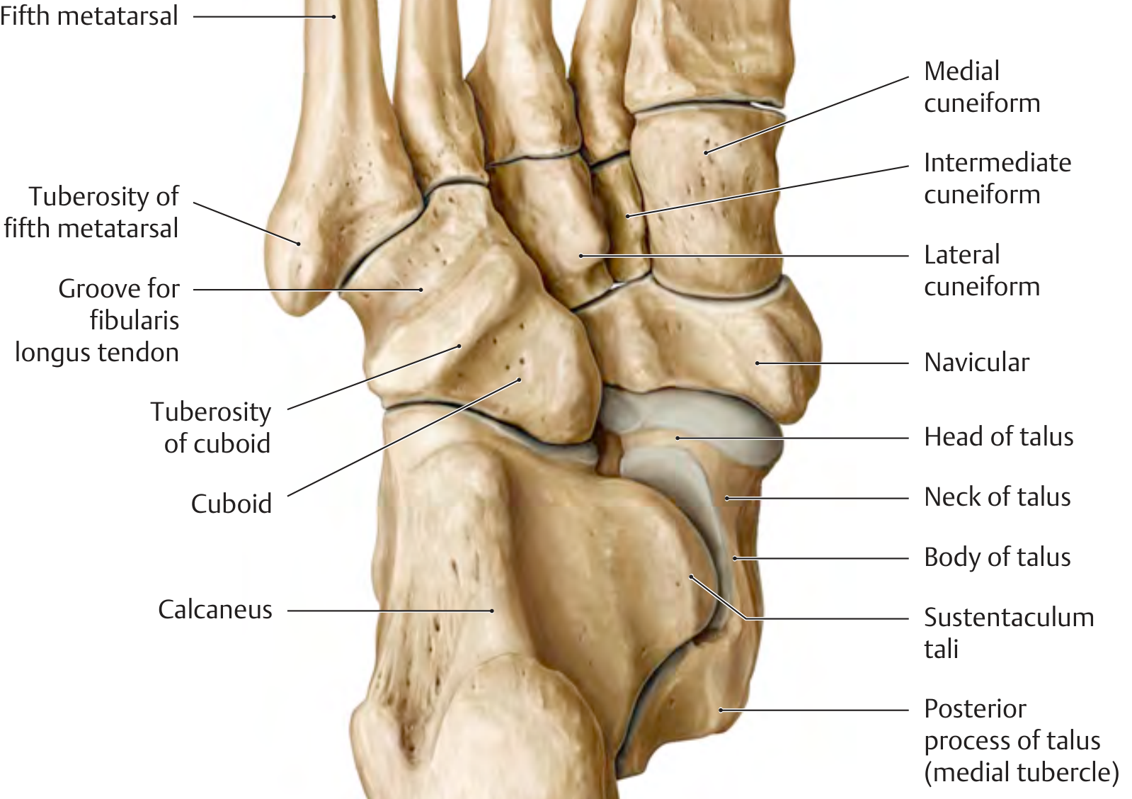
Leg bones
- Tibia: the large, medial, weight-bearing bone of the leg. Its lower end forms the medial malleolus.
- Fibula: the slender, lateral bone. Its lower end forms the lateral malleolus.
- The tibia and fibula articulate with the talus to make the ankle joint.
Foot bones: 26 in each foot
| Region | Bones |
|---|---|
| Tarsals (7) | Talus, calcaneus, navicular, cuboid, medial cuneiform, intermediate cuneiform, lateral cuneiform |
| Metatarsals (5) | Metatarsals I-V, numbered from the great-toe side to the little-toe side |
| Phalanges (14) | Great toe: proximal and distal phalanges. Other toes: proximal, middle, and distal phalanges |
Simple arrangement, posterior to anterior:
Tibia + Fibula
      ↓
    Talus
      ↓
 Calcaneus (heel)

Talus → Navicular → 3 Cuneiforms → Metatarsals I-V → Toes
Calcaneus → Cuboid → Metatarsals IV-V → Toes
- Talus transfers weight from the tibia to the foot.
- Calcaneus is the heel bone and largest tarsal.
- The sesamoid bones are usually found under the head of the first metatarsal.
The foot is commonly divided into:
- Hindfoot: talus and calcaneus
- Midfoot: navicular, cuboid, and three cuneiforms
- Forefoot: metatarsals and phalanges
The diagram is from General Anatomy and Musculoskeletal System, p. 444.
This is a shared conversation. Sign in to Orris to start your own chat.