
heart chambers and supply by which coronary artery , help me learn and make na flow chart pictorial to remember with pneumonic
coronary artery supply heart chambers diagram
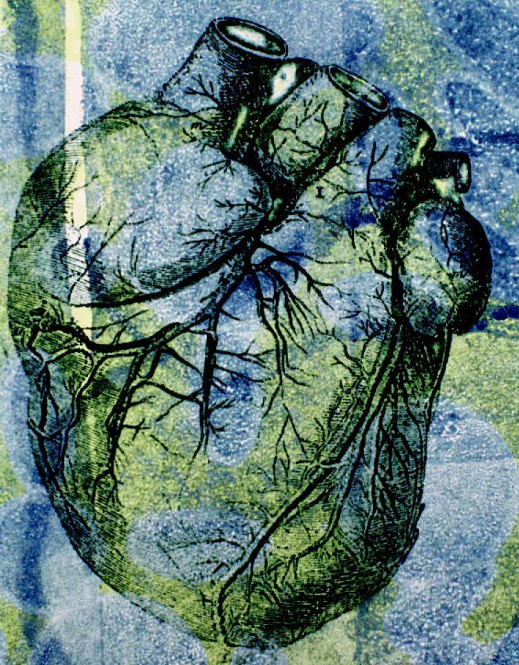
This stylized anatomical illustration depicts a human heart in an anterior view, emphasizing its gross external morphology and coronary vasculature. The organ exhibits its characteristic ovoid shape with superiorly positioned great vessels, including cross-sectional views of the aorta and pulmonary trunk. The surface anatomy highlights the intricate branching patterns of the coronary arteries and veins, demonstrating their distribution across the myocardium toward the apex. The image uses an artistic, high-contrast color palette of green, blue, and yellow to delineate anatomical boundaries and the etched texture of the cardiac tissue. Educationally, the diagram serves to illustrate the relationship between the major cardiac chambers and the superficial vascular network, particularly in the context of cardiovascular health and hemodynamic pressures. The visualization emphasizes the complexity of the organ's blood supply, which is critical for understanding conditions like hypertension and coronary artery disease.
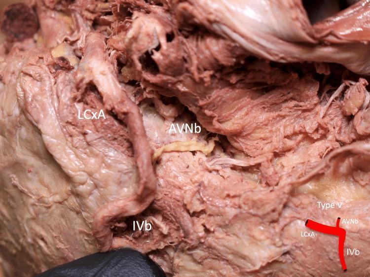
This clinical photograph of a formalin-fixed human heart cadaver specimen illustrates a Type V anatomical variation of the coronary arterial supply to the atrioventricular (AV) node. The image displays the inferior aspect of the heart, focusing on the inferior pyramidal space. Key vessels are labeled: the circumflex branch of the left coronary artery (LCxA), the atrioventricular nodal branch (AVNb), and the inferior interventricular branch (IVb). In this specific anatomical configuration, the AVNb is seen as a distinct, thinner vessel with a pale-yellow appearance originating directly from the LCxA, which has a larger diameter and more prominent reddish-brown coloration. The IVb is positioned inferiorly, and the AVNb acts as a connecting vessel between the LCxA and the distal myocardial structures. A small schematic inset in the bottom right corner provides a simplified diagram of this branching pattern. This material is significant for cardiovascular anatomy and surgical planning, demonstrating the variability of coronary dominance and blood supply to the cardiac conduction system.
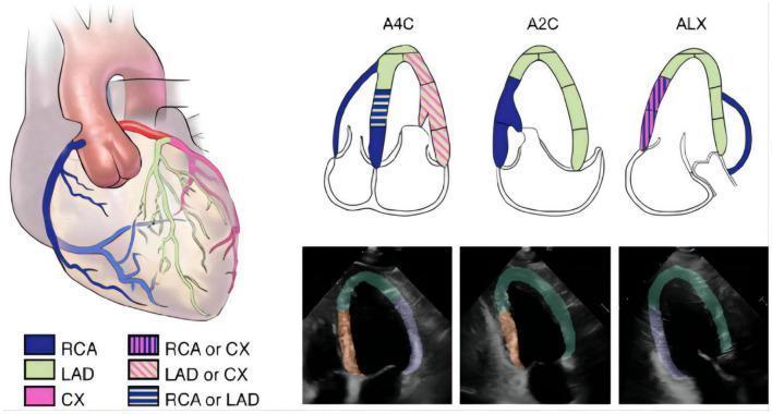
Educational medical diagram and diagnostic imagery illustrating left ventricular (LV) segmentation and coronary artery distribution based on 2015 ASE guidelines. The graphic includes an anatomical heart illustration, schematic apical views, and corresponding echocardiographic frames. The anatomical diagram color-codes the Right Coronary Artery (RCA, blue), Left Anterior Descending artery (LAD, green), and Circumflex artery (CX, pink), with striped patterns indicating overlapping territories. Three schematic diagrams represent standard echocardiographic planes: Apical Four-Chamber (A4C), Apical Two-Chamber (A2C), and Apical Long-Axis (ALX), mapping coronary blood supply to specific LV myocardial segments. Below these, three transthoracic echocardiogram frames demonstrate automated segmentation masks: Region A (apical, anterior, and anteroseptal walls) in green, Region F (inferior and inferoseptal walls) in orange, and Region L (anterolateral and inferolateral walls) in purple. This visual resource is designed to teach the correlation between coronary anatomy and regional wall motion territories in cardiac imaging.
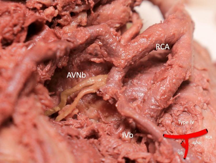
This clinical photograph of a formalin-fixed human heart specimen demonstrates a Type IV anatomical variation of the coronary arterial supply. The image focuses on the inferior pyramidal space, showing the Right Coronary Artery (RCA) and its distal branches. Key anatomical structures labeled include the RCA, the Inferior Interventricular Branch (IVb), and a duplicated Atrioventricular Nodal Branch (AVNb). The AVNb exhibits a distinct pale, yellowish-beige color and smooth texture compared to the deeper reddish-brown, striated appearance of the RCA and IVb. The duplicated AVNb originates from the RCA in close proximity to the origin of the IVb, illustrating a specific branching pattern relevant to cardiac electrophysiology and surgical anatomy. A schematic diagram in the lower right corner provides a simplified representation of the Type IV branching configuration. The specimen highlights developmental arterial variations that are clinically significant for procedures involving the AV node or during coronary revascularization.
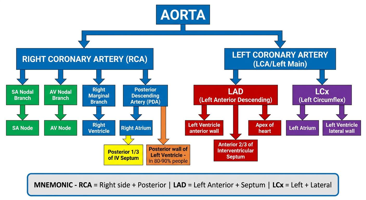
A detailed educational medical flowchart showing coronary artery supply to each heart chamber. The chart flows from top: "AORTA" at the top center, splitting into two main branches: "RIGHT CORONARY ARTERY (RCA)" on the left side and "LEFT CORONARY ARTERY (LCA/Left Main)" on the right side. Under RCA, show branches: "SA Nodal Branch", "AV Nodal Branch", "Right Marginal Branch", "Posterior Descending Artery (PDA)". Under LCA, show two branches: "LAD (Left Anterior Descending)" and "LCx (Left Circumflex)". Each artery branch should connect with arrows to the chambers/structures they supply using colored boxes: RCA supplies: Right Atrium (blue box), Right Ventricle (blue box), SA Node (green box), AV Node (green box), Posterior 1/3 of IV Septum (yellow box), Posterior wall of Left Ventricle - in 80-90% people (orange box). LAD supplies: Left Ventricle anterior wall (red box), Anterior 2/3 of Interventricular Septum (red box), Apex of heart (red box). LCx supplies: Left Atrium (purple box), Left Ventricle lateral wall (purple box). Include a mnemonic box at the bottom: "MNEMONIC - RCA = Right side + Posterior | LAD = Left Anterior + Septum | LCx = Left + Lateral". Use clean white background, bold colored arrows, clear readable font, professional medical diagram style.
❤️ Coronary Artery Supply to Heart Chambers
📊 Visual Flowchart (Generated for You)
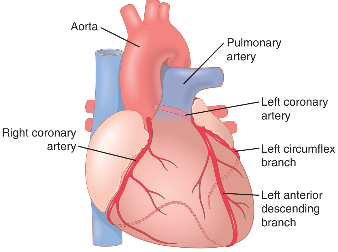
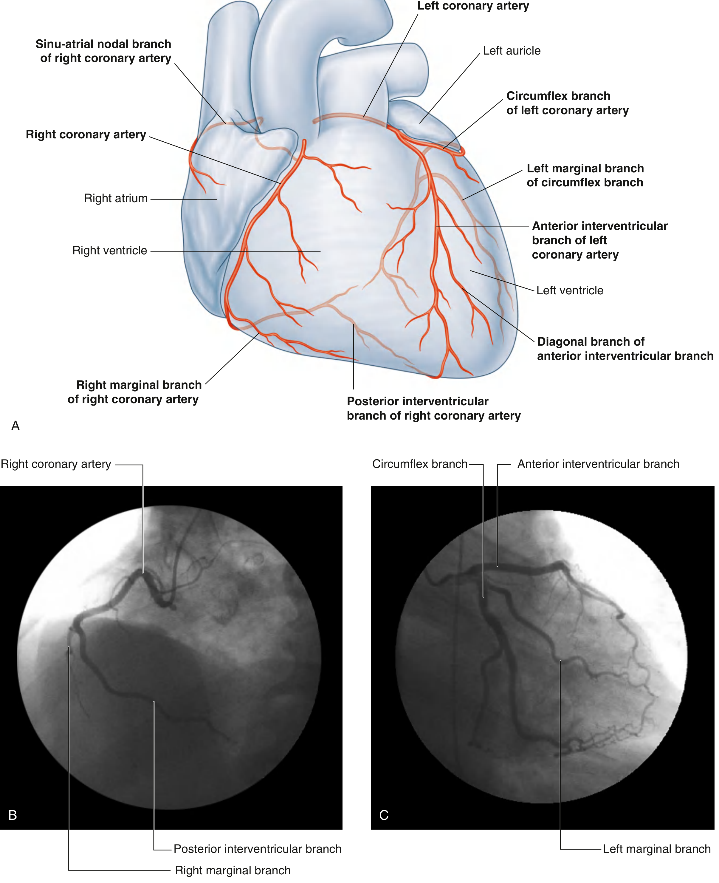
📚 Textbook Diagrams
🗺️ Complete Coronary Supply Table
| Artery | Branches | Structures Supplied |
|---|---|---|
| RCA (Right Coronary A.) | SA nodal branch | SA Node (60% of people) |
| AV nodal branch | AV Node (80-90% of people) | |
| Right marginal branch | Right Ventricle (anterior + inferior) | |
| Posterior Descending A. (PDA) | Right Atrium, Posterior LV, Posterior 1/3 of IV Septum | |
| LCA → LAD (Left Anterior Descending) | Diagonal branches, Septal perforators | Anterior wall of LV, Anterior 2/3 of IV Septum, Apex of heart |
| LCA → LCx (Left Circumflex) | Left marginal branch | Left Atrium, Lateral + Posterior LV wall |
🧠 MNEMONICS TO REMEMBER
Mnemonic 1 — "RCA = Right + Rear"
Right Coronary Artery = Right side + Rear (Posterior)
- Right Atrium ✓
- Right Ventricle ✓
- Rear (Posterior) LV wall ✓
- Rhythm nodes: SA + AV nodes ✓
Mnemonic 2 — "LAD = Leads Anteriorly Down"
LAD = Left Anterior Descending
- Left Ventricle - Anterior wall ✓
- Anterior 2/3 of Septum ✓
- Down to the Apex ✓
Mnemonic 3 — "LCx = Left Comes Around to the Lateral Side"
Circumflex wraps around the left AV groove (like a crown)
- Left Atrium ✓
- Lateral LV wall ✓
- In left dominant hearts: also supplies posterior LV + PDA
Mnemonic 4 — "LASS" for Left Coronary Territory
LAD + Anterior + Septum + Systolic pump (LV apex)
Quick Recall Card:
          AORTA
         /      \
       RCA       LCA (Left Main)
      /            /        \
 Right side     LAD          LCx
 + Posterior  (Anterior    (Lateral +
   (nodes,     LV, Ant.     Left Atrium)
   RV, RA)     Septum,
               Apex)
🏥 Coronary Dominance (Important Exam Point)
| Type | % Population | Who supplies PDA? |
|---|---|---|
| Right dominant | 80-90% | RCA gives PDA |
| Left dominant | 10-15% | LCx gives PDA |
| Co-dominant | ~5% | Both contribute |
"PDA determines dominance" - whichever artery gives off the Posterior Descending Artery is the dominant one.
⚡ Nodes & Conduction System Supply
| Structure | Primary Supply | Mnemonic |
|---|---|---|
| SA Node | RCA (60%) or LCx (40%) | "Right usually STARTS the beat" |
| AV Node | RCA (80-90%) | "Right controls the A-V delay" |
| Bundle of His | Dual (LAD + RCA) | |
| Right Bundle Branch | LAD (septal perforators) | |
| Left Bundle Branch | LAD (anterior) + RCA (posterior) |
Clinical Pearl: Inferior MI (RCA territory) → heart block (AV node ischemia). Anterior MI (LAD territory) → bundle branch blocks.
💡 MI Territory Quick Summary
| Coronary Blocked | MI Location | ECG Changes |
|---|---|---|
| LAD | Anterior MI | V1-V4 ST elevation |
| RCA | Inferior MI | II, III, aVF ST elevation |
| LCx | Lateral MI | I, aVL, V5-V6 ST elevation |
📖 Sources
- Guyton and Hall Textbook of Medical Physiology - "The left coronary artery supplies mainly the anterior and left lateral portions of the left ventricle, whereas the right coronary artery supplies most of the right ventricle, as well as the posterior part of the left ventricle in 80% to 90% of people."
- Gray's Anatomy for Students - Cardiac vasculature, Fig. 3.79-3.81
Make a comparison poster: Right dominant vs Left dominant hearts
A professional medical comparison poster titled "Coronary Dominance: Right vs Left Dominant Heart" with a clean white background and two clearly separated columns side by side. LEFT COLUMN - "RIGHT DOMINANT HEART" with a blue header banner and blue color theme: - Large bold text: "80-90% of population" - Subtitle: "Most Common" - Heart diagram outline showing RCA as the dominant, larger artery on the right side - Bullet points listing: • RCA gives off Posterior Descending Artery (PDA) • RCA supplies: Right Atrium, Right Ventricle • RCA supplies: Inferior wall of LV • RCA supplies: Posterior 1/3 of IV Septum • RCA supplies: SA Node (60%) • RCA supplies: AV Node (80-90%) • LAD supplies: Anterior LV + Anterior Septum • LCx supplies: Left Atrium + Lateral LV - Clinical note box: "Inferior MI (RCA block) = Heart block risk" RIGHT COLUMN - "LEFT DOMINANT HEART" with a red header banner and red color theme: - Large bold text: "10-15% of population" - Subtitle: "Less Common" - Heart diagram outline showing LCx as the dominant, larger artery wrapping around - Bullet points listing: • LCx gives off Posterior Descending Artery (PDA) • LCx supplies: Left Atrium + Lateral LV • LCx supplies: Inferior wall of LV (posterior) • LCx supplies: Posterior 1/3 of IV Septum • LCx may supply: SA Node + AV Node • RCA only supplies: Right Atrium + Right Ventricle • LAD still supplies: Anterior LV + Anterior Septum - Clinical note box: "LCx block = Larger territory at risk, worse prognosis" CENTER divider with text: "KEY RULE: The artery that gives off the PDA determines dominance" BOTTOM SECTION: Comparison table with 3 columns (Feature | Right Dominant | Left Dominant) showing: PDA origin, Population %, AV Node supply, Posterior LV supply, Clinical significance. MNEMONIC BOX at very bottom in yellow: "Remember: Most people are RIGHT - Right dominant in 80-90%! PDA = determines Dominance" Style: Clean, modern medical education poster, bold typography, color-coded anatomy, professional and easy to read.
Error calling model 'gemini-3-pro-image' (RESOURCE_EXHAUSTED): 429 RESOURCE_EXHAUSTED. {'error': {'code': 429, 'message': 'Resource has been exhausted (e.g. check quota).', 'status': 'RESOURCE_EXHAUSTED'}}
We encountered an error while processing your request. Please try again or reach out to us for help.