
Incisional hernia
Reading File
Reading File
Finding Sources
Finding Sources
Searching PubMed
"incisional hernia" AND repair
Searching the Web
European Hernia Society midline incisional hernia guidelines 2023 diagnosis management
Reading File
Reading File
Reading Web Page
https://pubmed.ncbi.nlm.nih.gov/37727928
Reading File
Incisional hernia
An incisional hernia is protrusion of abdominal contents through a weakness or defect in the muscle-fascial layer at the site of a previous surgical scar. It can occur after open abdominal surgery or, less often, at a laparoscopic port site. Bailey and Love's Short Practice of Surgery, p. 1098.
Causes and risk factors
It results from impaired healing or failure of fascial closure. Important factors include:
- Surgical-site infection or wound breakdown
- Obesity
- Smoking
- Diabetes, especially if poorly controlled
- Malnutrition, steroid treatment, immunosuppression
- Chronic cough/COPD, constipation, or other causes of raised intra-abdominal pressure
- Poor tissue quality, closure under tension, or inadequate closure technique
- Previous laparotomy and a midline incision
Bailey and Love's Short Practice of Surgery, pp. 1098-1099. In particular, postoperative surgical-site infection, smoking, obesity, uncontrolled diabetes, and immunosuppression are associated with increased risk. Fischer's Mastery of Surgery, p. 5851.
Clinical features
- A swelling or bulge at or near an operative scar
- Becomes more noticeable while coughing, standing, straining, or lifting
- May reduce when lying down or with gentle pressure
- Discomfort, dragging pain, poor cosmesis, difficulty with activity or clothing
- It may enlarge over months or years
- Multiple defects can exist along one scar
Intestinal obstruction can occur because of adhesions. Strangulation is less common than in narrow-neck hernias, but is possible, especially with a small defect and a large sac. Bailey and Love's Short Practice of Surgery, p. 1099.
Diagnosis
Diagnosis is usually clinical, with examination both standing and supine, including cough impulse and assessment of reducibility.
CT abdomen is often used when the diagnosis is uncertain or for operative planning, particularly in large, recurrent, obese, or complex hernias. It shows the defect size, contents, multiple defects, and loss of domain.
Management
Urgent surgical assessment is needed if there is:
- Sudden severe or persistent pain
- An irreducible, tender, firm swelling
- Redness or skin discoloration over the hernia
- Vomiting, abdominal distension, inability to pass stool or flatus
- Fever or systemic illness
These features can indicate obstruction or strangulation.
Elective/non-emergency management
- A small, painless, stable hernia can sometimes be managed with observation after discussion of risks and preferences.
- An abdominal binder may reduce symptoms but does not repair the defect.
- Optimize before elective repair: weight management, smoking cessation, diabetes control, nutrition, fitness, and treatment of chronic cough or constipation.
- Surgery is generally considered for pain, functional limitation, progressive enlargement, recurrent obstruction, skin problems, or patient preference.
For most clinically significant incisional hernias, repair is with mesh reinforcement, using an open, laparoscopic, robotic, or combined approach depending on defect size, previous surgery, adhesions, contamination risk, and surgeon expertise. Complex or very large defects may require abdominal-wall reconstruction and specialist referral. Bailey and Love's Short Practice of Surgery, p. 1099.
The 2023 European Hernia Society guideline addresses adult midline incisional hernias and supports shared decision-making plus optimization of modifiable risk factors before repair.
Quick answer
Please provide the question you want answered.
Para umblical hernia treatment
Reading File
Finding Sources
Finding Sources
Searching PubMed
"umbilical hernia" AND repair
Searching the Web
European Hernia Society guidelines umbilical epigastric hernia treatment mesh repair
Paraumbilical hernia treatment
A paraumbilical hernia is a type of primary ventral hernia occurring next to the umbilicus. Management depends on symptoms, size, reducibility, general health, and whether there are complications.
Seek emergency care now if
- The lump becomes painful, hard, tender, or cannot be pushed back
- Vomiting, abdominal swelling, inability to pass stool or gas
- Red, purple, or dark skin over the lump
- Fever or severe persistent abdominal pain
These may indicate bowel obstruction or strangulation and can need emergency surgery.
Non-operative management
Appropriate in selected people with a small, painless, reducible, stable hernia or when operative risk is high.
- Observation with medical review if it enlarges or becomes symptomatic
- Reduce strain: treat constipation or chronic cough, avoid heavy lifting where possible
- Weight reduction and smoking cessation can reduce operative and recurrence risks
- An abdominal belt/binder can provide comfort temporarily but will not close or cure the hernia
Elective surgery
Repair is usually advised for a hernia that is painful, enlarging, difficult to reduce, causing activity limitation, or cosmetically troublesome.
Options:
-
Suture repair
- The defect is closed with stitches alone.
- May be considered for very small defects.
- Has a higher risk of recurrence as the defect size increases.
-
Mesh repair
- A mesh reinforces the abdominal wall, commonly placed in the preperitoneal plane during open repair.
- Generally preferred for many adult symptomatic paraumbilical hernias because it lowers recurrence risk compared with stitches alone.
- The European and American Hernia Society guideline recommends mesh repair for symptomatic umbilical/epigastric hernias, usually by an open approach with a preperitoneal flat mesh. A laparoscopic approach may be chosen for a large defect or increased risk of wound problems, such as obesity. See the EHS/AHS guideline.
-
Laparoscopic or robotic repair
- Often considered for larger hernias, recurrent hernias, or patients at higher risk of wound complications.
- The appropriate approach is individualized based on defect size, contents, previous operations, body habitus, and surgeon experience.
Recent systematic reviews also support lower recurrence with mesh versus suture in elective adult umbilical hernia repair, although the balance of risks and benefits should be individualized (2024 meta-analysis).
Before planned repair
Optimizing health improves outcomes:
- Stop smoking
- Aim for weight reduction if overweight
- Control diabetes
- Improve nutrition and fitness
- Manage chronic cough and constipation
A general surgeon or abdominal-wall hernia specialist should examine the hernia. Ultrasound or CT may be used if the diagnosis is uncertain or the hernia is large/complex.
Onlay inlay repair
Reading File
Finding Sources
Finding Sources
Searching PubMed
"incisional hernia" AND onlay mesh
Searching the Web
onlay versus inlay mesh incisional ventral hernia repair surgical definition outcomes guidelines
Searching Images
diagram of abdominal wall mesh placement onlay inlay sublay retrorectus incisional hernia repair
Reading File
Reading File
Reading File
Onlay versus inlay mesh repair
These terms describe where the mesh is placed during open ventral, incisional, or paraumbilical hernia repair.
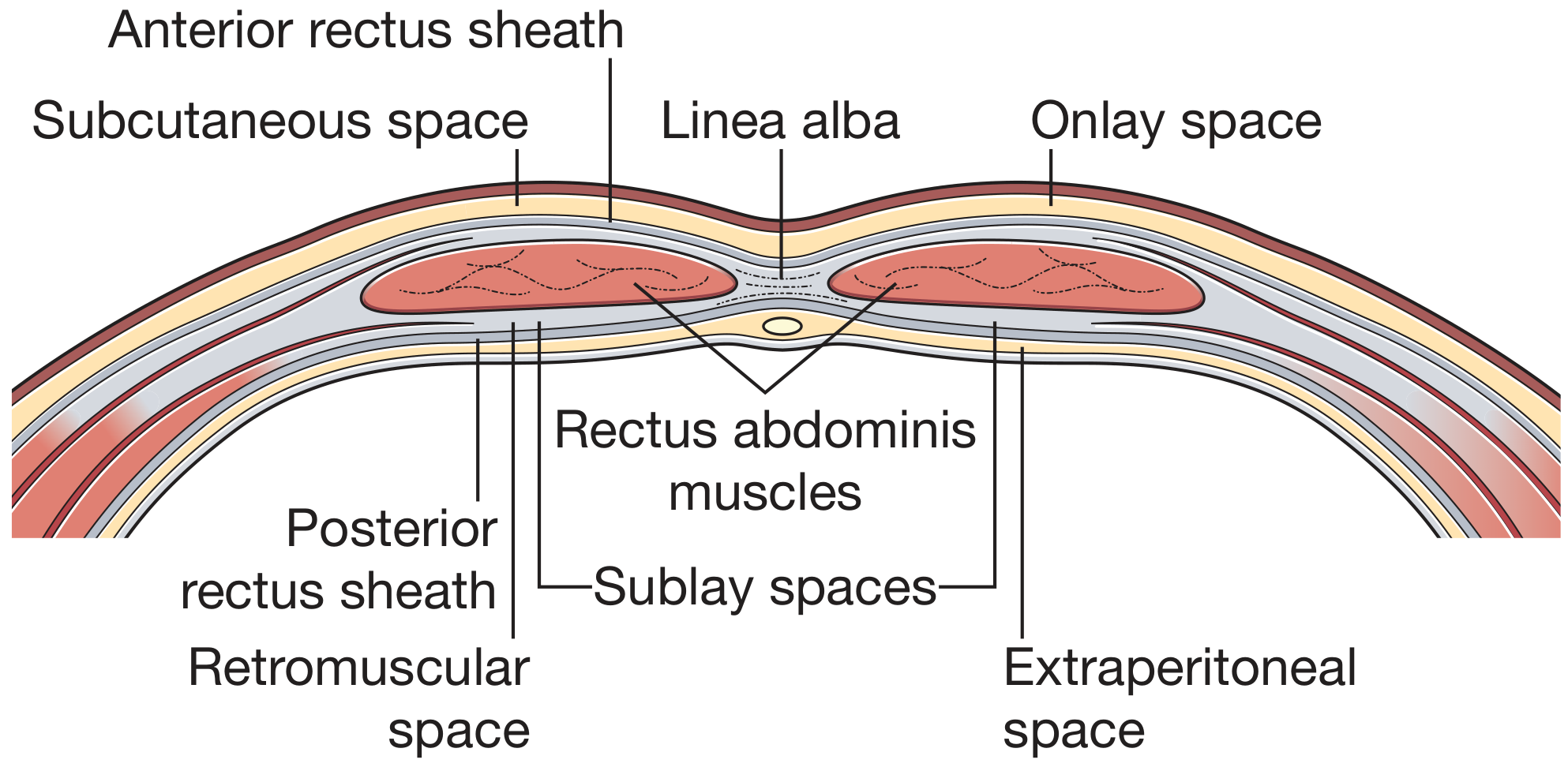
1. Onlay repair
The fascial defect is first closed, then mesh is placed on top of the anterior rectus sheath/fascia, beneath the subcutaneous tissue.
Basic steps
- Dissect the hernia sac and reduce its contents.
- Close the fascial defect with sutures.
- Create space superficial to the fascia.
- Place a mesh with wide overlap around the repaired defect.
- Fix mesh to fascia. A drain is often used.
Advantages
- Technically relatively straightforward.
- Useful if deeper planes are scarred or difficult to access.
- Mesh does not contact bowel.
Disadvantages
- Requires raising skin and subcutaneous flaps.
- Higher risk of seroma, wound infection, skin-flap ischemia/necrosis, and mesh exposure if the wound breaks down.
- Generally less mechanically favorable than a retromuscular/preperitoneal mesh plane.
2. Inlay repair, also called bridging repair
Mesh is placed within the fascial gap, with its edges sutured directly to the margins of the defect. The fascia is not closed over it.
Basic principle
fascial edge - mesh bridge - fascial edgeAdvantages
- May be necessary when the fascia cannot be approximated without excessive tension.
- Can serve as a temporary or damage-control option in selected emergency or very complex situations.
Disadvantages
- Higher recurrence because tension is concentrated at the mesh-fascia junction.
- It does not restore normal abdominal-wall continuity or function as well as fascial closure with reinforcement.
- For elective hernia repair, it is generally avoided whenever possible.
Practical comparison
| Feature | Onlay | Inlay/bridging |
|---|---|---|
| Mesh location | Superficial to anterior fascia | Within fascial defect |
| Fascia closed first? | Yes | No |
| Main concern | Seroma and wound complications | Recurrence and failure at mesh edge |
| Elective use | Possible, selected cases | Usually avoided |
| Better alternative when feasible | Retrorectus/preperitoneal sublay | Fascial closure plus retromuscular/preperitoneal mesh |
For elective ventral or incisional hernia repair, retromuscular or preperitoneal sublay placement, with primary closure of the defect when feasible, is often preferred because it has lower recurrence and surgical-site infection risk in available surgical evidence. Fischer's Mastery of Surgery, p. 5908. Inlay meshes are not recommended for routine elective repair. Bailey and Love's Short Practice of Surgery, p. 1086.
Terminology note: “IPOM” means intraperitoneal onlay mesh, but it is actually mesh placed inside the abdominal cavity against the peritoneal surface, not a conventional superficial onlay.
This is a shared conversation. Sign in to Orris to start your own chat.