
How to approach pancytopenia , and proceed with history and physical exams to make differential diagnosis
Reading File
How to approach pancytopenia , and proceed with history and physical exams to make differential diagnosis..especially in wards , and also for my mrcp and fcps part 2 exam
Finding Sources
Finding Sources
Searching the Web
pancytopenia approach history physical examination differential diagnosis MRCP FCPS
Good, I have strong leads. Let me now read the key textbook sections and the best web sources in parallel.
Reading File
Reading File
Reading Web Page
https://pmc.ncbi.nlm.nih.gov/articles/PMC11399640
Let me now fetch the Frameworks for Internal Medicine sections and the key diagram, plus the Frameworks ineffective hematopoiesis and hypersplenism sections.
Reading File
Loading Image
Reading File
Excellent material gathered. Now let me get the hypersplenism section and the Frameworks flowchart image, plus a quick pubmed search for recent reviews.
Reading File
Loading Image
Searching PubMed
pancytopenia approach diagnosis evaluation
I now have comprehensive material from Harrison's, Frameworks for Internal Medicine, Family Medicine textbook, and current literature. Let me compile the complete, exam-level answer.
Approach to Pancytopenia - Ward & Exam (MRCP/FCPS Part 2)
Definition
Pancytopenia = simultaneous reduction in all three cell lines:
- Haemoglobin < 13 g/dL (men) / < 12 g/dL (women)
- WBC < 4,000/µL (or ANC < 1,500/µL)
- Platelets < 150,000/µL
The Pathophysiological Framework (Memorise This First)
This is the single most important conceptual map for MRCP/FCPS:
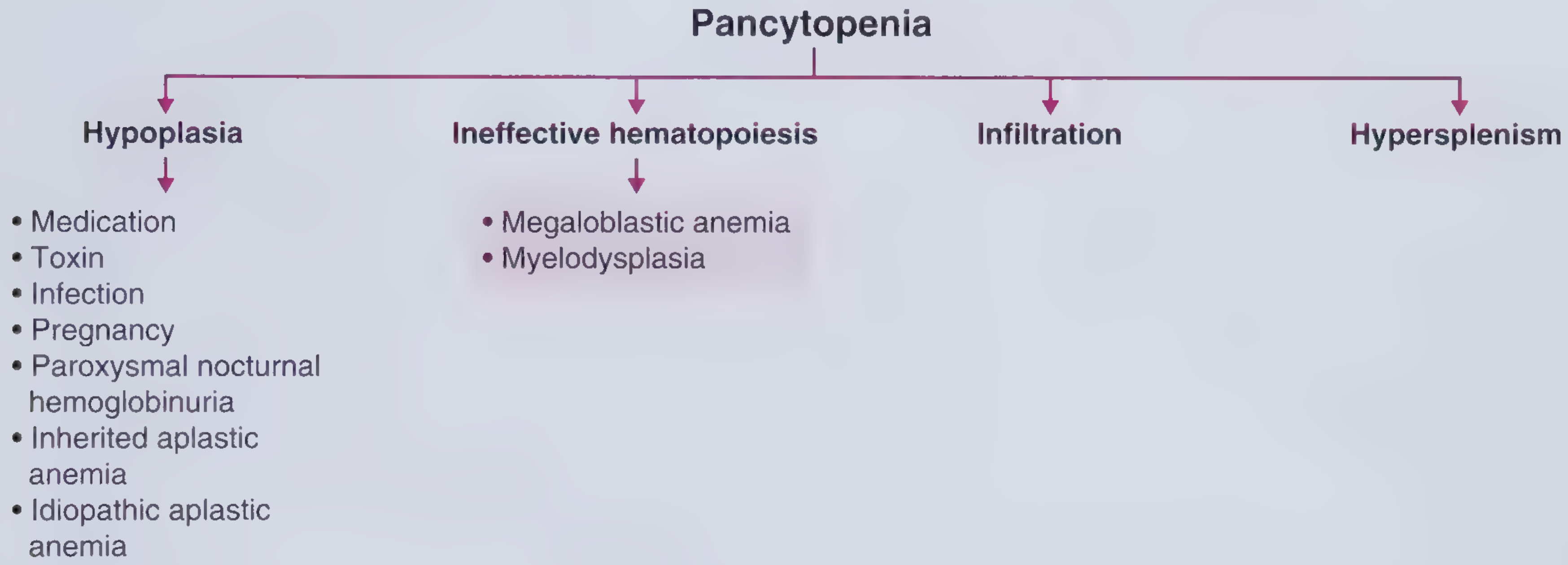
Every cause of pancytopenia falls into one of four mechanisms:
| Mechanism | Bone Marrow | Reticulocytes | Key Causes |
|---|---|---|---|
| Hypoplasia (↓ production) | Hypocellular | Low | Aplastic anaemia, drugs, radiation, PNH, Fanconi |
| Ineffective haematopoiesis | Normo/Hypercellular | Low | Megaloblastic anaemia (B12/folate), MDS |
| Infiltration (myelophthisis) | Infiltrated | Low/variable | Leukaemia, lymphoma, myelofibrosis, metastatic carcinoma, TB, Gaucher's |
| Hypersplenism | Normal/Hypercellular | Normal/High | Cirrhosis, portal hypertension, lymphoma, leishmaniasis, Felty syndrome |
Frameworks for Internal Medicine - p. 361-366; Harrison's 22e - Chapter 107
History Taking - Systematic Approach
1. Symptoms of Pancytopenia Itself
Anaemia symptoms (ask about):
- Fatigue, exertional dyspnoea, reduced exercise tolerance
- Palpitations, chest pain (cardiovascular compromise in severe anaemia)
- Pallor noticed by patient/family
Neutropenia symptoms:
- Recurrent/severe infections, fevers, rigors
- Oral ulcers, mucositis
- Specific infective symptoms (chest, urinary, skin)
Thrombocytopenia symptoms:
- Easy bruising, petechiae, purpura
- Prolonged bleeding from cuts
- Epistaxis, gum bleeding
- Heavy menstrual periods (women)
- Haematuria, melaena (more severe)
2. Aetiology-Directed History
Drug and Toxin History (CRITICAL - often missed)
Ask specifically about:
- Chemotherapy or radiation - most obvious
- Antibiotics: chloramphenicol (bone marrow toxicity, unique vacuoles on BM biopsy), linezolid, cotrimoxazole
- Antithyroid drugs: methimazole, propylthiouracil (agranulocytosis + aplasia)
- NSAIDs: indomethacin, phenylbutazone
- Anti-epileptics: carbamazepine, phenytoin
- Methotrexate, azathioprine, mycophenolate
- Alcohol: direct marrow toxin, associated folate deficiency, causes reversible pancytopenia or selective thrombocytopenia
- Arsenic/heavy metals: ask about recurrent GI symptoms, peripheral neuropathy, hair loss - a classic MRCP/FCPS vignette (arsenic poisoning by spouse!)
Nutritional History
- Dietary habits - strict vegan/vegetarian diet → B12 deficiency
- Alcohol use → folate deficiency + direct toxicity
- Malabsorption symptoms: diarrhoea, steatorrhoea, weight loss → coeliac disease, Crohn's
Infective History
- HIV risk factors (sexual history, IV drug use, transfusions)
- TB contacts, travel to endemic areas, night sweats, weight loss
- Viral infections: recent EBV (infectious mononucleosis), CMV, parvovirus B19
- Leishmaniasis: months of fever, weight loss, splenomegaly in a traveller from endemic region (Mediterranean, South Asia, East Africa) - classic MRCP vignette
Travel History
- Tropical travel raises suspicion for leishmaniasis, malaria, brucellosis, enteric fever
Past Medical History
- Autoimmune conditions: SLE (pancytopenia is a diagnostic criterion!), rheumatoid arthritis
- Known liver disease → cirrhosis + hypersplenism
- Prior haematological conditions
- HIV status
- Pregnancy (ask in all women of reproductive age)
- Thyroid disease and treatment
Family History
- Inherited bone marrow failure syndromes in children/young adults:
- Fanconi anaemia: short stature, radial ray defects, café-au-lait spots
- Dyskeratosis congenita: nail dystrophy, oral leucoplakia, skin pigmentation
- Shwachman-Diamond syndrome: pancreatic exocrine insufficiency
Constitutional/B-symptoms
- Fever > 38°C, drenching night sweats, weight loss > 10% in 6 months → lymphoma, leukaemia, TB
Social History
- Occupational exposures: benzene, pesticides, radiation (industrial)
- Alcohol intake (quantify)
Physical Examination - System by System
General Inspection
- Pallor (conjunctival, palmar)
- Jaundice → haemolysis, liver disease, megaloblastic anaemia
- Petechiae, purpura, bruising
- Weight (cachexia → malignancy, TB)
- Nutritional status
Skin and Nails
| Finding | Suggests |
|---|---|
| Petechiae/purpura | Thrombocytopenia |
| Jaundice | Megaloblastic, haemolysis, liver disease |
| Café-au-lait spots | Fanconi anaemia |
| Nail dystrophy + oral leucoplakia + hyperpigmentation | Dyskeratosis congenita |
| Butterfly rash, discoid rash | SLE |
| Spider naevi, palmar erythema, leukonychia | Chronic liver disease → cirrhosis |
| Alopecia | SLE, arsenic poisoning, chemotherapy |
Mouth and Oropharynx
- Oral ulcers → neutropenia
- Angular stomatitis, glossitis → B12/folate deficiency
- Oral leucoplakia → dyskeratosis congenita
- Thrush → immunosuppression
Lymph Nodes
- Lymphadenopathy is NOT a feature of aplastic anaemia - its presence should prompt you to think infection or lymphoma/leukaemia instead
- Generalised lymphadenopathy + elevated LDH → lymphoma
- Cervical nodes + tonsillar enlargement + splenomegaly → EBV/infectious mononucleosis
Abdomen
- Splenomegaly - most important finding
- Massive spleen: leishmaniasis, myelofibrosis, CML
- Moderate: lymphoma, portal hypertension, storage disorders (Gaucher's)
- Signs of portal hypertension (ascites, caput medusae) + splenomegaly → cirrhosis with hypersplenism
- Hepatomegaly: liver disease, lymphoma, leukaemia, infiltrative disorders
- Hepatosplenomegaly: lymphoma, leukaemia, leishmaniasis, sarcoidosis, myelofibrosis
Neurological
- Peripheral neuropathy + subacute combined degeneration (dorsal column + corticospinal tract) → B12 deficiency
- Peripheral neuropathy + GI symptoms + alopecia → arsenic poisoning
Musculoskeletal
- Symmetrical polyarthritis + splenomegaly + neutropenia → Felty syndrome (RA triad)
- Radial ray defects (absent thumbs) → Fanconi anaemia
Eyes
- Fundoscopy: flame haemorrhages, Roth spots → severe anaemia, infective endocarditis
- Uveitis → sarcoidosis, lymphoma
Putting It Together: Clue Clusters for Differentials
| Clinical Cluster | Most Likely Diagnosis |
|---|---|
| Young patient + no organomegaly + no lymphadenopathy + insidious onset | Aplastic anaemia |
| Vegan diet / elderly / glossitis / subacute combined degeneration | B12 deficiency megaloblastic anaemia |
| Elderly + macrocytosis + dysplastic features on blood film | Myelodysplastic syndrome (MDS) |
| Fever + night sweats + weight loss + lymphadenopathy | Lymphoma / leukaemia / TB |
| Chronic liver disease signs + massive splenomegaly | Cirrhosis + hypersplenism |
| Traveller from tropics + massive splenomegaly + months of fever | Visceral leishmaniasis (kala-azar) |
| RA + splenomegaly + neutropenia | Felty syndrome |
| Young woman + malar rash + arthralgia + serositis | SLE |
| PNH triad: pancytopenia + haemolysis + thrombosis (especially portal/hepatic) | Paroxysmal nocturnal haemoglobinuria (PNH) |
| Methimazole use + hyperthyroid symptoms | Antithyroid drug-induced aplasia |
| Hair loss + peripheral neuropathy + recurrent GI symptoms in spouse | Arsenic poisoning |
| Blasts on blood film | Acute leukaemia - urgent haematology referral |
| Child + short stature + radial ray defects + café-au-lait | Fanconi anaemia |
| Child + pancreatic exocrine insufficiency + short stature | Shwachman-Diamond syndrome |
Initial Investigations (Ward-Level)
Tier 1 - Order Immediately
- CBC with differential - repeat to confirm
- Peripheral blood film - single most important bedside investigation
- Blasts → leukaemia (urgent)
- Hypersegmented neutrophils + oval macrocytes → megaloblastic
- Teardrop cells (dacrocytes) + nucleated RBCs → myelofibrosis
- Abnormal lymphocytes → lymphoma/viral
- Microangiopathic changes → TTP/HUS
- Reticulocyte count
- Low → production defect (aplasia, megaloblastic, MDS, infiltration)
- High/normal → peripheral destruction (hypersplenism, haemolysis)
- LFTs, renal function, LDH, uric acid
- B12 and folate levels
- Coagulation screen (PT, APTT) - DIC, liver disease
- HIV serology
- Viral serology: EBV, CMV, hepatitis B and C
- ANA, dsDNA, complement → SLE screen
- Abdominal ultrasound - spleen size (>10 cm = splenomegaly), liver morphology, portal hypertension
Tier 2 - Guided by Clinical Context
- TSH (thyroid disease)
- Haematinics: iron studies, copper level (copper deficiency can mimic MDS)
- Blood cultures if febrile
- Bone marrow aspirate and trephine biopsy (BMAT) - indicated in most cases where cause is not obvious
- Flow cytometry (PNH screen, leukaemia/lymphoma immunophenotyping)
- Cytogenetics (MDS, leukaemia)
- Vitamin B12 absorption tests / intrinsic factor antibodies → pernicious anaemia
- Ferritin, serum iron (haemophagocytic lymphohistiocytosis - very elevated ferritin)
- Leishmaniasis serology / bone marrow for Leishman-Donovan bodies if travel history
When to Call Haematology Urgently
- Blasts on blood film (acute leukaemia)
- Severe aplastic anaemia (ANC < 200/µL, platelets < 20,000, reticulocytes < 20,000)
- Suspected PNH
- Lymphoma/myelofibrosis on blood film
- Haemophagocytic lymphohistiocytosis (fever + massive splenomegaly + very high ferritin + cytopenias)
MRCP/FCPS Exam High-Yield Points
- Aplastic anaemia: no organomegaly, no lymphadenopathy, hypocellular bone marrow - diagnosis by exclusion after thorough workup
- PNH: pancytopenia + Coombs-negative haemolytic anaemia + venous thrombosis at unusual sites (Budd-Chiari, portal vein) - diagnosed by flow cytometry (absent CD55/CD59)
- Megaloblastic anaemia: most common cause in developing world; macrocytosis + hypersegmented neutrophils + neurological symptoms; B12 is associated with subacute combined degeneration of the cord (NOT folate)
- MDS: elderly patient, macrocytic anaemia, dysplastic cells on film, may evolve to AML
- Felty syndrome: RA + splenomegaly + neutropenia (not full pancytopenia typically, but can be)
- Hairy cell leukaemia: middle-aged man, massive splenomegaly, no lymphadenopathy, "fried egg" cells on film, TRAP-positive; classic MRCP topic
- Leishmaniasis: do not miss in a traveller - bone marrow biopsy shows Leishman-Donovan bodies (amastigotes within macrophages)
- Bone marrow biopsy is usually indicated unless the cause is obvious (e.g., clear portal hypertension with cirrhosis)
- Lymphadenopathy in pancytopenia = infection or malignancy, NOT aplastic anaemia
- Reticulocyte count is your first branch point - low = production failure; high = destruction/sequestration
Sources: Harrison's Principles of Internal Medicine 22e (2025), Ch. 107 | Frameworks for Internal Medicine, pp. 361-366 | PMC review: "Approach to pancytopenia: From blood tests to the bedside" | "Practical Approach to Pancytopenia"
How to approach to a case of pancytopenia in fcps part 2 , short or long case ,, what questions can be asked in exam by professors .. and tricky concepts to remember , give relevant and medically fit mnemonics to remember the information
Finding Sources
Finding Sources
Finding Sources
Reading File
Reading File
Finding Sources
Finding Sources
Finding Sources
I have all the content I need from the textbooks. Now let me write the comprehensive, exam-focused answer with mnemonics.
Pancytopenia - FCPS Part 2 Short/Long Case Master Guide
The Examiner's Mental Model
Professors in FCPS/MRCP vivas are testing two things:
- Can you safely assess a sick patient (structured, systematic)
- Can you distinguish common from rare, benign from malignant
Your job in the case is to anchor on three branch points that guide everything:
MASTER MNEMONIC: "HIIH" - The 4 Mechanisms
Hypoplasia - Ineffective hematopoiesis - Infiltration - Hypersplenism
Every cause of pancytopenia lives in one of these four boxes. Internalize this framework - the examiner will push you there every time.
The SHORT CASE Approach (10-15 min)
Step 1 - Opening Statement (30 seconds)
"This is a middle-aged / elderly / young patient who appears pale / cachectic / jaundiced / well..."
Then immediately scan for:
- Pallor (anaemia)
- Petechiae / purpura / bruising (thrombocytopenia)
- Jaundice (megaloblastic, haemolysis, liver disease)
- Lymphadenopathy (malignancy, infection)
- Splenomegaly (the single most informative finding)
Step 2 - The Spleen is Your Pivot
| Spleen Size | Think First |
|---|---|
| No splenomegaly | Aplastic anaemia, megaloblastic, MDS |
| Mild-moderate splenomegaly | Lymphoma, portal hypertension, viral infections |
| Massive splenomegaly | Leishmaniasis, myelofibrosis, CML, hairy cell leukaemia |
The LONG CASE Approach
History Structure - Mnemonic: "PAID FACTS"
P - Presenting symptoms (anaemia / bleeding / infection features) A - Associated symptoms (B-symptoms, jaundice, joint pains) I - Iatrogenic / drugs (chemotherapy, chloramphenicol, MTX, antithyroids, carbamazepine) D - Dietary & nutritional (vegan diet, alcohol, malabsorption) F - Family history (inherited bone marrow failure in young patients) A - Autoimmune history (SLE, RA) C - Constitutional symptoms (fever, night sweats, weight loss → B-symptoms) T - Travel history (tropics → leishmaniasis, malaria, brucellosis) S - Social history (alcohol, occupational benzene/radiation exposure)
Physical Examination Structure - Mnemonic: "SCALP-N"
S - Skin/Mucous membranes (pallor, petechiae, jaundice, butterfly rash, café-au-lait, spider naevi) C - Cervical/Axillary/Inguinal nodes (lymphadenopathy → malignancy/infection; ABSENCE → aplastic anaemia) A - Abdomen (liver size, spleen size, ascites, caput medusae) L - Limbs (peripheral neuropathy, radial ray defects, nail dystrophy) P - Peripheral blood smear findings to correlate N - Neurological (subacute combined degeneration → B12)
Classic Examination Clue Clusters - Exam Favourites
Mnemonic for Causes by Age: "YVAM"
Young patient → aplastic anaemia, inherited marrow failure, PNH, ALL Vegan / elderly → B12 megaloblastic Aged (>60) → MDS, AML, lymphoma, myeloma Middle-aged traveller → leishmaniasis, brucellosis, viral
Questions Professors WILL Ask in FCPS Part 2
Tier 1 - They Will Always Ask These
- "What is your single most important bedside investigation?"
- Answer: Peripheral blood film - it costs nothing and tells you everything
- "You see no lymphadenopathy and no splenomegaly - what is your top diagnosis?"
- Answer: Aplastic anaemia (lymphadenopathy in pancytopenia = NOT aplastic)
- "What is the single most discriminating investigation you'd order first?"
- Answer: Reticulocyte count - it splits production failure from destruction/sequestration
- "How do you classify severity of aplastic anaemia?"
- Severe AA (SAA): BM cellularity < 25% + any 2 of: (neutrophils < 0.5 × 10⁹/L, platelets < 20 × 10⁹/L, reticulocytes < 20 × 10⁹/L)
- Very severe AA (VSAA): same as SAA but neutrophils < 0.2 × 10⁹/L
- "What is the triad of PNH?"
- Pancytopenia + Haemolysis (Coombs-negative) + Venous thrombosis at unusual sites
- "How do you diagnose PNH?"
- Flow cytometry - absent CD55 and CD59 on RBCs and granulocytes
- "What organism do you look for in the bone marrow of a patient with massive splenomegaly returning from South Asia?"
- Leishman-Donovan bodies (amastigotes of Leishmania donovani within macrophages)
- "What blood film finding tells you this patient needs urgent haematology?"
- Blast cells → acute leukaemia, do not delay
Tier 2 - Tricky Professor Traps
- "A patient has B12 deficiency. Should you give folate?"
- NOT before replacing B12 - giving folate alone in B12 deficiency can precipitate or worsen subacute combined degeneration of the cord (SCD)
- "Hairy cell leukaemia - what is specific on the blood film?"
- Dry tap on aspirate (fibrosis), cells with "fried egg" / hairy cytoplasmic projections, TRAP stain positive, CD103+
- "Your patient has pancytopenia and is COOMBS NEGATIVE with haemolysis - what does that tell you?"
- Think PNH (complement-mediated, not antibody-mediated, hence Coombs-negative)
- "What is Felty syndrome?"
- RA + splenomegaly + neutropenia (the triad) - can give pancytopenia
- "What is the most common cause of pancytopenia in the developing world?"
- Megaloblastic anaemia (B12/folate deficiency)
- "What is the relationship between PNH and aplastic anaemia?"
- They overlap - PNH clones can emerge from aplastic anaemia; both share immune-mediated stem cell destruction. 5-10% of aplastic patients develop PNH over time
- "Name an infection that causes pancytopenia via haemophagocytosis"
- EBV, CMV, parvovirus B19, TB - all can trigger Haemophagocytic Lymphohistiocytosis (HLH)
TRICKY CONCEPTS TO REMEMBER
1. The "No Lymphadenopathy" Rule
- Aplastic anaemia = NO lymphadenopathy, NO splenomegaly
- If you see either → reconsider (lymphoma, leukaemia, infection)
2. PNH Thrombosis is "Backwards"
- Thrombosis typically at unusual venous sites: hepatic vein (Budd-Chiari), portal vein, cerebral sinuses
- A young patient with Budd-Chiari + no liver disease = PNH until proven otherwise
3. B12 vs Folate Neurological Rule
"B12 causes B(rain) and Spinal cord disease - Folate does NOT"
- Subacute combined degeneration = B12 ONLY
- Folate deficiency can cause NTDs in pregnancy but NOT SCD
4. Reticulocyte Count Interpretation
| Reticulocyte Count | Marrow Response | Mechanism |
|---|---|---|
| Low (< 20 × 10⁹/L) | Failure | Aplasia, MDS, megaloblastic, infiltration |
| Normal/High (> 80 × 10⁹/L) | Responding | Hypersplenism, haemolysis |
5. Drug Causes - Mnemonic "CALM-ABC"
Chloramphenicol - Antithyroid (methimazole/PTU) - Linezolid - Methotrexate - Anticonvulsants (carbamazepine) - Benzene - Chemotherapy
6. Bone Marrow is Hypocellular vs Hypercellular - Key Decision
| Bone Marrow | Think |
|---|---|
| Hypocellular (< 25% cellularity) | Aplastic anaemia, drugs, radiation, Q fever, anorexia |
| Normocellular / Hypercellular | MDS, megaloblastic, infiltration, hypersplenism, PNH |
This is why bone marrow biopsy (trephine) is essential - aspirate alone is not enough
7. Hepatitis + Aplastic Anaemia
- Seronegative hepatitis (unknown viral agent) is a well-recognized trigger for aplastic anaemia
- Acute hepatitis → aplastic anaemia within weeks to months
- Examiner loves this because it surprises candidates
8. MDS vs Megaloblastic - How to Tell Apart
| Feature | Megaloblastic | MDS |
|---|---|---|
| Age | Any (vegan) / elderly | Elderly (usually > 60) |
| B12/folate | Low | Normal |
| Response to treatment | Rapid (days) | No response |
| Bone marrow | Hypercellular, nuclear-cytoplasmic dissociation | Dysplastic changes, ring sideroblasts |
| Natural history | Curable | Progressive, may → AML |
9. HLH - The Killer Diagnosis Not to Miss
- Fever + massive splenomegaly + pancytopenia + ferritin > 500 ng/mL (often > 10,000)
- Bone marrow shows macrophages engulfing blood cells (haemophagocytosis)
- Triggered by EBV, CMV, TB, lymphoma
- HScore - used to calculate probability of HLH
10. Fanconi Anaemia Classic FCPS Vignette
- Child with pancytopenia + short stature + absent thumbs / radial aplasia + café-au-lait spots
- Confirmed by chromosomal breakage test (DEB/MMC test)
- High risk of AML and solid tumours
Causes Mnemonic - "MIAMI BLAST" (for ALL causes)
M - Megaloblastic anaemia (B12/folate) I - Infiltrative (leukaemia, lymphoma, myelofibrosis, metastases, TB, Gaucher's) A - Aplastic anaemia (idiopathic, drugs, toxins, infections) M - Medications / toxins (chloramphenicol, benzene, MTX, alcohol) I - Infections (HIV, EBV, CMV, TB, leishmaniasis, HLH) B - B12/folate already covered (reinforcer) L - Liver disease / hypersplenism (cirrhosis, portal hypertension) A - Autoimmune (SLE, Felty, AIHA) S - Storage / systemic (Gaucher's, sarcoidosis, PNH) T - Tropical (leishmaniasis, malaria, brucellosis)
Blood Film Findings - Rapid Recognition Table
| Blood Film Finding | Diagnosis |
|---|---|
| Hypersegmented neutrophils + oval macrocytes | Megaloblastic anaemia |
| Blast cells (large immature cells) | Acute leukaemia - URGENT |
| Teardrop cells (dacrocytes) + nucleated RBCs | Myelofibrosis |
| "Fried egg" / hairy cytoplasmic projections | Hairy cell leukaemia |
| Bilobed / hypolobated neutrophils (Pelger-Huet) | MDS |
| Microangiopathic haemolytic anaemia (MAHA) + thrombocytopenia | TTP/HUS (not true pancytopenia) |
| Smear cells (smudge cells) | CLL (can cause hypersplenism) |
| Ring sideroblasts on iron stain | MDS with ring sideroblasts |
| Amastigotes within macrophages | Leishmaniasis |
Summary Algorithm for the Viva
PANCYTOPENIA confirmed on CBC
        |
        ↓
PERIPHERAL BLOOD FILM + RETICULOCYTES
        |
  ________|________
  |               |
BLASTS?        No blasts
  |               |
Leukaemia    Reticulocytes low?
(URGENT)      YES → Production failure
              NO → Hypersplenism / destruction
                    |
              BONE MARROW
              Hypocellular → Aplasia
              Hypercellular + dysplasia → MDS/Megaloblastic
              Infiltrated → Malignancy/TB/Gaucher's
One-Line Exam Closers (Impress the Examiner)
- "In pancytopenia, lymphadenopathy essentially rules out aplastic anaemia."
- "PNH is the only condition that causes Coombs-negative haemolysis with thrombosis at unusual venous sites."
- "A dry tap on bone marrow aspirate in a middle-aged man with massive splenomegaly = hairy cell leukaemia."
- "Seronegative hepatitis is an underappreciated but well-established trigger for aplastic anaemia."
- "Megaloblastic anaemia is the most common cause of pancytopenia in the developing world - B12 and folate must always be checked before bone marrow biopsy."
- "Very high ferritin (> 10,000) in a febrile patient with pancytopenia = HLH until proven otherwise."
Sources: Harrison's Principles of Internal Medicine 22e (2025), Ch. 107 | Frameworks for Internal Medicine, pp. 361-366 | Quick Compendium of Clinical Pathology 5e | PMC: Approach to pancytopenia - blood tests to bedside
This is a shared conversation. Sign in to Orris to start your own chat.