
3 in 1 block , PENG block , Lumbosacral block techniques with diagramatic view explanation in MD Anaesthesia. With possible,e viva questions
3 in 1 femoral nerve block ultrasound anatomy diagram
PENG pericapsular nerve group block hip ultrasound
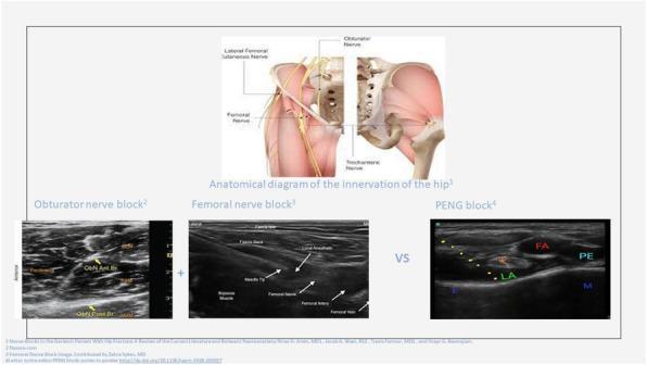
This educational medical graphic illustrates the innervation and regional anesthesia techniques for the hip. The top section features an anatomical diagram of the pelvis and upper thigh in two views: an anterior superficial view showing the Lateral Femoral Cutaneous, Femoral, and Obturator nerves, and a deep view showing the Trochanteric nerve. The bottom section presents three ultrasound images demonstrating specific regional nerve blocks. 1) Obturator Nerve Block: Shows the anterior and posterior branches of the obturator nerve (ObN Ant. Br. and ObN Post Br.). 2) Femoral Nerve Block: Displays the needle tip approaching the femoral nerve, adjacent to the femoral artery, femoral vein, and psoas muscle under the fascia iliaca. 3) PENG (Pericapsular Nerve Group) Block: Highlights landmarks including the Femoral Artery (FA), Pectineus muscle (PE), iliopectineal ligament (LA), and iliacus muscle (M). The diagram serves as a clinical guide for anesthesiology and pain management, comparing traditional nerve blocks with the newer PENG technique for hip analgesia.
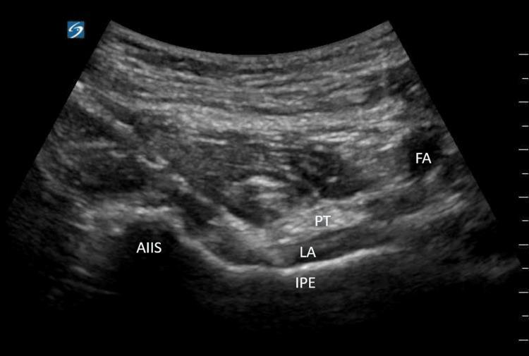
This diagnostic ultrasound image demonstrates the ultrasound-guided technique for a Pericapsular Nerve Group (PENG) block, a regional anesthesia procedure used for hip analgesia. The transverse view identifies key musculoskeletal and vascular landmarks. On the left, the anterior inferior iliac spine (AIIS) appears as a hyperechoic bony prominence. Centrally, the iliopubic eminence (IPE) is visible as a deep bony landmark. Superior to the IPE, the psoas tendon (PT) is visualized as an oval hyperechoic structure. To the right, the femoral artery (FA) appears as an anechoic circular lumen. A significant hyperechoic area labeled LA (local anesthetic) is seen deposited in the fascial plane between the psoas tendon and the iliopubic eminence, which is the target site for the PENG block to provide sensory blockade of the articular branches of the femoral, obturator, and accessory obturator nerves.
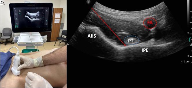
This composite educational image illustrates the Pericapsular Nerve Group (PENG) block, a regional anesthesia technique for hip analgesia. Panel A shows the clinical setup, including the patient’s supine position and the transverse orientation of a curved-array ultrasound probe at the level of the inguinal crease. Panel B presents the corresponding sonographic anatomy. Key landmarks include the hyperechoic bony contours of the anterior inferior iliac spine (AIIS) and the iliopubic eminence (IPE). The psoas tendon (PT) is identified as a hypoechoic oval structure resting between these bony prominences. The femoral artery (FA) is visualized medially as an anechoic, pulsatile vessel. A dashed red arrow demonstrates the in-plane needle trajectory, targeting the fascial plane between the psoas tendon and the pubic ramus. This block specifically targets the articular branches of the femoral nerve, obturator nerve, and accessory obturator nerve to provide sensory blockade of the anterior hip capsule without significant motor impairment.
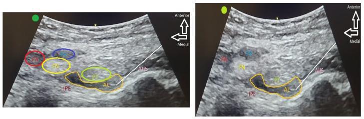
Diagnostic ultrasound images illustrating the Pericapsular Nerve Group (PENG) block for regional anesthesia. The composite contains two panels showing the cross-sectional anatomy of the anterior hip region. The left panel identifies key landmarks: the femoral artery (FA), femoral vein (FV), and femoral nerve (FN) located medially and superiorly. Deep to these lie the psoas tendon (PT), iliopubic eminence (IPE), and anterior inferior iliac spine (AIIS). The right panel demonstrates the clinical implementation of the block, where hypoechoic local anesthetic (AL) is seen spreading within the fascial plane between the psoas tendon and the underlying bony cortex (IPE and AIIS). The images highlight the anatomical plane containing sensory branches of the femoral, obturator, and accessory obturator nerves targeted for hip fracture analgesia. Orientation arrows indicate anterior and medial directions, emphasizing the deep approach required to reach the periarticular space while avoiding superficial neurovascular structures.
Regional Anaesthesia: 3-in-1 Block, PENG Block & Lumbosacral Block
MD Anaesthesia Reference Guide with Viva Questions
PART 1: THREE-IN-ONE (3-IN-1) BLOCK
Concept & Rationale
- Femoral nerve (L2-L4, posterior divisions)
- Lateral femoral cutaneous nerve (L2-L3)
- Obturator nerve (L2-L4, anterior divisions)
"The femoral nerve runs down the thigh in a fascial sheath that is continuous with the nerve sheath that contains all three nerves more proximally. If a large amount of local anesthetic is injected into this sheath, it will track proximally, medially, and laterally and thereby block all three nerves."
- Roberts and Hedges' Clinical Procedures in Emergency Medicine
Anatomy
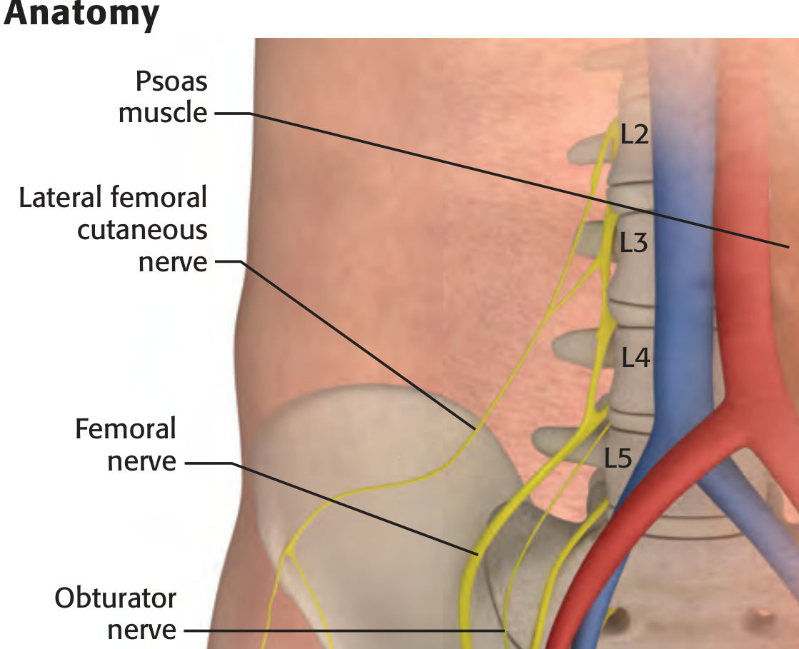
| Nerve | Origin | Course | Sensory Territory |
|---|---|---|---|
| Femoral | L2-L4 (posterior div.) | Lateral to femoral artery under inguinal ligament | Anterior thigh, femoral shaft, medial leg (saphenous) |
| Lateral femoral cutaneous | L2-L3 | 1-2 cm medial to ASIS, under inguinal ligament | Anterolateral thigh to knee |
| Obturator | L2-L4 (anterior div.) | Medial border of psoas, obturator canal | Medial thigh, hip joint capsule |
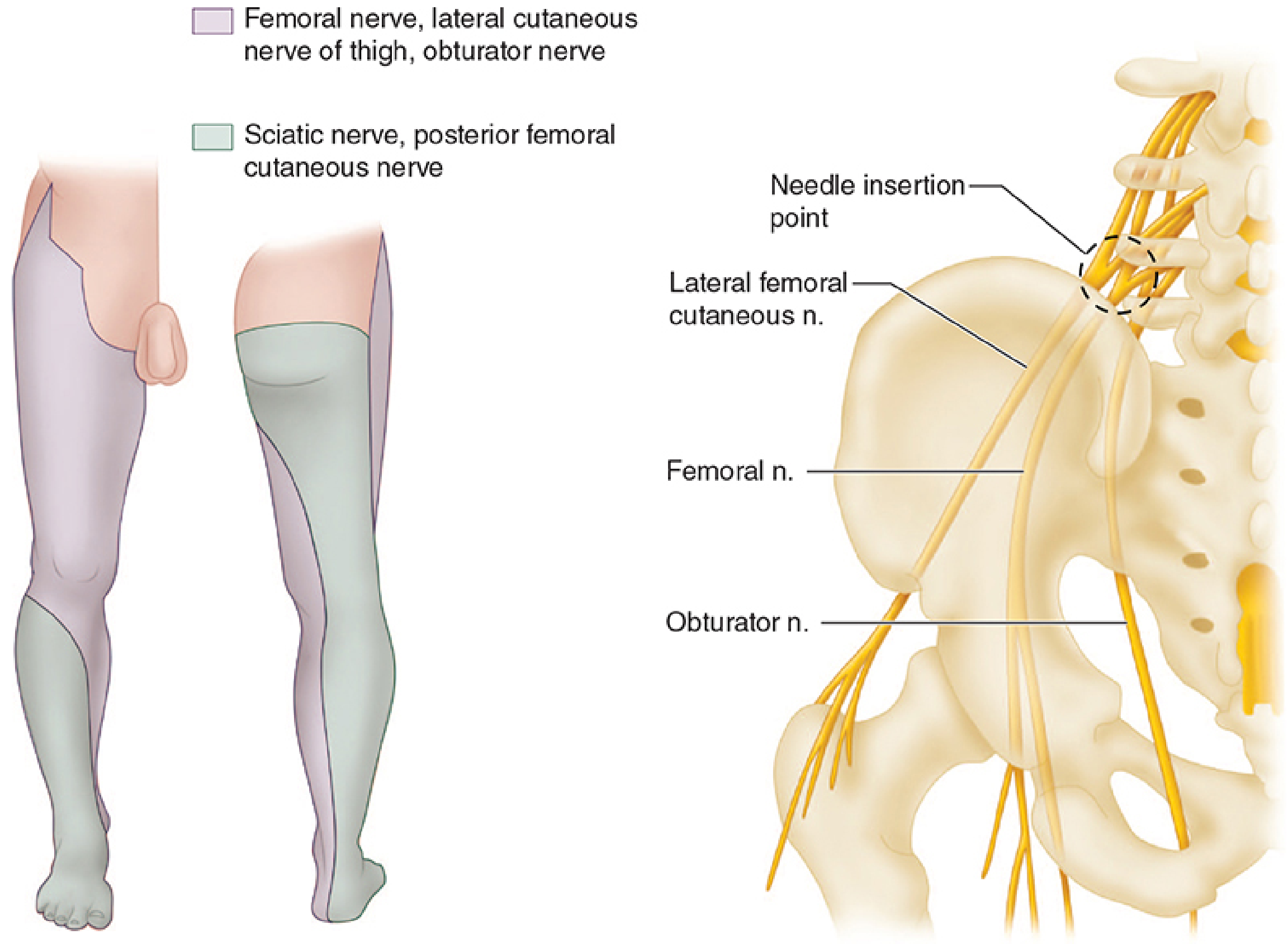
Indications
- Primary: Hip fracture analgesia (NOF fractures), especially elderly patients
- Anterior thigh and knee surgeries
- ORIF of femoral shaft
- Supplement to general/neuraxial anaesthesia for knee arthroplasty
- Patients with respiratory compromise (reduces opioid requirement)
Technique
- Patient supine
- Palpate femoral artery 2 cm distal to the inguinal ligament
- Mark injection point 1-2 cm lateral to the femoral artery (NAVEL: Nerve-Artery-Vein-Empty space-Lymphatics, lateral to medial)
- Raise a subcutaneous wheal of local anaesthetic
- Insert a 22-25G, 3.75 cm needle at 45-60° angle to skin, directed cephalad
- Advance until:
- "Pop" / loss of resistance as fascia lata penetrated, OR
- Paraesthesia elicited, OR
- Needle pulsates laterally (proximity to nerve)
- Inject 25-30 mL of local anaesthetic (larger volume than femoral block alone)
- Apply digital pressure distal to injection site to force fluid to track proximally
- High-frequency linear probe placed transversely below the inguinal ligament
- Identify femoral artery, vein, fascia iliaca, and femoral nerve (hyperechoic, lateral to artery)
- Insert needle in-plane (lateral to medial)
- Confirm local anaesthetic spread under fascia iliaca, circumferentially around femoral nerve
Local Anaesthetic Doses
| Drug | Concentration | Volume |
|---|---|---|
| Bupivacaine | 0.375-0.5% | 25-30 mL |
| Ropivacaine | 0.5% | 25-30 mL |
| Levobupivacaine | 0.375-0.5% | 25-30 mL |
| Lidocaine (with epinephrine) | 1.5-2% | 25-30 mL |
Complications
- Femoral artery/vein puncture (hence ASIS lateral landmark important)
- Local anaesthetic systemic toxicity (LAST)
- Nerve injury
- Haematoma
- Infection
- Quadriceps weakness (motor block - fall risk in elderly)
- Incomplete obturator block (common - in ~50% cases the obturator remains unblocked)
PART 2: PENG (PERICAPSULAR NERVE GROUP) BLOCK
Concept & Background
- Femoral nerve (articular branches)
- Obturator nerve (articular branches via accessory obturator nerve)
- Accessory obturator nerve
Anatomy
| Structure | US Appearance | Role |
|---|---|---|
| Anterior inferior iliac spine (AIIS) | Hyperechoic bony prominence (lateral) | Lateral landmark |
| Iliopubic eminence (IPE) | Hyperechoic bony prominence (central) | Medial landmark, target plane above |
| Psoas tendon (PT) | Oval hyperechoic structure | Superficial to IPE, needle goes lateral to it |
| Femoral artery (FA) | Anechoic, pulsatile (medial) | Safety landmark - avoid |
| Target plane | Between PT superiorly and pubic ramus | Site of local anaesthetic deposition |
Indications
- Hip fracture (NOF, intertrochanteric, subtrochanteric) - pre-op and post-op analgesia
- Total hip arthroplasty
- Hip arthroscopy
- When motor preservation is essential (early mobilisation, fall risk)
- Alternative when fascia iliaca block is contraindicated
Technique
- Patient supine
- Place curved-array (low-frequency) or linear (high-frequency) probe at the level of the inguinal crease, parallel to the inguinal ligament
- Probe orientation: from ASIS laterally to pubic tubercle medially
- Identify on US: AIIS, IPE (bony prominences), psoas tendon (oval, hyperechoic, between AIIS and IPE), femoral artery (pulsatile, medial)
- Insert 22G, 10 cm needle in-plane, lateral to medial
- Advance until needle tip contacts the pubic ramus lateral to psoas tendon
- Negative aspiration
- Inject 20 mL of local anaesthetic (e.g., 0.2-0.5% ropivacaine, 3 mg/kg up to 20 mL)
- Confirm spread: hypoechoic pool should lift the psoas tendon off the pubic ramus - "hydrodissection" of the fascial plane
Ultrasound Anatomy (Labeled Image)
Local Anaesthetic
- Ropivacaine 0.2-0.5%, 20 mL (3 mg/kg in children)
- Bupivacaine 0.25-0.5%, 20 mL
Advantages over 3-in-1 / Femoral Block
| Feature | Femoral / 3-in-1 | PENG |
|---|---|---|
| Motor block | Yes (quadriceps) | No (motor sparing) |
| Fall risk | High | Low |
| Obturator coverage | Variable | Articular branches covered |
| Technique | Relatively simple | Requires US skills |
| Evidence level | Strong (RCTs) | Emerging (case series, small RCTs) |
Complications
- Haematoma (femoral vessels adjacent)
- Intramuscular injection into psoas
- Incomplete analgesia (only articular branches, not all hip pain pathways)
- Femoral nerve palsy (if injected too superficially/laterally)
- LAST
PART 3: LUMBOSACRAL BLOCK
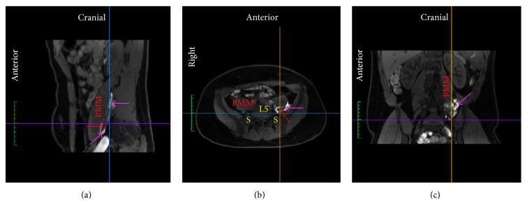
A. POSTERIOR LUMBAR PLEXUS BLOCK (Psoas Compartment Block)
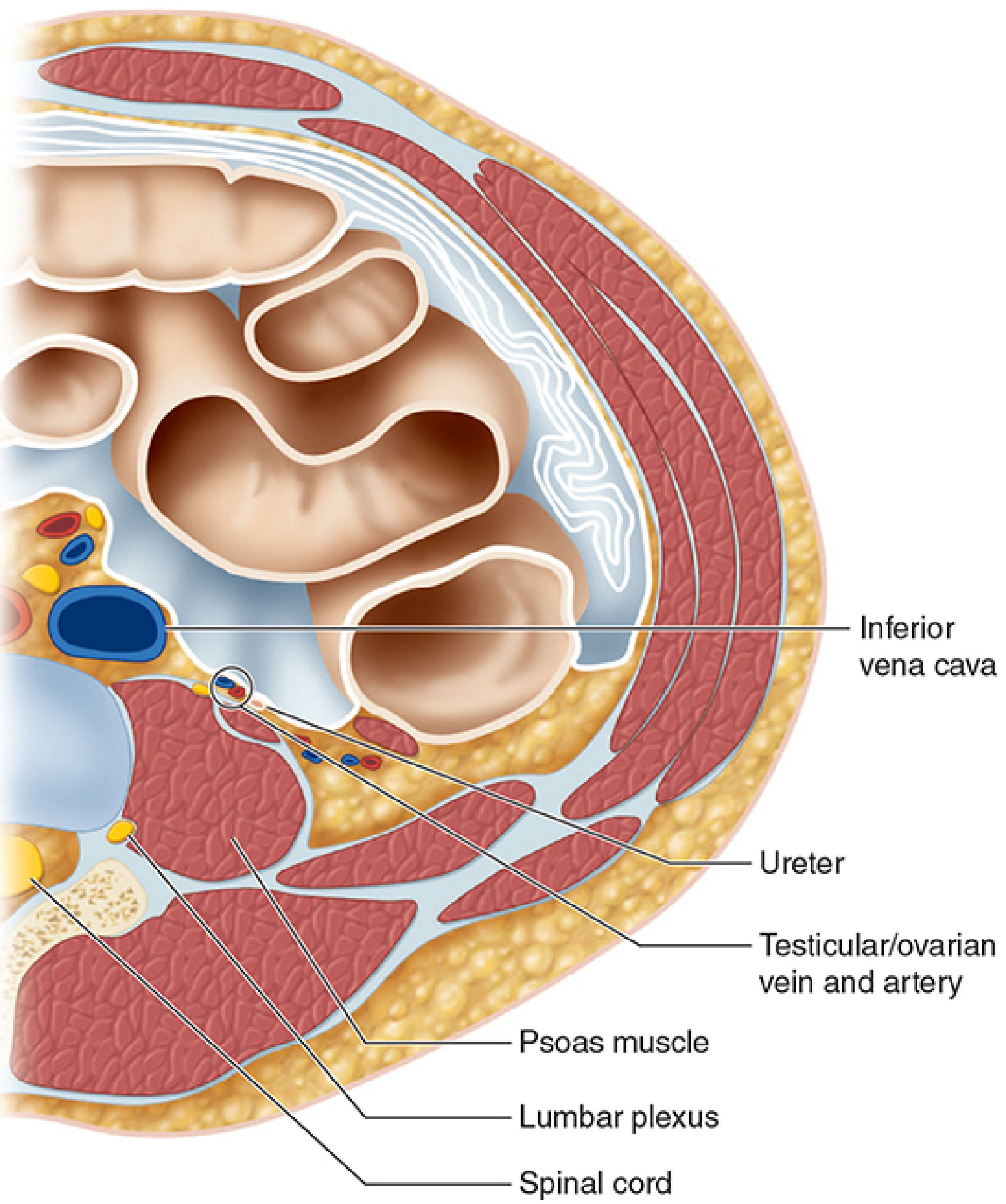
- Hip surgery (arthroplasty, ORIF)
- Femoral shaft fracture
- Anterior thigh surgery
- Combined with sciatic block: complete lower limb anaesthesia
- Patient lateral decubitus (operative side up) or prone
- Draw a line connecting both iliac crests (intercristal line ≈ L4 level)
- Draw a parasagittal line through the posterior superior iliac spine (PSIS)
- Injection point: intersection of the intercristal line with the lateral third of the distance between midline and PSIS
- Insert a long (10-15 cm), insulated stimulating needle perpendicular to the skin
- Advance until transverse process is contacted, then "walk off" caudally
- Never insert >3 cm past the transverse process depth
- Endpoint: quadriceps contraction (femoral motor response)
- Inject 20-30 mL local anaesthetic
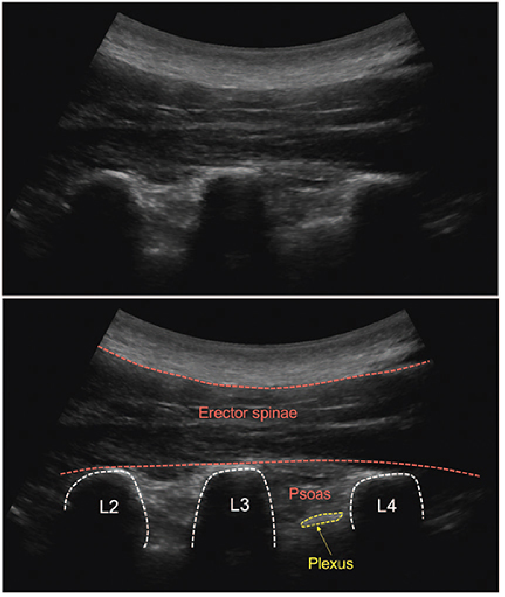
- Large curvilinear probe, parasagittal orientation, lateral to lumbar spinous processes
- Identify the "trident sign": acoustic shadows of L2, L3, L4 transverse processes
- Psoas muscle visible between shadows (striated appearance)
- Lumbar plexus = hyperechoic density in posterior part of psoas
- Needle in-plane, between L3-L4 transverse processes
- Inject 20 mL into the nerve root plane
- Ropivacaine 0.5%, 20-30 mL
- Bupivacaine 0.375-0.5%, 20-30 mL
- Retroperitoneal haematoma (most serious - cannot compress)
- Epidural / intrathecal spread (bilateral block, total spinal)
- Intravascular injection (aorta, inferior vena cava, renal vessels proximity)
- Renal capsular puncture
- Nerve injury
- Contraindicated in anticoagulated patients
B. SACRAL PLEXUS BLOCK (PARASACRAL BLOCK)
- Combined with lumbar plexus block for complete lower limb anaesthesia (hip disarticulation, extensive thigh surgery)
- Perineal surgeries
- When PFCN coverage is needed
- Patient lateral decubitus, operative side up
- Draw a vertical line from PSIS to ischial tuberosity
- Mark a point 6 cm inferior to PSIS along this line
- Insert long stimulating needle perpendicular to skin (parasagittal orientation)
- Advance until motor response in leg or foot is elicited (usually at ~7 cm depth)
- Inject 15-20 mL local anaesthetic
- Caution: Pelvic viscera are at risk at this depth
Combined Lumbosacral Block: Coverage Summary
| Block Component | Nerves Blocked | Coverage |
|---|---|---|
| Posterior lumbar plexus | Femoral, LFCN, Obturator, genitofemoral | Anterior + medial thigh, hip |
| Parasacral / sciatic | Sciatic (tibial + common peroneal), PFCN | Posterior thigh, entire leg below knee |
| Combined | All major lower limb nerves | Complete lower limb (except perineum) |
QUICK COMPARISON TABLE
| Feature | 3-in-1 Block | PENG Block | Lumbar Plexus Block |
|---|---|---|---|
| Target | Femoral, LFCN, Obturator (main trunks) | Articular branches of femoral/obturator/AON | Lumbar plexus (L1-L4) within psoas |
| Approach | Anterior (inguinal) | Anterior (inguinal crease) | Posterior (paravertebral) |
| Motor block | Yes (quadriceps) | No (motor sparing) | Yes |
| Volume | 25-30 mL | 20 mL | 20-30 mL |
| US needed | Helpful, not mandatory | Mandatory | Strongly recommended |
| Key complication | Quadriceps weakness/fall risk | Haematoma, intramuscular injection | Retroperitoneal haematoma, epidural spread |
| Evidence | Strong (RCTs) | Emerging | Strong |