

Diffrentials & prognosis
pediatric intestinal lymphoma ileum differential diagnosis
Burkitt lymphoma starry sky pattern histology
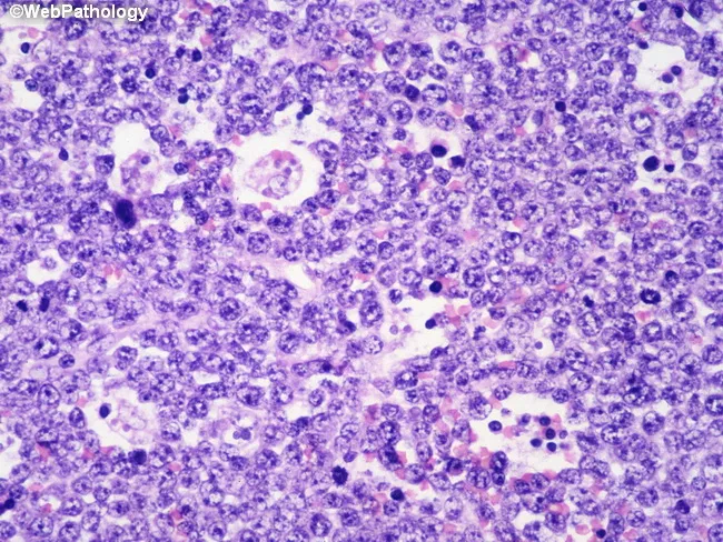
Imaging modality and technique: Light microscopy of hematoxylin and eosin stained renal tissue section. The specimen reveals a densely cellular infiltrate composed of medium-sized lymphoid cells with round to oval nuclei, finely clumped chromatin, and multiple nucleoli. The tumor cells are arranged in diffuse sheets with high mitotic activity and numerous apoptotic figures. Interspersed histiocytes actively phagocytose apoptotic debris, producing scattered tingible-body macrophages that contribute to a characteristic 'starry-sky' pattern. In addition to uniform cytology, occasional mitotic figures are evident, underscoring the tumor’s aggressive biology. The background kidney parenchyma shows limited residual tubular architecture, with infiltration disrupting normal architecture. Immunophenotypic context (if tested) would typically show B-cell markers with a very high proliferation index; c-MYC rearrangement is a common molecular feature of Burkitt lymphoma. The lesion is localized within the renal parenchyma, a recognized, though less common, site of extranodal Burkitt lymphoma. Clinically, this histology corresponds to an aggressive B‑cell lymphoma requiring prompt oncologic management; differential considerations include other high-grade B-cell lymphomas (DLBCL), Burkitt-like lymphoma, and lymphoblastic lymphoma. The image is valuable for educational purposes, illustrating classic cytologic features, proliferative activity, and the hallmark starry-sky pattern in a renal presentation. This histology supports rapid diagnosis and urgent multidisciplinary treatment planning.
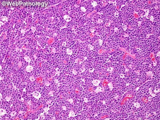
Comprehensive description of Burkitt lymphoma histology. This is a bright-field light microscopy image of an H&E stained lymph node biopsy. The tissue demonstrates the hallmark 'starry sky' pattern produced by numerous pale tingible-body macrophages interspersed among a dense population of medium-sized lymphoid cells. The malignant cells are small to medium with round nuclei, coarse chromatin, multiple small nucleoli, and scant cytoplasm, creating a high nuclear-to-cytoplasmic ratio. Mitotic activity is brisk, contributing to marked cellular turnover. The macrophages ingest apoptotic debris, appearing as clear spaces that resemble stars against a deep blue background of neoplastic lymphocytes. The overall architecture is diffuse, with little stromal preservation, consistent with a high-grade B-cell lymphoma. Immunophenotype is typically CD20+, CD10+, and BCL6+, with a near-100% Ki-67 proliferation index; genetic hallmark is c-MYC translocation, most commonly t(8;14)(q24;q32). Clinically, this pattern is characteristic of Burkitt lymphoma (aggressive non-Hodgkin lymphoma) and guides rapid, intensive chemotherapy. The image supports differential considerations including diffuse large B-cell lymphoma and lymphoblastic lymphoma; however, the starry-sky morphology remains highly specific in context. This description is relevant for medical education, pathology teaching, and research datasets to illustrate classic Burkitt histology, high-grade lymphoid neoplasia, and the diagnostic utility of abundant tingible-body macrophages.
Bright-field light microscopy of a renal biopsy stained with Hematoxylin and Eosin demonstrates infiltration of the renal parenchyma by a uniform population of medium-sized lymphoid cells with high nuclear-to-cytoplasmic ratio, round nuclei, finely dispersed chromatin, and prominent nucleoli. The neoplastic cells arrange in diffuse sheets, with scattered macrophages containing apoptotic debris that create a conspicuous starry-sky pattern. Mitotic activity is brisk, reflecting the high proliferative index typical of Burkitt lymphoma. The background renal architecture is variably preserved but overall shows tumor effacement of the cortex with scant residual tubules. The hallmark starry-sky appearance arises from numerous tingible-body macrophages amid the dense cellularity. Immunophenotype favors mature B-cell lineage: CD19/CD20 positive, CD10 and BCL6 positive; Ki-67 nearly 100% underscores extreme proliferative rate. Cytogenetics commonly reveal MYC rearrangement, most often t(8;14)(q24;q32). Clinically, this histology indicates an aggressive, highly chemosensitive non-Hodgkin lymphoma with potential primary renal involvement or secondary infiltration. Differential diagnoses include diffuse large B-cell lymphoma and other high-grade lymphomas, but the extreme proliferative index and starry-sky pattern support Burkitt lymphoma. Management requires prompt systemic therapy, staging, CNS assessment, and molecular testing for MYC rearrangement. This image serves as an educational resource for hematopathology, surgical pathology, and correlates with radiologic assessment.
Pathology Report Summary
Differentials
1. Burkitt Lymphoma (Most Likely)
| Feature in Report | Classic Burkitt Finding |
|---|---|
| 12-year-old male | Affects mainly children & young adults |
| Ileal location | Sporadic Burkitt most commonly presents as ileocecal mass |
| Focal "starry-sky" pattern | Hallmark - tangible body macrophages phagocytosing apoptotic debris |
| ~30/10 HPF mitoses | Among the fastest-growing human tumors |
| Small-to-medium cells, coarse chromatin, inconspicuous nucleoli | Classic Burkitt morphology |
| Indian-file/sheet/cord arrangement | Diffuse growth, no follicular architecture |
| No granulomas | Excludes Crohn's |
"Sporadic Burkitt lymphoma most often appears as a mass involving the ileocecum and peritoneum." - Robbins & Kumar Pathologic Basis of Disease
"The tumor exhibits a high mitotic index and contains numerous apoptotic cells...creating a characteristic 'starry sky' pattern." - Robbins & Kumar Pathologic Basis of Disease
2. Diffuse Large B-Cell Lymphoma (DLBCL) - with MYC/BCL2/BCL6 "Double/Triple Hit" variant
- The report specifically notes cytologic variation in deeper areas with medium-to-large cells, irregular nuclear contours, and prominent nucleoli - this is more in keeping with DLBCL morphology than classic Burkitt
- Recommended IHC panel includes BCL2, BCL6, and MYC - the classic "double/triple-hit" lymphoma workup
- DLBCL is the most common high-grade NHL overall
- High-grade B-cell lymphoma with MYC + BCL2/BCL6 rearrangements ("double/triple-hit") is a separate WHO category that can morphologically mimic Burkitt
3. Lymphoblastic Lymphoma (B-cell or T-cell precursor)
- The report recommends TdT and CD34 staining - these are the definitive markers of lymphoblastic lymphoma (precursor lymphoid neoplasm)
- Presents predominantly in children/adolescents
- Can involve the GI tract
- The cells can be small-to-medium with scant cytoplasm and inconspicuous nucleoli
4. Other Considerations (Less Likely)
- Mantle Cell Lymphoma (rare in children, usually CD5+, cyclin D1+)
- Enteropathy-associated T-cell lymphoma (EATL) (extremely rare at age 12, requires prior celiac disease; CD3+ T-cell phenotype)
- EBV-associated B-cell lymphoproliferative disorder (EBER-ISH included in panel to exclude this)
IHC Panel Interpretation Guide
| Marker | Burkitt | DLBCL | Lymphoblastic |
|---|---|---|---|
| CD20, PAX5 | + | + | + (B-cell type) |
| CD10 | + | +/- | +/- |
| BCL6 | + | + | - |
| BCL2 | Negative | + | +/- |
| MYC | + (nearly 100%) | 30-40% | - |
| Ki67 | ~100% | 60-90% | High |
| TdT | Negative | Negative | Positive |
| CD34 | Negative | Negative | Positive |
| CD3 | Negative | Negative | + (T-cell type) |
| EBER-ISH | +/- (15-20%) | +/- | - |
Prognosis
If Burkitt Lymphoma:
- Highly favorable with intensive chemotherapy despite aggressive biology
- Children and young adults: >90% cure rate with intensive regimens (e.g., CODOX-M/IVAC, R-CHOP protocols modified for pediatric use)
-
"Burkitt lymphoma is very aggressive but responds well to intensive chemotherapy. Most children and young adults can be cured." - Robbins & Kumar Pathologic Basis of Disease
- Stage matters: localized (Stage I/II) resected disease - cure rates approach 98%; advanced (Stage III/IV with marrow/CNS) - ~80-85% with aggressive therapy
- The fact that this tumor was surgically resected at a localized ileal site is a favorable prognostic sign
If DLBCL (including double/triple-hit):
- Standard DLBCL in children responds well to chemotherapy (event-free survival ~80%)
- "Double/triple-hit" lymphoma (MYC + BCL2/BCL6 co-rearrangement): significantly worse prognosis, ~40-50% long-term survival; requires intensified regimens (DA-R-EPOCH or similar)
If Lymphoblastic Lymphoma:
- Treated like ALL (ALL-style protocols); pediatric complete remission rates >80-90%
- CNS prophylaxis is mandatory
Immediate Clinical Action
- Rush IHC on the existing tissue block (no re-biopsy needed) for the recommended panel
- Urgent pediatric oncology referral - time-sensitive given 30/10 HPF mitotic rate
- Staging workup: CT chest/abdomen/pelvis, bone marrow biopsy, LP for CSF cytology (CNS involvement), LDH (tumor burden marker)
- FISH studies for MYC, BCL2, BCL6 rearrangements - will guide chemotherapy protocol selection