
My dad has got melasma... How long it takes to get cure
melasma treatment duration how long to cure 2025
melasma facial hyperpigmentation clinical photo
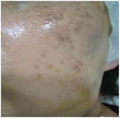
This clinical photograph displays the facial skin of a patient with melasma, specifically focusing on the lateral cheek and temple area. The image reveals numerous hyperpigmented macules and patches of varying sizes and shapes. These pigmented lesions range in color from light tan to dark brown, demonstrating a patchy, heterogeneous distribution typical of dermal or mixed melasma. The skin surface appears smooth and hydrated with a visible reflective sheen, and there are no signs of scaling, erythema, or post-inflammatory hyperpigmentation (PIH). The visual evidence represents a follow-up state after multiple treatment modalities, including Q-switched 1064 nm Nd:YAG laser sessions and adjuvant vitamin C therapy. The educational focus is on identifying clinical patterns of facial hyperpigmentation and evaluating treatment response in dermatologic practice for disorders of pigmentation.
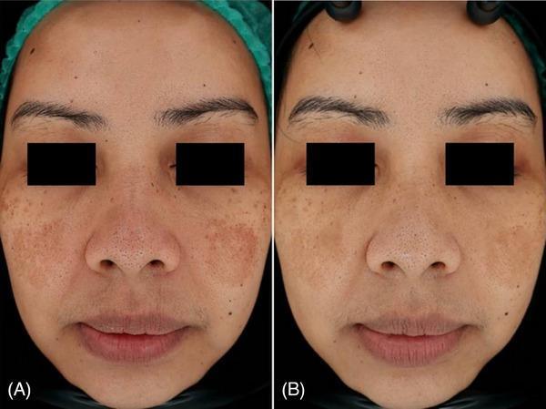
This clinical photograph comparison shows the face of a 39-year-old female patient diagnosed with facial melasma, highlighting a before-and-after treatment progression over three months. Image (A) displays baseline hyperpigmentation characterized by confluent, brownish patches with irregular borders, symmetrically distributed across the malar regions (cheeks) and the nasal bridge. The pigmentation appears dense with a mottled texture. Image (B), taken three months after laser therapy, demonstrates a significant clinical improvement. There is a visible reduction in the size, color intensity, and overall density of the melasma patches. The skin tone appears more uniform and brightened, with less prominent follicular or pore visibility in the affected areas. This comparison illustrates the effectiveness of dermatological laser intervention for managing epidermal and dermal hyperpigmentation disorders in adult patients.
Melasma: How Long Does It Take to Improve?
The Honest Answer: Manageable, Not Permanently "Cured"
Timeline by Treatment Type
| Treatment | Visible Improvement |
|---|---|
| Topical creams (hydroquinone, tretinoin) | 8-12 weeks |
| Triple combination cream (Kligman's formula) | 8-12 weeks, often used in cycles |
| Chemical peels (glycolic, salicylic acid) | 4-8 weeks per session |
| Laser/IPL therapy | 3-6 sessions over weeks to months |
| Oral tranexamic acid | 8-12 weeks with doctor's monitoring |
| Sun protection alone | Gradual; prevents worsening |
The Gold Standard Treatment
- Hydroquinone 4% (inhibits melanin production)
- Tretinoin cream (speeds up skin cell turnover)
- Mild topical corticosteroid (reduces inflammation)
Important Factors That Affect How Fast He Improves
- Skin type: Darker skin types (common in Asians, Hispanics, South Asians) respond more slowly and need gentler treatments to avoid further darkening
- Depth of pigment: "Epidermal" melasma responds faster than "dermal" melasma, which sits deeper in the skin and is harder to treat
- Sun protection: This is NON-NEGOTIABLE. Without daily SPF 50+ sunscreen, no treatment will work well. Sun exposure is the #1 trigger - as per Fitzpatrick's Dermatology, melasma always worsens in summer
- Hormones: If caused by oral contraceptives or hormone therapy, switching or stopping them (with a doctor's advice) helps - but the pigment can still persist for years
- Triggers: If the cause is a medication like phenytoin or finasteride, addressing it speeds recovery
Key Practical Tips for Your Dad
- Use broad-spectrum SPF 50+ sunscreen every single day, even indoors (visible light also triggers melasma). Reapply every 2 hours outdoors
- See a dermatologist for a prescription-strength hydroquinone cream - over-the-counter 2% versions are weaker
- Avoid self-treating with harsh bleaching agents - overuse of hydroquinone can cause a permanent darkening called exogenous ochronosis
- Be patient: Expect 2-3 months before seeing a real difference. Most people need maintenance therapy long-term
- Avoid laser treatments without expert supervision - Q-switched Nd:YAG laser in particular can actually increase pigmentation if used incorrectly
Quick Summary
- First visible results: 6-8 weeks
- Significant improvement: 3-6 months of consistent treatment
- Full control: Ongoing; may require indefinite maintenance
- Recurrence risk: High without strict sun protection
Why tracrolimus ointment used
tacrolimus ointment melasma mechanism of action why used
Why Is Tacrolimus Ointment Used in Melasma?
What Is Tacrolimus?
How Does It Work? (Mechanism of Action)
- Binding to FK506 binding protein (FKBP) inside immune cells
- This complex then inhibits calcineurin, a calcium-dependent enzyme
- This blocks the movement of NFAT (Nuclear Factor of Activated T-cells) into the cell nucleus
- Result: T-cell activation is disrupted and pro-inflammatory cytokines like IL-2, IL-3, IL-4, IL-12, and TNF-α are NOT produced
Why Specifically in Melasma?
| Reason | Explanation |
|---|---|
| Anti-inflammatory effect | Reduces local skin inflammation that stimulates melanocytes (pigment cells) to produce excess melanin |
| Steroid-sparing alternative | The standard Kligman's triple combination uses a corticosteroid to control inflammation - tacrolimus can replace this steroid, avoiding long-term steroid side effects (skin thinning, telangiectasia, perioral dermatitis) |
| Blocks cytokine-driven pigmentation | Cytokines like IL-1 and TNF-α stimulate melanogenesis; by blocking them, tacrolimus slows down pigment production |
| Used in combination formulas | In pilot studies, tacrolimus combined with tazarotene, azelaic acid, and zinc oxide showed effective melanin reduction by targeting multiple pathways at once |
Why Is It Preferred Over Topical Steroids?
- Skin atrophy (thinning)
- Stretch marks
- Telangiectasia (spider veins)
- Perioral dermatitis
- Rebound pigmentation when stopped
Is It Effective for Melasma?
- The patient cannot tolerate steroids long-term
- Used as a maintenance therapy after initial clearing with Kligman's formula
- Combined with hydroquinone, tretinoin, or azelaic acid