
Reason for mcv drop in cbc
MCV (Mean Corpuscular Volume) measures the average size of red blood cells. A low MCV indicates microcytic anemia. The main causes are iron deficiency, thalassemia, anemia of chronic disease, and sideroblastic anemia.
Searching sources...
Reading File
Finding Sources
Searching PubMed
microcytic anemia low MCV causes
Finding Sources
Reading File
Reading File
Reading File
Loading Image
Here is a thorough breakdown of the causes of a low MCV (microcytosis) on CBC, drawn from authoritative medical textbooks.
Causes of Low MCV (Microcytic Anemia)
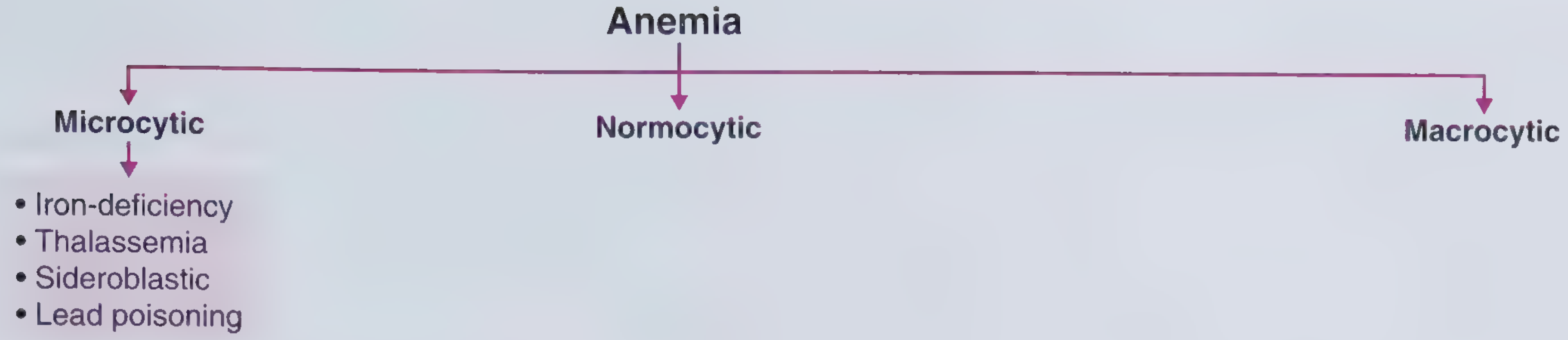
The mnemonic TAILS covers the main causes:
1. Iron Deficiency Anemia (IDA) - Most Common
The most common cause of anemia worldwide. Iron is required for hemoglobin synthesis; when iron stores are depleted, the bone marrow produces smaller, paler RBCs.
Causes of iron deficiency itself:
- Chronic blood loss - GI bleeding (ulcers, colorectal cancer, IBD), menstrual loss (most common in premenopausal women)
- Insufficient intake - malnutrition, poor diet
- Decreased absorption - celiac disease, post-gastrectomy
- Increased demand - pregnancy, infancy, rapid growth
Key labs: Low serum ferritin (<15 ng/mL confirms; >100 ng/mL rules out), low serum iron, high TIBC, low transferrin saturation, microcytosis + hypochromia on smear.
- Frameworks for Internal Medicine, p. 331
2. Thalassemia
An inherited (autosomal recessive) condition with decreased or absent synthesis of the alpha or beta globin chains of hemoglobin. The unmatched chains form abnormal hemoglobin tetramers, leading to ineffective erythropoiesis and hemolysis.
- Alpha-thalassemia: caused by deletions in the HBA1/HBA2 genes (common in Southeast Asian, African populations)
- Beta-thalassemia: caused by point mutations (common in Mediterranean, Middle Eastern populations)
Key clue: Microcytic anemia with a normal or elevated RBC count, target cells and teardrop cells on peripheral smear, basophilic stippling, nucleated RBCs.
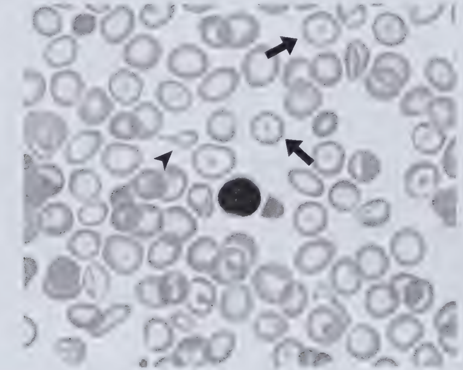
Peripheral blood smear from a patient with beta-thalassemia minor. (From Wintrobe's Atlas of Clinical Hematology, 2nd ed.)
3. Anemia of Chronic Disease (ACD)
Also called anemia of inflammation. Caused by defective iron utilization/metabolism due to upregulation of hepcidin (an acute-phase reactant), which sequesters iron in macrophages. The iron is present but unavailable for RBC production.
Associated with:
- Chronic infections (TB, HIV, osteomyelitis)
- Connective tissue disorders (rheumatoid arthritis, lupus)
- Malignancy
- Renal, thyroid, and pituitary disorders
Key distinction from IDA: ACD has normal-to-elevated ferritin (iron is stored but trapped), low serum iron, low TIBC. MCV is often only mildly low or even normal.
- Henry's Clinical Diagnosis and Management by Laboratory Methods, p. 129
4. Sideroblastic Anemia
A group of inherited and acquired conditions with impaired heme biosynthesis. Iron accumulates in mitochondria of RBC precursors, forming ring sideroblasts visible on bone marrow biopsy (iron stain).
Acquired causes (more common):
- Toxins: alcohol (most common acquired cause)
- Medications: isoniazid (INH), chloramphenicol, linezolid
- Nutritional: copper deficiency, pyridoxine (B6) deficiency
- Myelodysplastic syndromes (MDS) - especially RARS subtype
Hereditary: X-linked (ALAS2 gene mutation)
Hb typically ranges 4-10 g/dL. Smear shows hypochromic microcytosis (though normocytosis or macrocytosis are also possible), ± basophilic stippling.
- Frameworks for Internal Medicine, p. 332
5. Lead Poisoning
Lead inhibits multiple enzymes in the heme synthesis pathway (notably ALA-dehydratase and ferrochelatase), resulting in microcytic, hypochromic anemia.
Classic finding: Basophilic stippling on peripheral blood smear (aggregated ribosomal RNA), along with microcytosis. Also causes elevated free erythrocyte protoporphyrin (FEP).
Quick Comparison Table
| Cause | Ferritin | Serum Iron | TIBC | RDW | Distinguishing Feature |
|---|---|---|---|---|---|
| IDA | Low | Low | High | High | Low ferritin, menstrual hx, GI bleed |
| Thalassemia | Normal/High | Normal | Normal | Normal/Low | High RBC count, target cells, family hx |
| ACD | Normal/High | Low | Low | Normal | Chronic illness, hepcidin up |
| Sideroblastic | High | High | Low/Normal | High | Ring sideroblasts on BM biopsy |
| Lead poisoning | High | High | - | - | Basophilic stippling, exposure hx |
Bottom line: The most common cause worldwide is iron deficiency. In clinical practice, always check ferritin first - a low ferritin is diagnostic of IDA, while a normal or elevated ferritin with low MCV should prompt consideration of thalassemia, ACD, or sideroblastic anemia.
This is a shared conversation. Sign in to Orris to start your own chat.