
DIFFERENTIATION AND FUNCTIONS OF CD8 D CYTOTOXIC T LYMPHOCYTES
Reading File
Now let me search the medical library for CD8+ cytotoxic T lymphocyte content in parallel.
Finding Sources
Finding Sources
Finding Sources
Reading File
Excellent - I found the main sections. Let me now read all the key sections from Cellular and Molecular Immunology and Janeway's in parallel.
Reading File
Reading File
Reading File
Now let me get the granule exocytosis/killing mechanism section and the Janeway's section on CD8 activation, and also fetch the key figures:
Reading File
Reading File
Now let me fetch the key diagrams and continue reading the granule killing and Fas-L sections:
Loading Image
Loading Image
Reading File
I now have comprehensive information. Let me fetch one more key figure showing the killing mechanisms:
Loading Image
I now have all the material needed for a comprehensive answer. Let me compose it.
CD8+ Cytotoxic T Lymphocytes (CTLs): Differentiation and Functions
1. Overview
CD8+ T cells are MHC class I-restricted lymphocytes that differentiate into cytotoxic T lymphocytes (CTLs) - the principal adaptive immune effectors against intracellular pathogens and tumor cells. Naive CD8+ T cells are incapable of killing and must first undergo antigen-driven proliferation and differentiation to acquire cytotoxic machinery.
Induction and Effector Phases of CD8+ T Cell Responses:
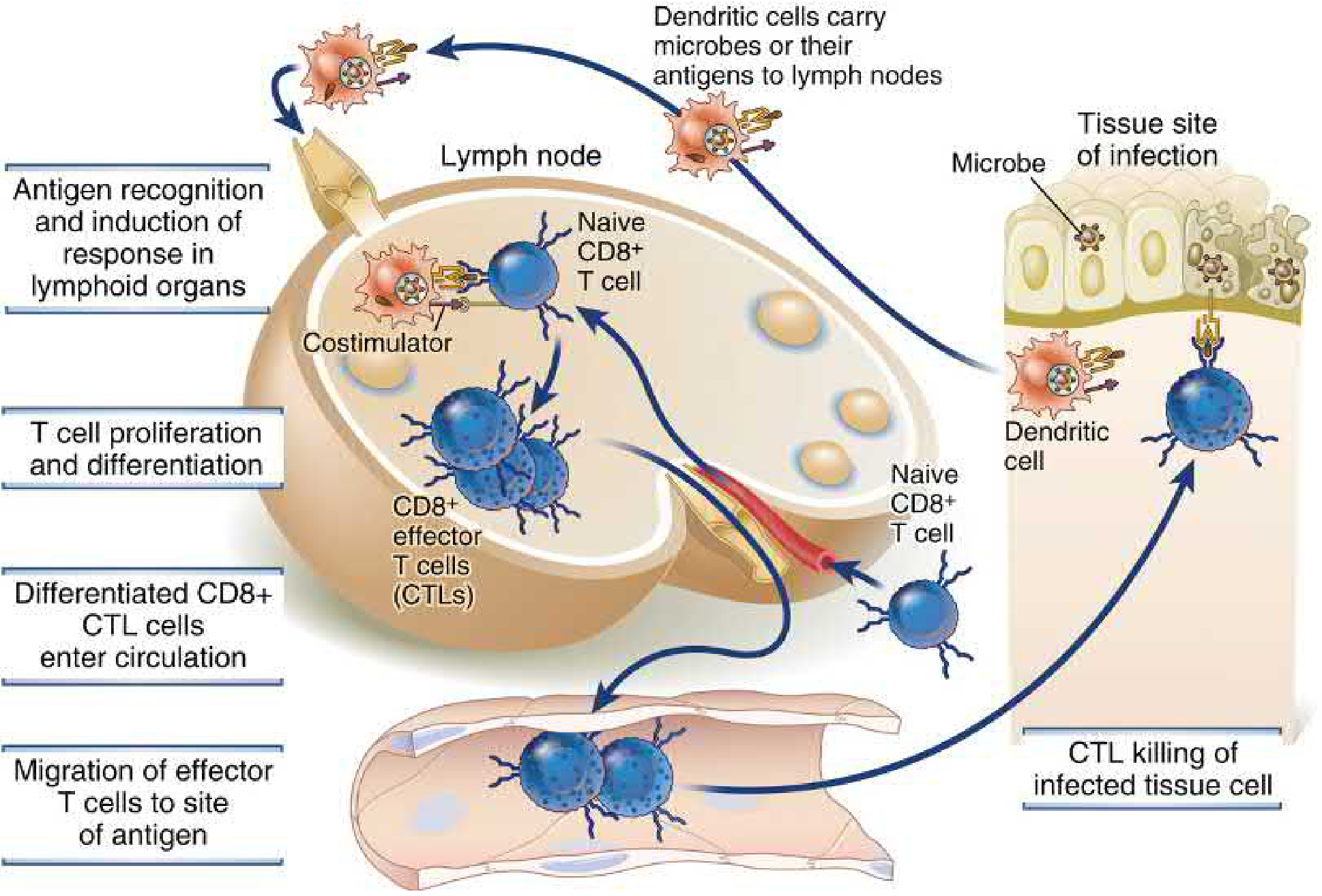
Dendritic cells carry microbial antigens to lymph nodes, where naive CD8+ T cells recognize antigen, receive costimulation, proliferate, and differentiate into CTLs. CTLs then enter circulation, migrate to sites of infection, and kill infected cells.
- Cellular and Molecular Immunology, Fig. 11.1
2. Differentiation of Naive CD8+ T Cells Into CTLs
2a. Antigen Presentation: Cross-Presentation
Naive CD8+ T cells require antigen displayed on MHC class I molecules. Since viruses and tumors arise from non-DC cells, a specialized subset of dendritic cells (the classical cDC1 subset) performs cross-presentation: ingesting infected/tumor cells, transferring protein antigens into the cytosol, and processing them through the proteasome-to-ER pathway for MHC class I loading. This is the essential first step for naive CD8+ T cell activation. - Cellular and Molecular Immunology, p. 728
2b. Three Signals for Activation
Like CD4+ T cells, naive CD8+ T cells need three signals:
| Signal | Molecule | Role |
|---|---|---|
| Signal 1 | TCR + peptide-MHC I | Antigen specificity |
| Signal 2 | CD28 + B7 (costimulation) | Prevents anergy |
| Signal 3 | Cytokines (IL-2, IL-12, IFN-γ, IL-27) | Drives proliferation and differentiation |
2c. Role of CD4+ T Helper Cells
In many infections, especially with latent viruses or non-immunogenic tumors, CD8+ T cell activation requires CD4+ T cell help. Two mechanisms operate:
- Licensing of DCs: CD4+ Th cells interact with DCs via CD40L-CD40 signaling, upregulating B7 costimulators so the DC can efficiently prime CD8+ T cells.
- Direct help: IL-2 produced by CD4+ Th cells supports CD8+ T cell proliferation and survival. - Cellular and Molecular Immunology, p. 535
2d. Transcriptional Control - T-bet and Eomes
The master transcription factor T-bet drives CTL differentiation, promoting expression of perforin, granzymes, and cytolytic machinery - similar to its role in Th1 differentiation. T-bet is activated by IL-12, IFN-γ, and TCR signals acting in concert.
After infection is cleared, most CTLs undergo apoptotic contraction. A small subset survives and differentiates into memory CD8+ T cells, preferentially expressing a related transcription factor, Eomes (Eomesodermin), which drives the memory phenotype. Memory cells are characterized by IL-7R expression, enabling long-lived survival and rapid recall responses. Genetic ablation of Eomes profoundly impairs memory generation while having little effect on primary CTL numbers. - Roitt's Essential Immunology, p. 219
3. Effector Functions of CD8+ CTLs
3a. Antigen Recognition and Immune Synapse Formation
CTLs recognize target cells via the TCR + CD8 coreceptor binding peptide-MHC class I. This triggers formation of the immunological synapse - a highly organized junction between the CTL and target cell:
- Signaling patch (central supramolecular activation cluster): contains TCR, CD8, PKC-θ, and LCK
- Secretory region: the site of directed granule exocytosis
- Outer ring: LFA-1 (on CTL) binding ICAM-1 (on target) stabilizes the synapse
The enclosed synaptic gap ensures that lethal molecules are delivered only to the target, not to bystander cells. - Cellular and Molecular Immunology, pp. 734-735
Steps in CTL-Mediated Killing:
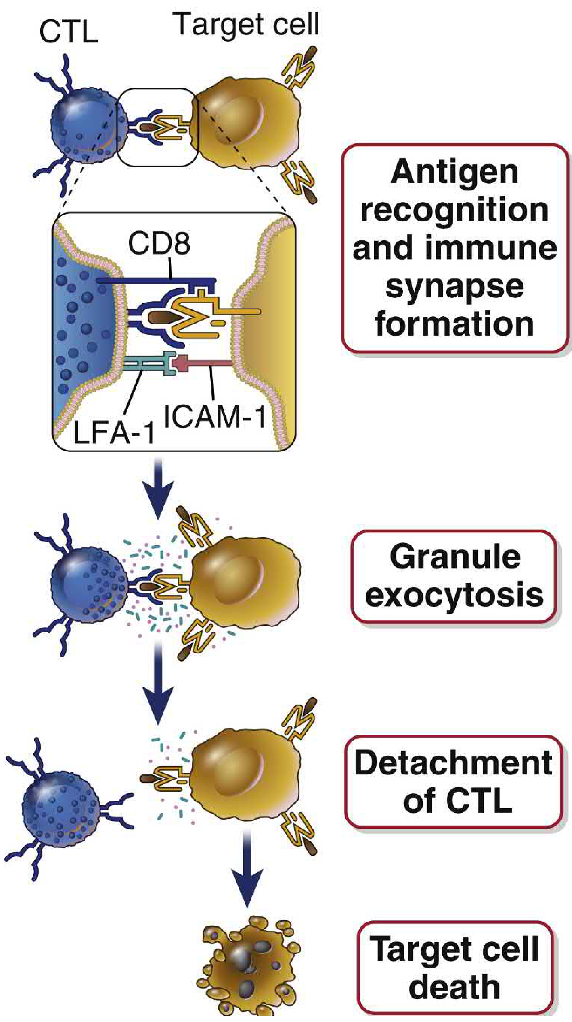
The CTL forms an immune synapse with the target cell, exocytoses granules directionally, then detaches - leaving the target to die by apoptosis.
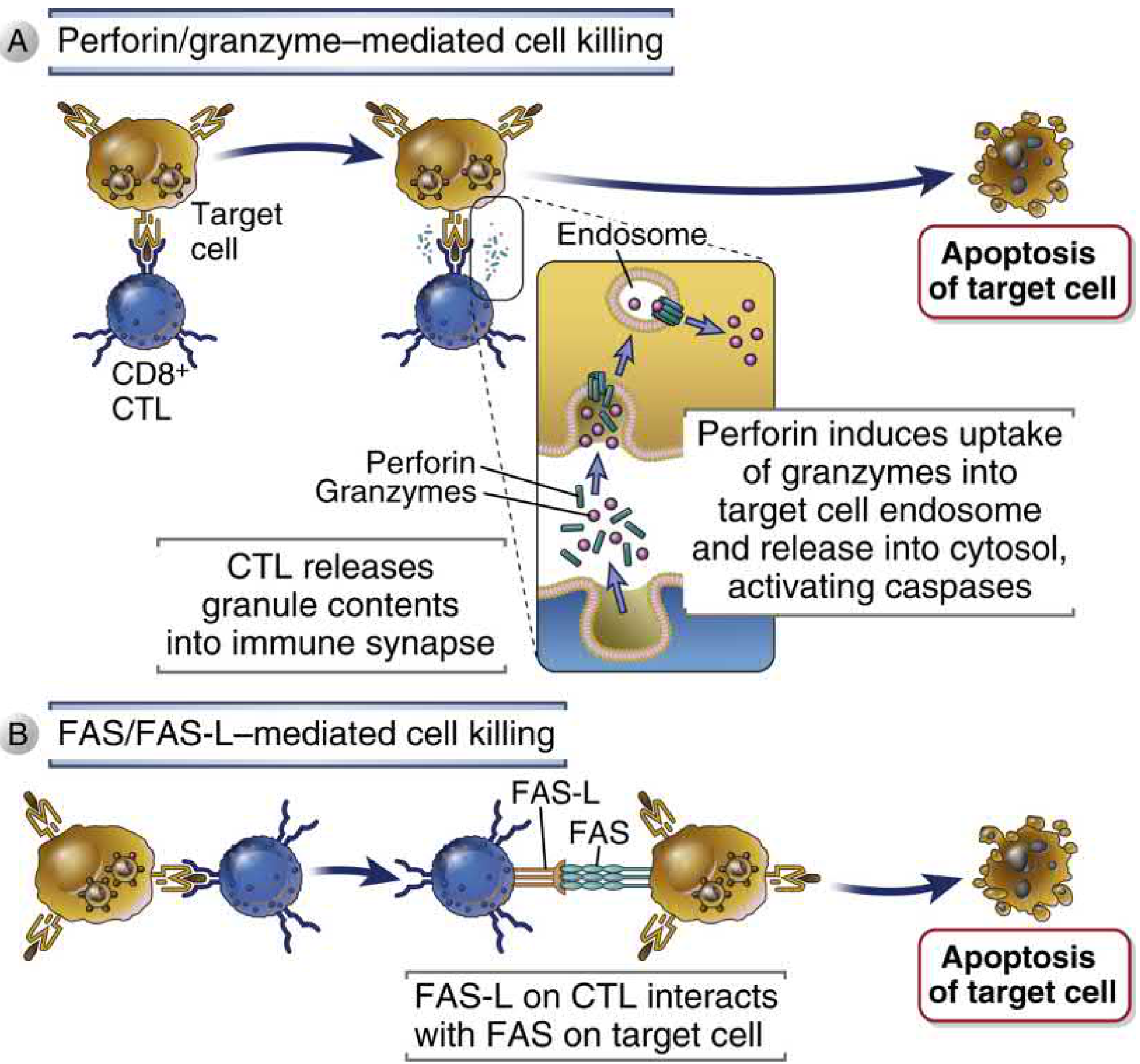
3b. Mechanism 1 - Granule Exocytosis (Perforin/Granzyme Pathway)
This is the principal killing mechanism.
Process:
- TCR activation triggers actin cytoskeleton reorganization
- The microtubule organizing center (MTOC) reorients toward the synapse
- Cytoplasmic granules (modified lysosomes) transport along microtubules to the synapse
- Granule membrane fuses with the CTL plasma membrane at the secretory region
- Granule contents are exocytosed into the synaptic cleft
- Death of the target cell occurs over the following 2-6 hours, even after CTL detachment ("lethal hit")
Key granule proteins:
| Protein | Function |
|---|---|
| Perforin | Homologous to C9 complement; facilitates granzyme entry into target cell cytosol |
| Granzyme A | Serine protease; induces caspase-independent apoptosis |
| Granzyme B | Serine protease; cleaves after Asp residues; directly activates caspases |
| Granzymes H, K | Additional serine proteases in CD8+ CTLs |
| Serglycin | Sulfated proteoglycan; holds granzymes and perforin inactive in granules |
Perforin enables uptake of granzymes into target cell endosomes, from which they escape into the cytosol to activate the caspase cascade and induce apoptosis. - Cellular and Molecular Immunology, pp. 738-740
CTL self-protection: CTLs are shielded from their own granule contents by:
- Cytosolic Spi6 (serine protease inhibitor that antagonizes granzyme B)
- Cathepsin B delivered to the CTL surface on exocytosis, which degrades any stray perforin near the CTL membrane
3c. Mechanism 2 - Fas/FasL Pathway
Activated CTLs express FasL (CD95L) on their surface, which binds Fas (CD95/APO-1) on the target cell. Fas-FasL interaction activates the extrinsic apoptosis pathway via FADD and caspase-8, leading to target cell death. This pathway is particularly important for:
- Killing activated T cells (immune contraction/peripheral tolerance)
- Killing targets that express Fas but may be resistant to granule-mediated killing - Cellular and Molecular Immunology, p. 667 (Fig. 11.6)
3d. Cytokine Production
CD8+ CTLs also secrete cytokines that mediate non-cytotoxic effector functions:
- IFN-γ: activates macrophages for enhanced microbicidal activity (classical activation); assay for frequency of antigen-specific CD8+ T cells
- TNF: contributes to inflammation and direct cytotoxicity
- IL-17: abundant in chronic skin inflammatory diseases (e.g., psoriasis)
CD8+ T cells arrive earlier and in greater numbers than CD4+ T cells in some reactions (e.g., contact sensitivity). - Cellular and Molecular Immunology, p. 745
4. Roles in Host Defense
| Context | Role of CTLs |
|---|---|
| Viral infections | Kill virus-infected cells (viruses live in non-phagosomal compartments inaccessible to antibodies or phagocytes) |
| Intracellular bacteria (M. tuberculosis, Listeria) | Destroy cells harboring cytosol-dwelling bacteria that escape from phagolysosomes |
| Tumor immunity | Kill transformed cells presenting tumor antigens on MHC I |
| DNA elimination | Caspases activated in target cells degrade both host and microbial DNA, preventing spread of infectious DNA |
| Latent viral infections | Keep EBV and other latent viruses in check; CTL defects cause reactivation |
The massive clonal expansion of CD8+ T cells after infection provides a large effector pool. - Cellular and Molecular Immunology, pp. 745-746
5. CTLs as Mediators of Tissue Injury
CTLs can cause harm in certain conditions:
- Hepatitis B and C: The liver damage is caused by CTL/NK killing of infected hepatocytes, not by the virus itself (viruses are non-cytopathic)
- Influenza and other viral infections: Immunopathological injury
- Autoimmune diseases: CD8+ CTLs contribute to tissue destruction (e.g., type 1 diabetes, multiple sclerosis)
- Graft rejection: Rejection of MHC-mismatched allografts
6. CD8+ T Cell Memory
After infection clearance:
- Most CTLs die by apoptosis (contraction phase)
- ~5-10% survive as long-lived memory CD8+ T cells
- Memory cells are functionally quiescent until re-exposed to antigen
- Characterized by IL-7R expression (survival signal) and Eomes-driven transcriptional program
- Tissue-resident memory T cells (T
RM): A subset that stays in non-lymphoid tissues (skin, gut, lung) for extended periods without recirculating, providing frontline protection at sites of prior infection - Cellular and Molecular Immunology, p. 744
7. Clinical Relevance - CTL Defects
Hemophagocytic Lymphohistiocytosis (HLH): Inherited mutations in perforin or granule exocytosis genes (e.g., Munc13-4, syntaxin-11) cause familial HLH - a macrophage activation syndrome. Virus-specific CTLs produce IFN-γ but cannot kill infected cells, leading to:
- Persistent viral antigen → chronic IFN-γ production
- Excessive macrophage activation
- Splenomegaly, hemophagocytosis, multiorgan failure
Treatment: Anti-IFN-γ antibody (emapalumab) is approved for this indication. - Cellular and Molecular Immunology, p. 746
Sources:
- Cellular and Molecular Immunology (Abbas, Lichtman, Pillai), Chapter 11, pp. 727-746
- Roitt's Essential Immunology, Chapter 7, pp. 219-220
- Janeway's Immunobiology 10e, Section 9-13, pp. 396-400
This is a shared conversation. Sign in to Orris to start your own chat.