
Snoring mechanism and why mostly in fat
snoring mechanism upper airway obstruction physiology obesity
upper airway anatomy snoring obstruction soft palate uvula pharynx diagram
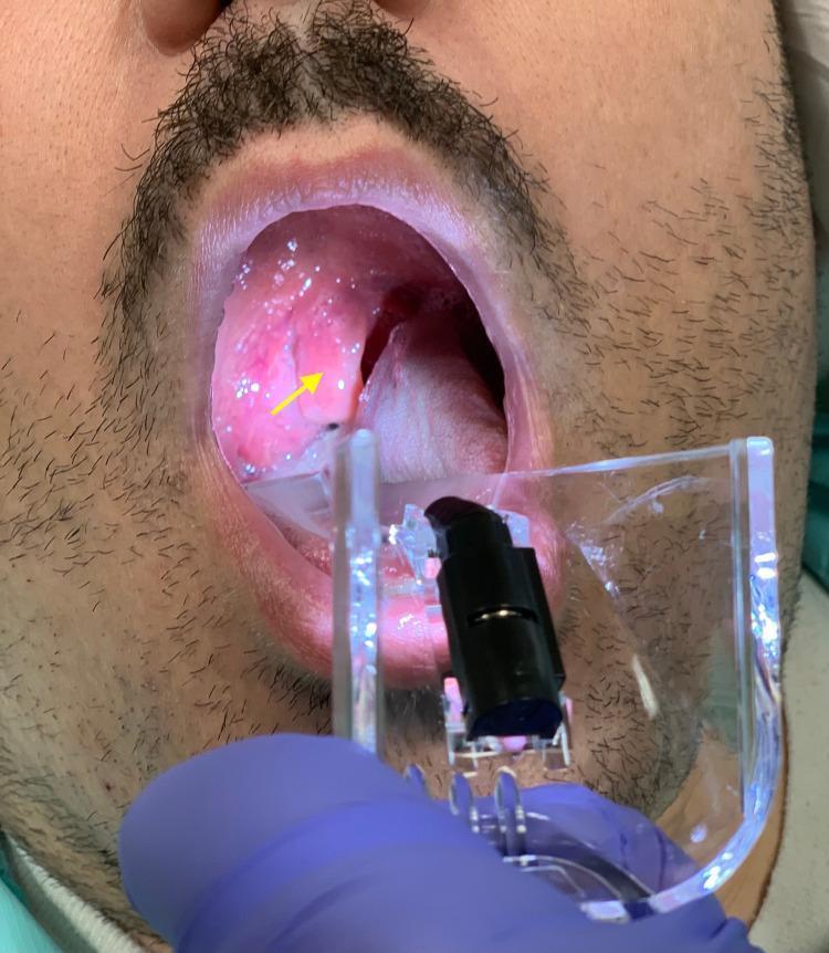
A clinical photograph captures an oropharyngeal examination of a patient using a clear plastic laryngoscope or tongue depressor. The primary finding, indicated by a yellow arrow, is a grossly edematous and erythematous uvula (uvulitis). The uvula is significantly enlarged and swollen, appearing bulbous and distorted, which contributes to significant narrowing of the oropharyngeal airway space. The surrounding mucosa of the soft palate and posterior pharynx exhibits generalized erythema, suggesting acute inflammation. This visual evidence is characteristic of conditions such as Quincke's edema (isolated uvular angioedema) or anaphylaxis-related airway involvement. The palatine tonsils and tongue are visible, with the latter being depressed to allow for a clear view of the upper airway. This image serves as a teaching tool for recognizing emergency airway obstruction signs in otolaryngology and emergency medicine.
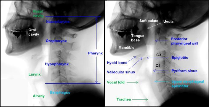
This lateral x-ray radiography shows two panels detailing the head and neck anatomy essential for the swallowing process. The diagram employs a color-coded labeling system to distinguish functional regions: white for the oral cavity, dark blue for the pharynx, green for the airway, and light blue for the esophagus. Key structures identified include the mandible, tongue base, soft palate, and uvula within the oral cavity. The pharynx is divided into three distinct segments: the nasopharynx, oropharynx, and hypopharynx, with specific landmarks such as the posterior pharyngeal wall, epiglottis, vallecular sinus, and pyriform sinus highlighted. The airway section identifies the larynx, vocal folds, and trachea. Vertebral landmarks C3 and C4 are indicated to provide spatial orientation for the pharyngeal structures and the upper esophageal sphincter. This clinical illustration serves as an educational reference for understanding the normal anatomical relationships required during a videofluoroscopic swallowing study (VFSS) and the transition of a bolus from the oral phase to the esophageal phase.
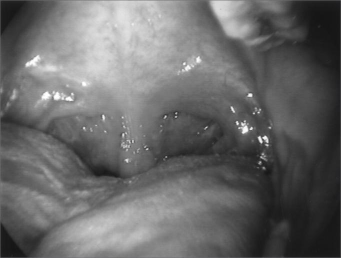
This clinical photograph provides an endoscopic view of the pharynx during inspiration, illustrating dynamic airway changes. The base of the tongue is positioned anteriorly in the lower foreground. The posterior pharyngeal wall is clearly visible, flanked by lateral pharyngeal walls that exhibit significant medial contraction, leading to a narrowing of the oropharyngeal space. Superiorly, the soft palate and uvula are visible. Deep within the airway, the laryngeal inlet is partially obscured, with the glottic opening appearing inappropriately constricted during the inspiratory phase. This presentation is characteristic of paradoxical laryngeal movement and pharyngeal dystonia, where the pharyngeal and laryngeal structures abnormally adduct during inhalation, causing upper airway obstruction. The mucous membranes appear moist and physiologically normal in texture, though the overall pharyngeal volume is reduced due to the active muscular constriction shown in this clinical case of respiratory-related dystonia.
Snoring: Mechanism and Why It Is More Common in Obese People
What is Snoring?
The Mechanism of Snoring
1. Normal wakefulness vs. sleep
2. Partial obstruction at key sites
- Soft palate and uvula - the most common source
- Base of tongue (falls posteriorly in the supine position)
- Lateral pharyngeal walls
- Epiglottis (less commonly)
3. Neuromechanical coupling
- Stable (normal breathing): neuromuscular reflexes maintain stiffness in response to negative inspiratory pressure
- Flutter (snoring): partial collapse with tissue vibration
- Full collapse (apnea): airway closes completely
4. Supine posture makes it worse
Why Is Snoring Predominantly Found in Obese People?
1. Parapharyngeal fat deposition
2. Increased neck circumference
3. Reduced pharyngeal muscle tone from fat infiltration
4. Reduced lung volumes (reduced tracheal traction)
- Abdominal and chest wall fat mass reduces functional residual capacity (FRC) and total lung volume
- Lower lung volumes = less "tracheal tug" - the natural caudal pull the lungs exert on the trachea and pharynx during breathing
- Without this tug, the pharynx is more prone to collapse
- Obese patients consume ~50% more oxygen and produce significantly more CO2 than healthy-weight individuals, further stressing the respiratory system. - Rosen's Emergency Medicine
5. Systemic inflammation
The Numbers
| Factor | Effect |
|---|---|
| BMI increase of 10% | ~6-fold increase in OSA risk |
| 60-90% of OSA patients | are overweight or obese |
| Neck circumference | strongest anatomical predictor |
| Weight loss of 10% | significantly reduces AHI and snoring severity |
Why Men Snore More Than Women
- Men deposit more fat in the neck region vs. women who tend to deposit abdominally
- Testosterone reduces upper airway muscle tone
- Progesterone in women is a mild respiratory stimulant that promotes pharyngeal dilator activity
- Post-menopause, the female snoring rate approaches the male rate
The Continuum: From Snoring to OSA
- Apneas (complete breath-holds >10 seconds)
- Oxygen desaturation
- Sleep fragmentation, daytime somnolence
- Long-term: hypertension, cardiovascular disease, insulin resistance