
Common causes of old age dysphagia And y do they use expanding metal stents
Common Causes of Dysphagia in Old Age
A. Age-Related Changes (Presbyphagia)
- Reduced muscle mass and strength of the tongue, pharynx, and upper oesophageal sphincter (sarcopenia)
- Slower reflexes and sensory decline in the oropharynx
- Reduced saliva production (dry mouth worsens bolus formation)
- Low-amplitude primary or secondary oesophageal contractions in older adults - these fail to propel the food bolus effectively, causing discomfort and dysphagia (Sleisenger & Fordtran's, Pathophysiology section)
B. Oropharyngeal Causes
| Cause | Notes |
|---|---|
| Stroke (CVA) | Most common neurological cause; bulbar or pseudobulbar palsy |
| Parkinson's disease | Bradykinesia of pharyngeal muscles; aspiration risk is high |
| Dementia | Late-stage cognitive impairment impairs voluntary swallowing initiation |
| Motor neuron disease (ALS) | Bulbar involvement |
| Myasthenia gravis | Fatigable pharyngeal weakness |
| Head & neck tumours | Pharyngeal/laryngeal carcinoma |
| Zenker's pharyngeal pouch | More common in elderly males; regurgitation of stale food is characteristic |
| Cervical osteophytes | Posterior pharyngeal compression from anterior cervical spurs |
| Post-radiation fibrosis | After head/neck radiotherapy |
C. Oesophageal Causes
| Cause | Notes |
|---|---|
| Carcinoma of the oesophagus | The most important cause in old age (peak 50-70 years); short history of progressive solid-then-liquid dysphagia, rapid weight loss (S Das Manual of Clinical Surgery) |
| Peptic (reflux) stricture | Long-standing GERD causes fibrotic narrowing |
| Achalasia | Can present late; difficulty with liquids first, then solids |
| Schatzki's ring | At the gastro-oesophageal junction; intermittent dysphagia for solids |
| External compression | Mediastinal lymph nodes, lung/gastric carcinoma, aortic aneurysm |
| Systemic sclerosis (scleroderma) | Smooth muscle atrophy of the lower oesophagus |
| Drug-induced oesophagitis | NSAIDs, bisphosphonates, doxycycline (very common in the elderly who take multiple drugs) |
Key clinical clue (S Das, Clinical Surgery): A comparatively short history of dysphagia in an elderly patient - a few months, progressive from solids to liquids - strongly suggests carcinoma of the oesophagus. A long, slow history with periods of remission suggests achalasia or a benign stricture.
Why Are Self-Expanding Metal Stents (SEMS) Used?
The Problem They Solve
- Advanced disease at presentation
- Poor performance status / comorbidities
- Age-related operative risk
How SEMS Work
- Made from special alloys, commonly nitinol (nickel-titanium), which has shape memory - it is compressed during delivery, then self-expands to its preset diameter once deployed in the lumen
- Placed by an experienced endoscopist under fluoroscopic guidance
- The stent should be long enough to cover the entire stricture, with an extra 4-6 cm on either end to prevent migration and tumour overgrowth at the edges (Yamada's Textbook of Gastroenterology)
Why Metal (Not Plastic)?
| Feature | SEMS | Plastic stents |
|---|---|---|
| Self-expansion | Yes - radial force keeps lumen open | No - passive |
| Migration | Lower (tissue anchors) | Higher |
| Palliation durability | Sustained | Short-lived; recurrent dysphagia common |
| Covered vs uncovered | Available in both | Not applicable |
Indications
- Palliative treatment of malignant dysphagia (oesophageal, gastric, or lung carcinoma causing extrinsic compression) - first-line when surgery/chemoradiation is not feasible
- Tracheo-oesophageal fistula - covered SEMS can seal the fistula
- Refractory benign strictures - covered/removable SEMS (though plastic stents are preferred here to allow removal)
- Malignant gastric outlet obstruction - duodenal SEMS for pancreatic cancer palliation
Efficacy and Complications
- ~95% successful at relieving malignant obstruction and restoring oral intake (Sleisenger & Fordtran's)
- Complications occur in ~30%:
- Early (< 1 week): chest pain, perforation, aspiration, airway compromise, malposition
- Late (> 1 week): stent migration, tumour in-growth causing re-obstruction, tracheo-oesophageal fistulisation, bleeding
- Stents placed across the gastro-oesophageal junction risk severe reflux, so anti-reflux stent designs or PPIs are added
Common causes of old age dysphagia And y do they use expanding metal stents
Common Causes of Dysphagia in Old Age
- Carcinoma of the oesophagus - #1 cause in elderly (50-70 yrs); short history, progressive solids → liquids
- Peptic/reflux stricture - from long-standing GERD
- Achalasia - liquids first, then solids
- Schatzki's ring, external compression (mediastinal tumour, aortic aneurysm)
- Drug-induced oesophagitis (NSAIDs, bisphosphonates - very common in elderly on polypharmacy)
- Stroke - most common neurological cause
- Parkinson's disease, dementia, MND
- Zenker's pharyngeal pouch
- Cervical osteophytes
- Sarcopenia of pharyngeal/oesophageal muscles
- Reduced reflexes, dry mouth, low-amplitude oesophageal contractions
Why Self-Expanding Metal Stents (SEMS)?
- Dilation - effect lasts only 12-28 days, high perforation risk
- Made of nitinol (shape-memory alloy) - compressed for delivery, then self-expands to maintain radial force against tumour
- ~95% effective at relieving malignant dysphagia
- Sustained palliation vs short-lived dilation
- Can seal tracheo-oesophageal fistulas (covered SEMS)
- Restores oral intake quickly, improving nutrition and quality of life
esophageal candidisasis
esophageal candidiasis endoscopy white plaques
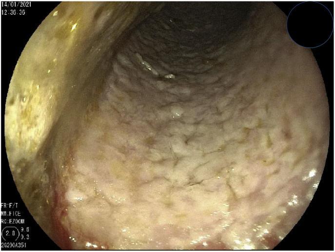
This diagnostic image is an upper gastrointestinal endoscopy photograph of the esophageal lumen. The image demonstrates a hallmark presentation of esophageal candidiasis (fungal infection). The esophageal mucosa is extensively covered by thick, whitish, cottage-cheese-like plaques. These deposits are confluent, merging to form a continuous, irregular layer that obscures the underlying vascular architecture of the esophageal wall. The lumen appears slightly dilated, and the texture of the plaque-covered mucosa is rough and granular. In areas where the white coating is less dense, there is evidence of erythema, suggesting underlying mucosal inflammation. This visual finding is classically associated with opportunistic fungal infections in immunocompromised patients or those with esophageal stasis. The clinical significance lies in the recognition of confluent exudative plaques as a diagnostic marker for infectious esophagitis, requiring further histopathological or microbiological confirmation via biopsy or brushings.
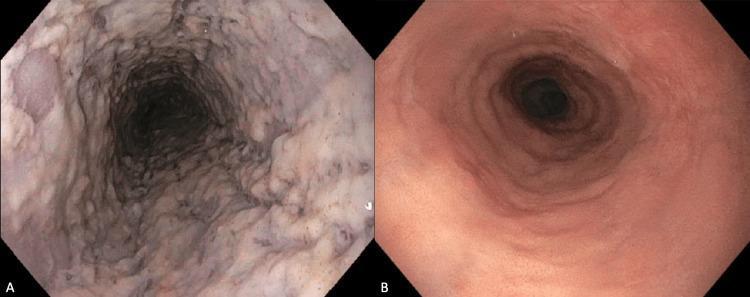
This side-by-side comparison displays two endoscopic images of the esophagus, illustrating a case of esophageal candidiasis before and after antifungal treatment. Image A is a clinical photograph from an upper gastrointestinal endoscopy showing severe esophageal candidiasis. The esophageal mucosa is extensively covered by thick, confluent, white-to-creamy plaques that give the lumen a shaggy and irregular appearance. These plaques cause apparent luminal narrowing and signify a fungal infection, commonly Candida albicans, often associated with immunocompromised states or corticosteroid use. Image B shows the same anatomical region 12 weeks following fluconazole therapy. The mucosa has returned to a healthy state, characterized by a smooth, glistening texture and a uniform pinkish-red color without evidence of exudates or plaques. The esophageal lumen appears patent and unobstructed. This comparison serves as an educational tool for identifying the classic endoscopic presentation of fungal esophagitis and the visual evidence of therapeutic resolution.
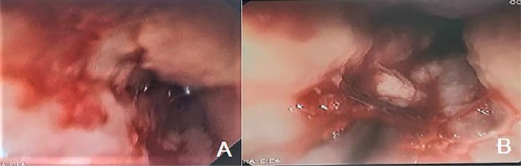
This composite diagnostic image displays two panels (A and B) of a digestive endoscopy within the esophagus, illustrating different stages or manifestations of esophageal pathology. Panel A shows esophageal candidiasis, characterized by confluent, elevated white plaques that are firmly adhered to the esophageal wall. The surrounding mucosa exhibits significant hyperemia (redness) and friable membranes, typical of fungal infection. Panel B demonstrates severe ulcerative esophagitis, featuring extensive, circumferential ulcerations. These lesions are covered with thick, white fibrin exudate and show active bleeding and signs of mucositis. The visual differences between the panels serve to educate on the endoscopic appearance of infectious candidal plaques versus severe erosive/ulcerative injury, which in this clinical context may be related to drug-induced hypersensitivity or secondary infection. The image is a critical tool for training in gastroenterology and internal medicine regarding the identification of esophageal mucosal diseases.
Esophageal Candidiasis
Aetiology & Organism
- Caused by Candida albicans in ~90% of cases
- Other species: C. glabrata, C. krusei, C. tropicalis (important because these may be azole-resistant)
- Candida is a normal oral commensal - disease occurs when host defences are impaired
Risk Factors / Predisposing Conditions
| Category | Examples |
|---|---|
| Immunosuppression | HIV/AIDS (most common overall), haematological malignancy, organ transplant |
| Drugs | Corticosteroids (inhaled or systemic), broad-spectrum antibiotics, immunosuppressants, chemotherapy |
| Endocrine | Diabetes mellitus, hypothyroidism, hypoparathyroidism |
| Oesophageal motility disorders | Achalasia, strictures (stasis promotes growth) |
| Nutritional | Malnutrition, iron deficiency |
| Miscellaneous | Advanced age, critical illness, prolonged nasogastric tube use |
In an otherwise immunocompetent patient presenting with esophageal candidiasis, always evaluate for occult HIV or other immune-compromising condition. - Current Surgical Therapy 14e
Clinical Features
| Symptom | Frequency |
|---|---|
| Odynophagia (painful swallowing) | +++ (common) |
| Dysphagia | +++ (common) |
| Heartburn / chest pain | + (rare) |
| Fever | 0 (not a feature - distinguishes from bacterial/viral oesophagitis) |
| Bleeding | + (rare) |
- Oral thrush (white plaques in mouth) is often present but 1 in 3 patients have no oral involvement - absence of thrush does NOT exclude oesophageal disease
- Can be asymptomatic (incidental finding at endoscopy)
Endoscopic Appearance
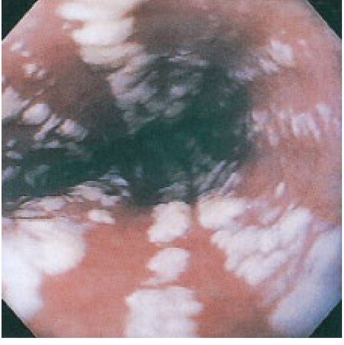
- Grade I: Few raised white plaques ≤2 mm, no oedema or ulceration
- Grade II: Multiple raised white plaques >2 mm, no ulceration
- Grade III: Confluent linear and nodular elevated plaques, hyperaemia
- Grade IV: Grade III + friability, ulceration, narrowing of the lumen ("shaggy oesophagus")
Pathological Findings Compared to Other Infections
| Feature | Candida | CMV/HSV | Bacteria |
|---|---|---|---|
| Plaque | ++++ | + | ++ |
| Ulcer | + | ++++ | +++ |
| Stricture | 0 | ++ | 0 |
| Fever | 0 | ++ | +++ |
Diagnosis
- Endoscopy - most accurate method; gross appearance is usually diagnostic
- Brushings/cytology - more sensitive than biopsy in mild disease (organisms can be washed off biopsy specimens during processing)
- Biopsy + histology - PAS stain or Gomori methenamine silver (GMS) stain highlights organisms; shows mycelia (pseudohyphae) and spores
- Barium swallow - suggestive but not confirmatory; normal result does not exclude diagnosis
- Serology/skin tests - play NO role
In HIV patients with thrush + odynophagia/dysphagia, empiric antifungal therapy is reasonable - endoscopy reserved for those who fail to respond. - Yamada's Textbook of Gastroenterology
Treatment
| Situation | Drug & Dose | Duration |
|---|---|---|
| Standard (first-line) | Oral fluconazole 200-400 mg/day | 14-21 days |
| Mild oropharyngeal | Nystatin suspension or clotrimazole troches 10 mg 5x/day | 7-14 days |
| Cannot tolerate oral | IV fluconazole or IV echinocandin (caspofungin, micafungin) | 14-21 days |
| Refractory/resistant | Voriconazole, itraconazole, or echinocandin; culture + susceptibility testing | Variable |
- Fluconazole 100 mg/day is also effective and may be used in less severe cases (Washington Manual)
- C. krusei is intrinsically fluconazole-resistant; C. glabrata may be resistant - suspect in refractory cases
- Suppressive therapy is NOT routinely indicated (even in HIV patients) - it does not reduce recurrence frequency and promotes resistance
Key Points to Remember
- Most common infectious oesophagitis; AIDS-defining illness
- C. albicans causes 90% - but consider resistant species in refractory disease
- Presents with odynophagia + dysphagia, no fever
- Endoscopy: white raised plaques (not ulcers) - "cottage cheese" appearance
- Diagnosis confirmed by brushings (cytology) > biopsy (histology) with PAS/GMS stain showing pseudohyphae
- First-line treatment: oral fluconazole 200-400 mg/day for 14-21 days
- All cases of oesophageal candidiasis require systemic therapy (topical agents like nystatin are insufficient)

Indenting the tnm staging
TNM staging bladder cancer diagram T stages
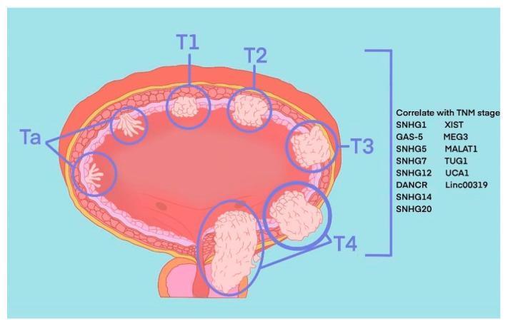
Anatomical diagram of a transverse section of the human urinary bladder illustrating the TNM staging system for bladder cancer (BLCA). The illustration depicts the progression of tumor invasion through the bladder wall layers. Ta stages are shown as small, papillary, finger-like projections confined to the inner urothelium. T1 represents a localized nodular mass invading the lamina propria. T2 shows deeper invasion into the muscularis propria (detrusor muscle). T3 demonstrates extension through the muscle layer into the surrounding perivesical adipose tissue. T4 illustrates advanced invasion into adjacent pelvic organs or the pelvic wall. To the right of the diagram, a list of molecular markers including various Small Nucleolar RNA Host Genes (SNHGs) and long intergenic non-coding RNAs (lincRNAs) such as SNHG1, SNHG14, SNHG20, MALAT1, and Linc00319 are provided, indicating their correlation with these pathological stages. The diagram serves as an educational tool for differentiating non-muscle-invasive bladder cancer (NMIBC) from muscle-invasive bladder cancer (MIBC).
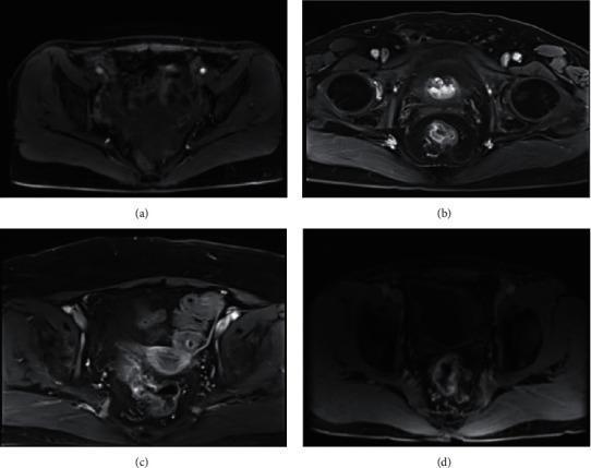
This diagnostic image set consists of four axial MRI scans (labeled a through d) demonstrating various TNM stages of rectal cancer (RC) in the pelvic region. Panel (a) shows stage T1 RC, characterized by a lesion with a high-signal submucosal layer intact and no visible involvement of the muscularis propria. Panel (b) illustrates stage T3 RC, where the tumor signal extends through the muscularis propria into the perirectal adipose tissue, though the mesorectal fascia appears clear. Panels (c) and (d) focus on nodal involvement (N staging); panel (c) shows stage N1 RC with a single hyperintense metastatic lymph node, while panel (d) demonstrates stage N2 RC with bilateral metastatic lymphadenopathy. The images utilize T2-weighted or diffusion-weighted characteristics to highlight variations in signal intensity between normal pelvic structures—such as the bladder, perirectal fat, and pelvic floor musculature—and malignant tissues. These scans serve as a comparison for assessing tumor depth (T-stage) and regional lymph node status (N-stage) in preoperative clinical staging.
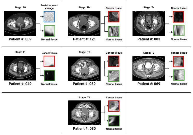
This diagnostic comparison chart displays seven panels of axial pelvic CT scans with intravenous contrast, each illustrating a specific TNM stage of urothelial carcinoma (T0, Tis, Ta, T1, T2, T3, and T4). For each stage, a full axial section of the pelvis is shown alongside two magnified Regions of Interest (ROIs) extracted from the bladder wall. The ROIs are categorized as either 'Cancer tissue' (highlighted in red or blue for post-treatment changes) or 'Normal tissue' (highlighted in green), providing a visual basis for histopathological and radiological correlation. The progression demonstrates key staging features: non-muscle invasive stages (Ta, Tis, T1) show localized bladder wall irregularities, while muscle-invasive stages (T2-T4) exhibit increasing wall thickening, loss of clear margins, and infiltration into the perivesical fat or adjacent structures, as seen in the T3 and T4 examples. This educational visual is designed for training in radiological staging of bladder cancer and development of machine-learning classification models for oncology.
TNM Staging of Bladder Cancer (AJCC 8th Edition)
Visual Overview
T - Primary Tumour
| Stage | Description | Key Point |
|---|---|---|
| TX | Cannot be assessed | - |
| T0 | No evidence of primary tumour | - |
| Ta | Non-invasive papillary carcinoma | Confined to urothelium; finger-like projections (as in your image) |
| Tis | Carcinoma in situ - flat tumour | High-grade; flat, not papillary; very aggressive biologically |
| T1 | Invades lamina propria (subepithelial connective tissue) | Crosses basement membrane but not muscle |
| T2a | Invades superficial muscularis propria (inner half) | MIBC begins here |
| T2b | Invades deep muscularis propria (outer half) | |
| T3a | Invades perivesical soft tissue - microscopically | Beyond bladder wall |
| T3b | Invades perivesical tissue - macroscopically (palpable extravesical mass) | |
| T4a | Invades prostatic stroma / uterus / vagina | Adjacent organs |
| T4b | Invades pelvic wall / abdominal wall | Fixed tumour |
Critical divide: Ta, Tis, T1 = Non-Muscle Invasive Bladder Cancer (NMIBC) - treated by TURBT ± intravesical therapy. T2 onwards = Muscle Invasive Bladder Cancer (MIBC) - requires radical cystectomy or chemoradiation.
N - Regional Lymph Nodes
| Stage | Description |
|---|---|
| NX | Cannot be assessed |
| N0 | No lymph node metastasis |
| N1 | Single node in true pelvis (obturator, internal/external iliac, perivesical, presacral) |
| N2 | Multiple nodes in true pelvis |
| N3 | Metastasis to common iliac lymph nodes |
M - Distant Metastasis
| Stage | Description |
|---|---|
| M0 | No distant metastasis |
| M1a | Distant lymph nodes beyond common iliac |
| M1b | Non-lymph node distant metastasis (lung, bone, liver) |
Anatomic Stage Groups
| Clinical Stage | T | N | M |
|---|---|---|---|
| Stage 0a | Ta | N0 | M0 |
| Stage 0is | Tis | N0 | M0 |
| Stage I | T1 | N0 | M0 |
| Stage II | T2a or T2b | N0 | M0 |
| Stage III | T3a, T3b, or T4a | N0 | M0 |
| Stage IVA | T4b OR any T | N1-3 | M0 |
| Stage IVB | Any T | Any N | M1 |
Memory Aid: Layers Invaded
Mucosa (urothelium)  →  Ta / Tis
      ↓
Lamina propria       →  T1       ← NMIBC boundary
      ↓
Muscularis propria   →  T2a/T2b  ← MIBC begins
      ↓
Perivesical fat      →  T3a/T3b
      ↓
Adjacent organs      →  T4a
      ↓
Pelvic/abdominal wall → T4b
Clinical Relevance of Staging
| Stage | Treatment |
|---|---|
| Ta, T1 | TURBT (transurethral resection) ± intravesical BCG or mitomycin C |
| Tis | Intravesical BCG (BCG-naive); high recurrence/progression risk |
| T2-T4 (M0) | Radical cystectomy + neoadjuvant cisplatin-based chemotherapy OR radical chemoradiation |
| M1 | Palliative systemic chemotherapy (gemcitabine + cisplatin) |
- Smith and Tanagho's General Urology, 19th Ed.
- Campbell-Walsh-Wein Urology, Table 137.2