
horner syndrome ptosis miosis anhidrosis and enopthalmos
Horner syndrome ptosis miosis enophthalmos clinical photo
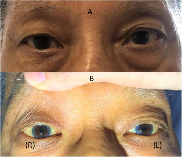
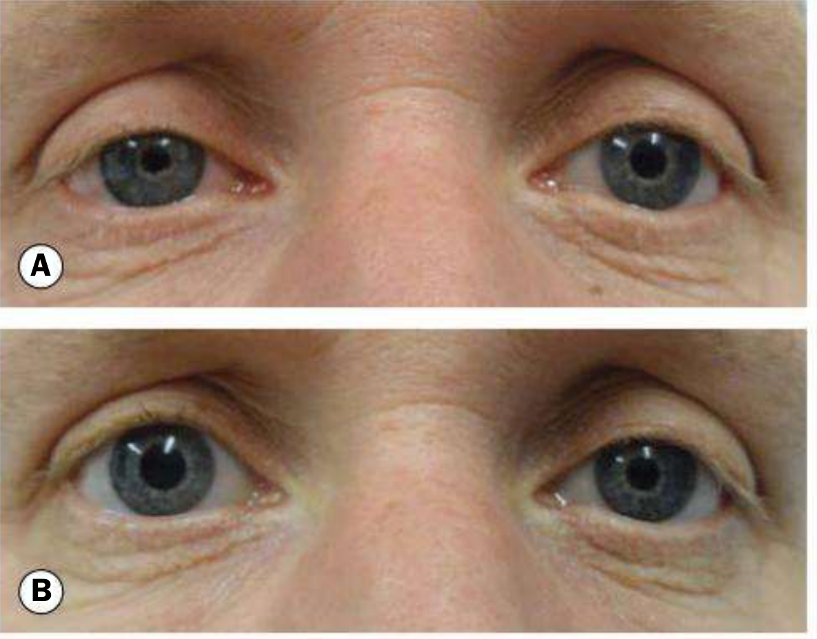
Two-panel clinical photograph demonstrating Horner’s syndrome in a patient. Panel A shows the patient's eyes at rest, highlighting a mild right-sided ptosis (drooping of the upper eyelid) and a subtle 'upside-down ptosis' (elevation of the right lower eyelid). Panel B provides a close-up view of the eyes with the upper eyelids manually retracted to reveal anisocoria; the right pupil (labeled R) is significantly smaller (miotic) compared to the left pupil (labeled L). The clinical findings illustrate the classic triad of sympathetic denervation: ptosis, miosis, and enophthalmos appearance due to narrowing of the palpebral fissure. This visual evidence supports a diagnosis of Horner’s syndrome, often associated with neurological or vascular pathology affecting the oculosympathetic pathway. The image serves as an educational tool for identifying autonomic ocular dysfunction in neurology and ophthalmology.
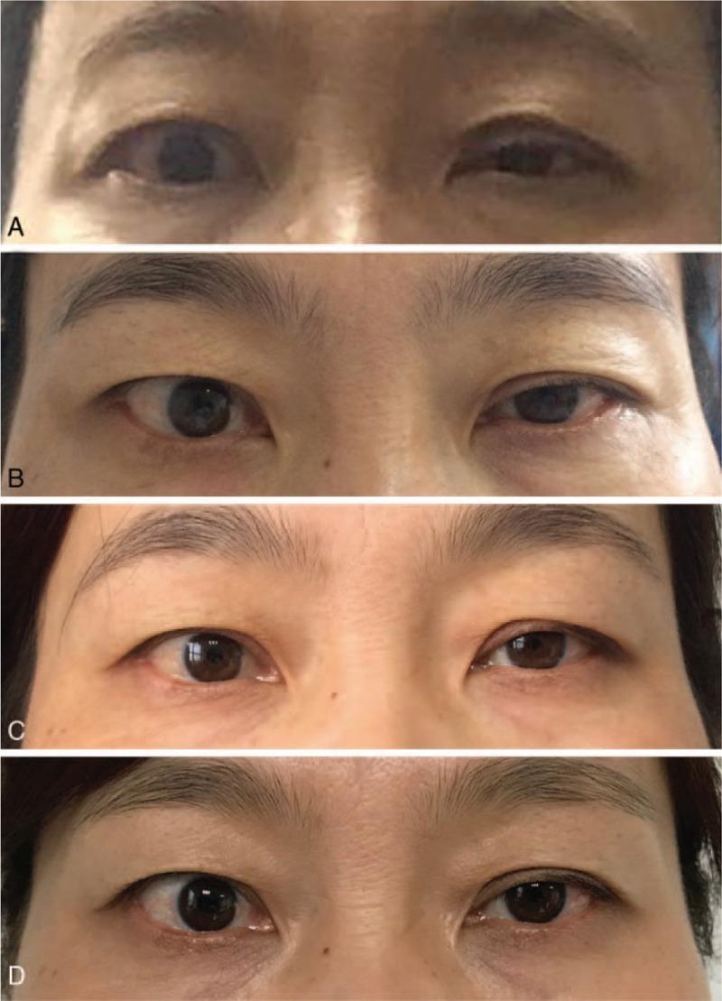
A series of four clinical photographs (A-D) demonstrating the longitudinal progression and recovery of left-sided Horner syndrome in a female patient over a one-year postoperative period. Image A (2 days postoperative) shows the classic triad of ptosis (drooping of the upper eyelid), miosis (constricted pupil), and apparent enophthalmos (sunken eye) on the left side compared to the normal right eye. Image B (one month) and Image C (six months) illustrate a gradual recovery of eyelid elevation and pupillary dilation. Image D (one year) displays a near-complete resolution of symptoms with restoration of ocular symmetry, though a slight residual ptosis may remain. The images serve as an educational timeline for the spontaneous or medically managed recovery of iatrogenic Horner syndrome following neck surgery, such as thyroidectomy. The progression highlights the clinical features of sympathetic pathway disruption and the objective markers used to track neurologic recovery of the oculosympathetic fibers.
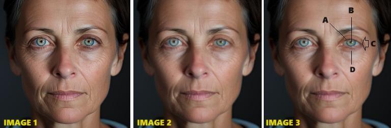
This composite educational illustration consists of three panels demonstrating the clinical manifestations of Horner syndrome through AI-generated and edited portraiture. Image 1 serves as a baseline portrait of an adult female. Image 2 shows the same face with edited features to demonstrate a pathological state. Image 3 provides clinical annotations of the classic triad. The anatomical focus is on the left eye region, where the following signs are labeled: (A) ptosis, visible as a drooping of the superior eyelid; (B) miosis, shown as a constricted pupil relative to the contralateral eye; (C) apparent enophthalmos, indicating a recessed appearance of the globe within the orbit; and (D) 'upside-down ptosis' or inverse ptosis, where the lower eyelid sits higher on the globe. The image set highlights key diagnostic features relevant to neurology and ophthalmology, specifically the sympathetic nerve pathway disruption that characterizes Horner syndrome. These visual cues are essential for medical students and clinicians in identifying autonomic dysfunction during a physical examination.
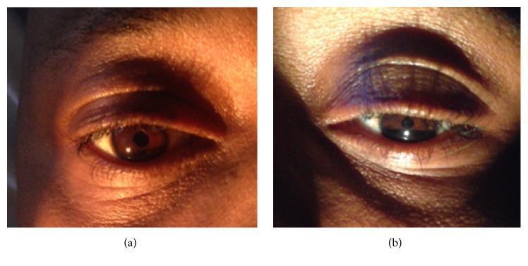
This clinical photograph consists of two side-by-side images (a and b) demonstrating the classic ophthalmic manifestations of Horner's syndrome. Image (a) shows a normal right eye with a typical upper eyelid position, palpebral fissure width, and pupil diameter. Image (b) displays the affected left eye, exhibiting a triad of clinical findings: ptosis (drooping of the upper eyelid), miosis (a constricted pupil compared to the contralateral side), and apparent enophthalmos (the appearance of a sunken globe within the orbit). The narrowing of the palpebral fissure is clearly visible in the left eye due to the superior eyelid malposition. This visual comparison is a definitive educational tool for identifying sympathetic pathway disruption, often associated with neurological or paraspinal pathology such as Pancoast tumors or leukemic infiltration (chloroma). The images are intended for intermediate to advanced medical learners focusing on neurology, ophthalmology, or oncology.
"Horner syndrome" diagnosis etiology
Horner Syndrome (Oculosympathetic Palsy)
- Ptosis - partial upper lid droop (1-2 mm)
- Miosis - small, poorly dilating pupil
- Anhidrosis - loss of sweating on the affected side of the face
- Enophthalmos - apparent recession of the eyeball (actually an optical illusion from narrowing of the palpebral fissure, not true backward displacement)
Anatomy of the Sympathetic Pathway (3 Neurons)
| Neuron | Path | Key Landmark |
|---|---|---|
| 1st order (central) | Posterior hypothalamus → descends uncrossed through brainstem → ciliospinal centre of Budge (C8-T2) | Brainstem and spinal cord |
| 2nd order (preganglionic) | Ciliospinal centre → over lung apex → superior cervical ganglion | Closely related to apical pleura; passes near subclavian artery |
| 3rd order (postganglionic) | Superior cervical ganglion → ascends along internal carotid artery → cavernous sinus → nasociliary branch of CN V1 → ciliary body and dilator pupillae | Along internal carotid artery |
- Müller's muscle (upper lid elevator) - loss causes ptosis
- Inferior tarsal muscle (lower lid retractor) - loss causes lower lid elevation
- Dilator pupillae - loss causes miosis
- Sudomotor and vasomotor fibres of the face - loss causes anhidrosis and facial flushing/warmth
Why Each Sign Occurs
| Sign | Muscle/Structure Affected | Mechanism |
|---|---|---|
| Ptosis | Müller's smooth muscle (upper lid) | Loss of sympathetic tone; levator palpebrae (CN III) still intact, so ptosis is partial (1-2 mm) |
| Miosis | Dilator pupillae | Sphincter pupillae (parasympathetic, CN III) acts unopposed; anisocoria is more obvious in the dark |
| Anhidrosis | Sudomotor fibres | Only present if lesion is proximal to the superior cervical ganglion (fibres running along the external carotid supply facial skin) |
| Enophthalmos | Orbital smooth muscle (of Müller) | True enophthalmos does not occur - it is an illusion from narrowed palpebral fissure |
| Lower lid elevation | Inferior tarsal muscle | Loss of sympathetic tone to the lower lid retractor |
- Adams and Victor's Principles of Neurology, 12th Ed., p. 3123
- Kanski's Clinical Ophthalmology, 10th Ed., p. 807
Causes by Level of Lesion
Central (1st-order neuron)
- Lateral medullary (Wallenberg) syndrome - stroke of the posterior inferior cerebellar artery (PICA); Horner is a key component alongside ipsilateral ataxia, contralateral spinothalamic sensory loss
- Brainstem tumour, demyelination (MS)
- Syringomyelia
- Cervical spinal cord lesion, high cervical trauma
Preganglionic (2nd-order neuron)
- Pancoast tumour (apical lung cancer) - classic cause; also causes T1 root compression (wasting of small hand muscles), shoulder/arm pain
- Aortic/carotid aneurysm or dissection
- Thoracic spine lesion
- Neck masses: thyroid tumour, cervical lymphadenopathy, neck surgery/trauma
Postganglionic (3rd-order neuron)
- Internal carotid artery dissection - acute painful Horner; an emergency
- Cavernous sinus mass, thrombosis
- Nasopharyngeal tumour
- Cluster headache (migrainous neuralgia)
- Otitis media
- Kanski's Clinical Ophthalmology, 10th Ed., Table 19.4
- Neuroanatomy through Clinical Cases, 3rd Ed., p. 608
Pharmacological Confirmation and Localization
Step 1 - Confirm Horner syndrome
Step 2 - Localize (pre- vs. postganglionic)
- Normal or central/preganglionic Horner: pupil dilates (intact 3rd-order neuron with noradrenaline stores)
- Postganglionic Horner: pupil does NOT dilate (no intact terminal from which to release noradrenaline)
- Kanski's Clinical Ophthalmology, 10th Ed., p. 808-809
Special Situations
- Acute painful Horner syndrome = emergency - must rule out carotid artery dissection (urgent MRI/MRA)
- Congenital Horner syndrome (perinatal brachial plexus/sympathetic chain injury): associated with heterochromia iridis (the affected iris remains lighter in colour due to lack of melanin stimulation)
- Bilateral Horner syndrome: rare; seen in autonomic neuropathies and high cervical cord transection
- Wallenberg syndrome (lateral medullary infarct): Horner + ipsilateral facial numbness + contralateral body sensory loss + dysphagia + ataxia
Summary: Localization Clues at a Glance
| Feature | 1st-order (central) | 2nd-order (preganglionic) | 3rd-order (postganglionic) |
|---|---|---|---|
| Anhidrosis | Ipsilateral face and body | Entire ipsilateral face | Absent or confined to medial face |
| Associated signs | Contralateral motor/sensory, cerebellar | Arm/hand wasting (Pancoast), cough, neck mass | Facial pain, headache, orbital signs |
| Carotid dissection risk | No | No | Yes (acute painful) |
| Hydroxyamphetamine | Dilates | Dilates | Does NOT dilate |
Clinical pearl: Enophthalmos in Horner syndrome is not true enophthalmos - the eyeball position does not actually change. The appearance is entirely due to narrowing of the palpebral fissure (ptosis + lower lid elevation), making the eye look sunken. Measuring the actual globe position with exophthalmometry will be normal. - Adams and Victor's Principles of Neurology, 12th Ed.
Please explain it in a simpler way for 1st year anatomy students
Horner Syndrome - Simple Explanation
What is it?
The Wire That Gets Cut - A Simple Analogy
BRAIN (Hypothalamus)
      ↓  [Wire 1 - through brainstem & spinal cord]
RELAY 1 (C8-T2 spinal cord)
      ↓  [Wire 2 - over lung apex, up the neck]
RELAY 2 (Superior cervical ganglion, behind jaw)
      ↓  [Wire 3 - along carotid artery to eye]
THE EYE
The 4 Signs - Simply Explained
1. Ptosis - Droopy Upper Eyelid
- The sympathetic nerve controls a small muscle called Müller's muscle in the upper eyelid
- When it's cut off, the upper lid drops slightly (about 1-2 mm)
- Remember: This is NOT a full droopy lid like in a nerve 3 palsy - just a small, partial droop
2. Miosis - Small Pupil
- Normally the sympathetic nerve keeps the pupil wide open (dilated) by pulling the dilator pupillae muscle open like a camera aperture
- When the sympathetic nerve is cut, the dilator muscle stops working
- The only muscle left working is the sphincter pupillae (controlled by the parasympathetic system), which squeezes the pupil shut
- Result: small pupil on the affected side
Easy memory trick: Sympathetic = Dilates (think: sympathetic means excited, big eyes when excited). When it's gone, the pupil shrinks.
3. Anhidrosis - No Sweating on That Side of the Face
- The sympathetic nerve also controls sweat glands on the face
- When it's cut, the skin on the affected side stops sweating
- That side of the face may also look slightly red/warm (blood vessels dilate without sympathetic tone)
4. Enophthalmos - Eye Looks Sunken
- This one is a trick - the eyeball doesn't actually move backward
- It just looks sunken because the eyelid is drooping down (from above) and the lower lid is slightly elevated (from below), squeezing the visible part of the eye
- The narrowed gap between the lids makes the eye appear to sit further back
One More Sign: Lower Lid Elevation (Inverse Ptosis)
- There's also a tiny muscle in the lower eyelid controlled by the sympathetic nerve
- When it fails, the lower lid rises slightly
- Together with the upper lid drooping, the whole eye opening (palpebral fissure) gets narrower - this is what creates the "sunken eye" appearance
Where Can the Wire Be Cut? - The 3 Locations
| Location | Wire Cut | Common Cause | Memory Trick |
|---|---|---|---|
| Central (1st wire) | Brain to spinal cord | Stroke in the brainstem | "Wallenberg stroke" |
| Preganglionic (2nd wire) | Spinal cord to neck | Lung tumor at the top of the lung (Pancoast tumor) | "Top of lung tumor" |
| Postganglionic (3rd wire) | Neck to eye | Carotid artery injury/dissection | "Neck artery torn" |
A Quick Picture to Remember It All
Normal Eye:              Horner Eye:

  ___________              _________
 |     O     |            |   o     |
 |___________|            |_________|

  Wide lid opening         Narrow lid opening
  Big pupil (O)            Small pupil (o)
  Lower lid down           Lower lid raised
  Normal sweating          No sweating on that side
The One-Line Summary
Horner syndrome = sympathetic nerve cut off from the eye → droopy upper lid + small pupil + no sweating + eye appears sunken (on the SAME side as the lesion)