
ts of forearm
anatomical transverse section of forearm showing flexor and extensor compartments radius ulna interosseous membrane
forearm anatomy
transverse section forearm anatomy flexor extensor compartments radius ulna
Transverse section of the forearm
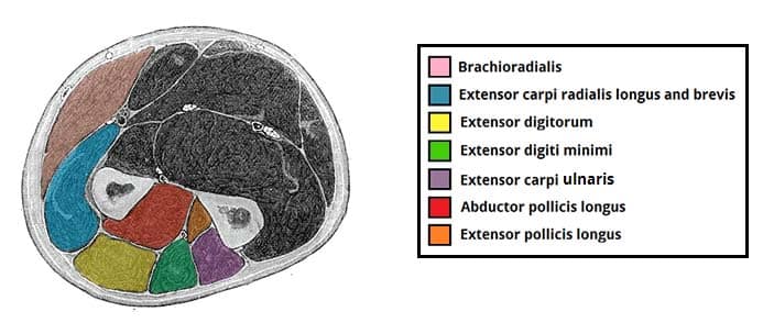
1. Bones and membrane
- Radius: lateral side, or thumb side
- Ulna: medial side, or little-finger side
- Interosseous membrane: strong fibrous sheet joining the two bones; separates deep flexor and extensor structures and transmits forces between radius and ulna.
2. Anterior compartment - flexor-pronator compartment
- Pronator teres
- Flexor carpi radialis
- Palmaris longus
- Flexor carpi ulnaris
- Flexor digitorum superficialis
- Flexor digitorum profundus
- Flexor pollicis longus
- Pronator quadratus, near the distal forearm
- Median nerve: usually between flexor digitorum superficialis and profundus
- Ulnar nerve and ulnar artery: medial side, near flexor carpi ulnaris
- Radial artery: lateral side, deep to brachioradialis
- Anterior interosseous nerve and artery: on the anterior surface of the interosseous membrane
3. Posterior compartment - extensor-supinator compartment
- Extensor digitorum
- Extensor digiti minimi
- Extensor carpi ulnaris
- Extensor carpi radialis longus and brevis
- Brachioradialis, on the lateral side
- Supinator
- Abductor pollicis longus
- Extensor pollicis brevis
- Extensor pollicis longus
- Extensor indicis
- Posterior interosseous nerve, the continuation of the deep branch of radial nerve
- Posterior interosseous artery
Easy orientation in a diagram
- Anterior side: bulk of flexor muscles, median and ulnar neurovascular bundles
- Posterior side: extensor muscle mass
- Lateral/radial side: brachioradialis and radial artery
- Medial/ulnar side: flexor carpi ulnaris with ulnar nerve and artery
Diagram of topographical anatomy of forearm with Regions.
An accurate educational labelled diagram of the topographical anatomy of the forearm, shown in two full-length views side by side: anterior (flexor) view and posterior (extensor) view, elbow at top and wrist at bottom. Clearly label regions: cubital fossa, anterior forearm flexor-pronator region, posterior forearm extensor-supinator region, lateral radial region, medial ulnar region, radial styloid, ulnar styloid, wrist. Include concise surface landmarks: brachioradialis, flexor carpi radialis, palmaris longus, flexor carpi ulnaris, extensor digitorum, extensor carpi ulnaris, and indicate radial artery and ulnar nerve/artery at the distal forearm. Use anatomically correct relative locations, clinical anatomy textbook style.
labelled surface anatomy regions of forearm anterior posterior diagram radial ulnar flexor extensor
forearm anatomy
forearm surface anatomy regions anterior posterior radial ulnar landmarks
Topographical anatomy of the forearm
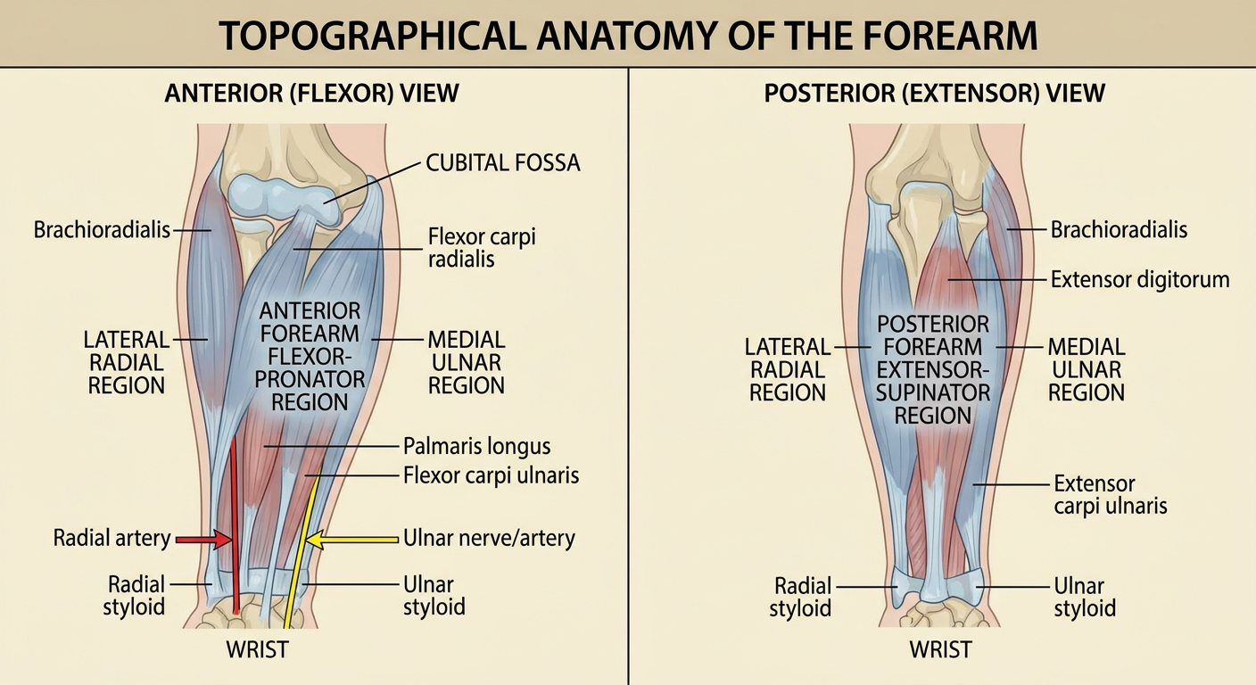
- Cubital fossa: triangular depression on the anterior aspect of elbow.
- Anterior antebrachial region: flexor-pronator compartment.
- Posterior antebrachial region: extensor-supinator compartment.
- Lateral or radial region: thumb-side border, marked proximally by brachioradialis and distally by the radial artery.
- Medial or ulnar region: little-finger-side border, with the ulnar nerve and artery becoming superficial near the distal forearm.
- Carpal or wrist region: distal boundary, with the radial and ulnar styloid processes as palpable landmarks.
Diagram of the puncture of the joints
Choose the joint(s) to show.
Transverse section of the forearm
1. Bones and membrane
- Radius: lateral side, or thumb side
- Ulna: medial side, or little-finger side
- Interosseous membrane: strong fibrous sheet joining the two bones; separates deep flexor and extensor structures and transmits forces between radius and ulna.
2. Anterior compartment - flexor-pronator compartment
- Pronator teres
- Flexor carpi radialis
- Palmaris longus
- Flexor carpi ulnaris
- Flexor digitorum superficialis
- Flexor digitorum profundus
- Flexor pollicis longus
- Pronator quadratus, near the distal forearm
- Median nerve: usually between flexor digitorum superficialis and profundus
- Ulnar nerve and ulnar artery: medial side, near flexor carpi ulnaris
- Radial artery: lateral side, deep to brachioradialis
- Anterior interosseous nerve and artery: on the anterior surface of the interosseous membrane
3. Posterior compartment - extensor-supinator compartment
- Extensor digitorum
- Extensor digiti minimi
- Extensor carpi ulnaris
- Extensor carpi radialis longus and brevis
- Brachioradialis, on the lateral side
- Supinator
- Abductor pollicis longus
- Extensor pollicis brevis
- Extensor pollicis longus
- Extensor indicis
- Posterior interosseous nerve, the continuation of the deep branch of radial nerve
- Posterior interosseous artery
Easy orientation in a diagram
- Anterior side: bulk of flexor muscles, median and ulnar neurovascular bundles
- Posterior side: extensor muscle mass
- Lateral/radial side: brachioradialis and radial artery
- Medial/ulnar side: flexor carpi ulnaris with ulnar nerve and artery
- Cubital fossa: triangular depression on the anterior aspect of elbow.
- Anterior antebrachial region: flexor-pronator compartment.
- Posterior antebrachial region: extensor-supinator compartment.
- Lateral or radial region: thumb-side border, marked proximally by brachioradialis and distally by the radial artery.
- Medial or ulnar region: little-finger-side border, with the ulnar nerve and artery becoming superficial near the distal forearm.
- Carpal or wrist region: distal boundary, with the radial and ulnar styloid processes as palpable landmarks.
Diagram of zone of kanavella
Accurate labelled medical anatomy diagram of Kanavel spaces of the hand, palmar view. Show the thumb radial bursa, little finger ulnar bursa, flexor tendon digital synovial sheaths of index, middle and ring fingers, the midpalmar space and thenar space, carpal tunnel, and Parona space in the distal forearm. Use color-coded translucent regions and arrows to show possible spread of infection from thumb and little finger through radial and ulnar bursae, including their potential communication. Clear concise labels, clean surgical anatomy textbook style.
Kanavel spaces hand diagram radial bursa ulnar bursa thenar midpalmar Parona space
Kanavel hand spaces anatomy
Kanavel spaces anatomy diagram radial ulnar bursae Parona space
Kanavel spaces of the hand
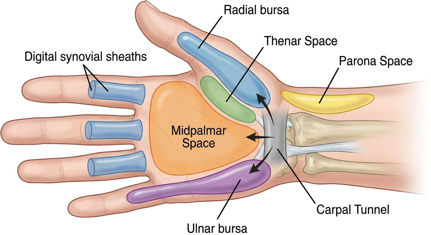
Key structures shown
- Radial bursa: synovial sheath surrounding the flexor pollicis longus tendon of the thumb. It continues proximally through the carpal tunnel.
- Ulnar bursa: common flexor synovial sheath in the palm, usually continuous with the flexor sheath of the little finger.
- Digital flexor tendon sheaths: sheaths of index, middle, and ring fingers usually end near the metacarpal neck, so they do not usually directly communicate with the ulnar bursa.
- Thenar space: deep potential space lateral in the palm, associated with the thumb and index-finger side.
- Midpalmar space: deep potential space medial in the palm, related to the middle, ring, and little-finger side.
- Space of Parona: potential space in the volar distal forearm, between the flexor tendons and pronator quadratus.
Clinical importance
Puncture of the joints
Educational multi-panel medical diagram titled Common Joint Aspiration (Arthrocentesis) Landmarks. Include labelled surface-anatomy panels for shoulder, elbow, wrist, knee and ankle. Show patient positioning, bony landmarks, and a needle icon pointing toward each joint space, without showing skin penetration, blood, or injection. Shoulder: posterior approach near posterolateral acromion. Elbow: posterolateral soft spot. Wrist: dorsal radiocarpal joint space. Knee: superolateral approach at patella. Ankle: anteromedial tibiotalar joint space. Add a clear note: trained clinician, sterile technique, ultrasound guidance where appropriate. Accurate clinical anatomy textbook style.
labelled diagram common joint aspiration arthrocentesis shoulder elbow wrist knee ankle landmarks
arthrocentesis ultrasound guidance systematic review
joint aspiration arthrocentesis shoulder elbow wrist knee ankle landmarks procedure guideline
Common joint puncture sites - arthrocentesis
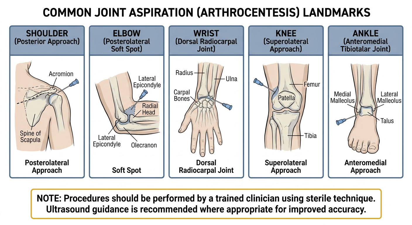
| Joint | Common landmark approach |
|---|---|
| Shoulder | Anterior approach just inferior and lateral to the coracoid process, or a posterior approach near the posterolateral acromion |
| Elbow | Posterolateral “soft spot” bounded by the olecranon, radial head, and lateral epicondyle |
| Wrist | Dorsal radiocarpal joint space, commonly between the extensor tendons |
| Knee | Superolateral approach adjacent to the patella, targeting the suprapatellar recess |
| Ankle | Anteromedial tibiotalar approach, medial to the tibialis anterior tendon |