
Stroke types etiology pathogenesis clinical feature signs investigation complications
Reading File
Now I'll search the library for comprehensive stroke content in parallel:
Finding Sources
Finding Sources
Searching PubMed
"stroke" AND "management"
Now let me read the main stroke sections from Bradley and Daroff's Neurology (the gold standard neurology reference) and Goldman-Cecil Medicine in parallel:
Reading File
Reading File
Reading File
The Goldman-Cecil result was about sickle cell disease stroke. Let me now search more specifically for stroke types, etiology, pathogenesis, and clinical features from the neurology and emergency medicine textbooks:
Finding Sources
Finding Sources
Finding Sources
Now let me read the key sections from Rosen's Emergency Medicine (comprehensive stroke chapter) and Frameworks for Internal Medicine (which has both ischemic and hemorrhagic stroke), plus the Plum and Posner ICH section:
Reading File
Reading File
Reading File
Excellent - I have very rich content. Let me now read the hemorrhagic stroke section from Rosen's (lines 2770-2910) and the remaining ischemic stroke details (clinical features and complications) in parallel:
Reading File
Reading File
I now have extensive material. Let me fetch the key stroke classification diagram and read a couple more sections for clinical syndromes and complications:
Loading Image
Reading File
I now have comprehensive material. Let me do one final parallel read to get the clinical features by stroke territory and complications:
Reading File
Reading File
I now have comprehensive data from multiple authoritative sources. Let me compile the full answer:
Stroke: Types, Etiology, Pathogenesis, Clinical Features, Signs, Investigations, and Complications
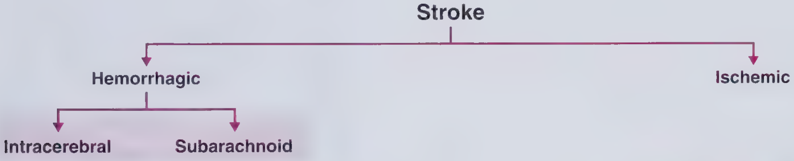
Stroke Classification - Frameworks for Internal Medicine
1. Definition
A stroke (cerebrovascular accident, CVA) is a sudden-onset focal neurological deficit caused by either ischemia (inadequate blood supply) or hemorrhage into brain tissue. It is the second leading cause of death worldwide and the leading cause of adult disability.
2. Types of Stroke
| Type | Subtypes | Proportion of All Strokes |
|---|---|---|
| Ischemic | Thrombotic (large vessel, small vessel/lacunar), Embolic (cardioembolic, artery-to-artery) | ~80-85% |
| Hemorrhagic | Intracerebral hemorrhage (ICH), Subarachnoid hemorrhage (SAH) | ~15-20% |
- ICH accounts for 10-15% of all strokes
- SAH accounts for ~5% of all strokes
3. Etiology
Ischemic Stroke
A. Thrombotic (~1/3 of ischemic strokes)
- Large-vessel occlusion: Atherosclerotic plaque at vessel bifurcations (especially internal carotid artery, MCA origin). Plaque ulceration + platelet adhesion leads to clot that occludes or embolizes distally.
- Small-vessel/Lacunar (~20%): Lipohyalinosis of small penetrating arteries due to hypertension or diabetes. Infarcts 2-20mm in basal ganglia, thalamus, pons, internal capsule.
B. Cardioembolic (~25% of ischemic strokes)
- Atrial fibrillation (most common): 5x increased stroke risk
- Mural thrombus (post-MI), valvular disease (rheumatic, prosthetic), cardiomyopathy, paradoxical embolism through PFO
- Artery-to-artery embolism (from carotid plaque)
C. Cryptogenic (>1/3 of all strokes) - no identifiable cause
D. Special causes (especially in young patients)
- Carotid/vertebral artery dissection (leading cause of stroke in young; follows trauma, sneezing, chiropractic manipulation)
- Hypercoagulable states: antiphospholipid antibodies, protein C/S deficiency, factor V Leiden
- Sickle cell disease (vasculopathy)
- Pregnancy/OCP use
- Cocaine, amphetamines (vasospasm + hypertension)
- Fibromuscular dysplasia
- Migraine-associated (prolonged vasospasm)
- Vasculitis (varicella, fungal meningitis, giant cell arteritis)
- Cerebral venous sinus thrombosis (CVST)
Hemorrhagic Stroke
Intracerebral Hemorrhage (ICH)
- Hypertensive vasculopathy (most common): Lipohyalinosis of small deep penetrating arteries → deep hemorrhages (putamen 44%, thalamus 13%, cerebellum 9%, pons 9%)
- Cerebral amyloid angiopathy (CAA): Amyloid deposits in cerebral vessel walls → lobar hemorrhages in elderly
- Arteriovenous malformations (AVMs)
- Saccular (berry) aneurysm rupture
- Drug intoxication (cocaine, sympathomimetics)
- Blood dyscrasias, anticoagulation
- Venous sinus thrombosis
- Hemorrhagic transformation of ischemic stroke
- Tumors (primary or metastatic)
Subarachnoid Hemorrhage (SAH)
- Ruptured saccular (berry) aneurysm (most common, ~85%)
- Arteriovenous malformation
- Trauma
- Cocaine use
4. Pathogenesis
Ischemic Stroke - Cerebral Blood Flow (CBF) Thresholds
Normal CBF: 40-60 mL/100g brain/min
| CBF Level | Effect |
|---|---|
| 15-18 mL/100g/min | Brain loses electrical activity ("electrically silent") - neuronal membrane intact, reversible deficit |
| < 10 mL/100g/min | Membrane failure: ↑extracellular K⁺, ↑intracellular Ca²⁺ → irreversible cell death (infarct core) |
Ischemic Penumbra: The zone surrounding the infarct core, sustained by collateral vessels. Electrically silent but metabolically viable. Target for reperfusion therapy. Maintained for hours - basis for the treatment window.
Cascade of events after ischemia:
- Loss of ATP → Na⁺/K⁺-ATPase failure → membrane depolarization
- Glutamate excitotoxicity → massive Ca²⁺ influx
- Free radical production, lipid peroxidation
- Inflammatory response: microglial activation, cytokines, BBB disruption, edema
- Impaired CMRO₂ (cerebral metabolic rate of oxygen) and glucose metabolism
Thrombotic mechanism: Atherosclerotic plaque at vessel bifurcation → plaque rupture + platelet aggregation → thrombus → vessel occlusion or distal embolization
Lacunar mechanism: Lipohyalinosis from chronic hypertension affects small terminal perforating arteries → microaneurysms (Charcot-Bouchard) or hyaline degeneration → occlusion → small deep infarcts
Embolic mechanism: Clot or debris from proximal source (heart, aorta, proximal vessels) travels distally and occludes cerebral vessel
Hemorrhagic Stroke Pathogenesis
ICH: Acute vessel rupture → hematoma formation → compressive mass effect on surrounding brain tissue + perihematomal edema → raised ICP → herniation (in severe cases). Secondary injury from hematoma expansion (early worsening), thrombin-mediated toxicity, hemoglobin breakdown products (delayed toxicity)
Venous stroke (CVST): Thrombosis of cerebral veins/sinuses → raised venous pressure → upstream capillary/venule congestion → interstitial edema → ischemia and/or hemorrhagic venous infarction
5. Clinical Features by Stroke Territory
Anterior Circulation (ICA / MCA / ACA)
Middle Cerebral Artery (MCA) - most common
- Contralateral hemiplegia (face and arm > leg)
- Contralateral hemisensory loss
- Contralateral homonymous hemianopia
- Gaze preference toward lesion side (eyes "look toward the lesion")
- Aphasia (dominant hemisphere - Broca's/Wernicke's/global)
- Anosognosia, neglect, constructional apraxia (non-dominant hemisphere)
Anterior Cerebral Artery (ACA)
- Contralateral weakness/sensory loss (leg > arm, face spared)
- Abulia, personality change, urinary incontinence
Internal Carotid Artery (ICA)
- Combination of MCA + ACA territory
- Amaurosis fugax (transient monocular blindness from ophthalmic artery embolus) - pathognomonic for carotid disease
Posterior Circulation (Vertebrobasilar)
Posterior Cerebral Artery (PCA)
- Contralateral homonymous hemianopia (macular sparing)
- Memory deficits, visual agnosias, cortical blindness (bilateral)
- Thalamic syndrome (central post-stroke pain)
Basilar Artery/Brainstem
- "Crossed" signs: ipsilateral cranial nerve palsy + contralateral hemiplegia
- Vertigo, diplopia, dysarthria, dysphagia
- Locked-in syndrome (bilateral ventral pontine infarction): quadriplegia, mutism, preserved vertical eye movements and consciousness
- Wallenberg syndrome (PICA infarct): ipsilateral facial numbness, contralateral body pain/temp loss, dysphagia, hoarseness, Horner's syndrome, ataxia
Cerebellar Infarction
- Ipsilateral limb ataxia, dysmetria, intention tremor
- Nystagmus, vertigo, nausea/vomiting
- Truncal ataxia
Lacunar Syndromes (Small vessel)
| Syndrome | Location | Features |
|---|---|---|
| Pure motor hemiplegia | Posterior limb of internal capsule, pons | Unilateral face/arm/leg weakness, no sensory loss |
| Pure sensory stroke | Thalamus (VPL nucleus) | Unilateral hemisensory loss only |
| Ataxic hemiparesis | Pons or internal capsule | Ipsilateral ataxia + contralateral hemiparesis |
| Dysarthria-clumsy hand | Pons or internal capsule | Dysarthria + ipsilateral hand clumsiness |
| Sensorimotor stroke | Thalamus + posterior internal capsule | Combined motor + sensory deficit |
Hemorrhagic Stroke - Location-Specific Features
Putamen hemorrhage (44% of hypertensive ICH)
- Contralateral motor/sensory deficits
- Eyes deviate toward lesion
Thalamic hemorrhage (13%)
- Contralateral hemisensory loss
- Skew deviation (lower eye on side of lesion)
- Wrong-way eye deviation (away from lesion)
- "Peering at tip of nose" - almost pathognomonic
- Loss of vertical gaze
Pontine hemorrhage (9%)
- Pinpoint pupils (bilateral)
- Quadriplegia
- Hyperthermia, coma
- Horizontal gaze palsy; eye movements become random ("bobbing")
Cerebellar hemorrhage (9%)
- Sudden onset occipital headache, vomiting
- Inability to walk (ataxia)
- No hemiplegia (important distinguishing feature)
- Rapidly progresses to coma if untreated - surgical emergency
Lobar hemorrhage (CAA, typically elderly)
- Severe headache
- Focal deficits depending on lobe
- Seizures (common)
- ~50% decreased consciousness; ~20% in coma at admission
Subarachnoid Hemorrhage (SAH)
- "Thunderclap headache" - worst headache of life, sudden onset
- Meningismus (nuchal rigidity, Kernig's, Brudzinski's signs)
- Nausea, vomiting, photophobia
- Focal deficits (may be absent initially)
- Loss of consciousness (at onset)
- Subhyaloid (preretinal) hemorrhages on fundoscopy
6. Physical Signs
| Sign | Significance |
|---|---|
| Hemiplegia/hemiparesis | Corticospinal tract involvement |
| Upper motor neuron facial palsy (lower face) | Contralateral hemispheric lesion |
| Homonymous hemianopia | Post-chiasmal lesion |
| Aphasia | Dominant (usually left) hemisphere |
| Neglect/anosognosia | Non-dominant parietal lobe |
| Gaze deviation toward lesion | Frontal eye field involvement |
| Ipsilateral CN palsy + contralateral hemiplegia | Brainstem (crossed sign) |
| Horner's syndrome (ptosis, miosis, anhidrosis) | Lateral medulla/brainstem (Wallenberg) |
| Nuchal rigidity | Subarachnoid blood |
| Papilledema | Raised ICP (severe stroke, large ICH) |
| Hyperdynamic BP | Cushing's response (↑BP + ↓HR): raised ICP |
| Irregular heart rhythm | Atrial fibrillation (cardioembolic source) |
| Carotid bruit | Carotid stenosis |
FAST Screen (prehospital):
- Facial droop
- Arm drift
- Speech disturbance
- Time to call emergency services
NIH Stroke Scale (NIHSS): Standardized 0-42 point assessment covering consciousness, gaze, visual fields, facial palsy, motor arm/leg, limb ataxia, sensation, language, dysarthria, extinction/inattention. Used to quantify stroke severity and guide thrombolysis decisions.
7. Investigations
Immediate / Emergency
| Investigation | Purpose |
|---|---|
| Non-contrast CT head | First line - distinguishes ICH from ischemia; sensitive for hemorrhage > 1cm; early ischemic changes visible in 67% within 3 hours (hyperdense artery sign, sulcal effacement, loss of insular ribbon, gray-white blurring) |
| Blood glucose | Hypoglycemia mimics stroke; must be excluded immediately |
| ECG | Detect AF (cardioembolic source); stroke can itself cause ECG changes |
| Serum electrolytes, renal function | Baseline; guide thrombolysis eligibility |
| FBC, PT/INR, APTT | Coagulopathy, anticoagulation status |
| Cardiac enzymes (troponin) | Concurrent ACS; neurogenic myocardial injury |
Neuroimaging
| Modality | Findings/Use |
|---|---|
| MRI DWI (Diffusion-Weighted Imaging) | Most sensitive for acute ischemia (detects within minutes); restricted diffusion = bright DWI + dark ADC |
| MRI FLAIR | Shows older infarcts; blood products |
| MRI PWI (Perfusion-Weighted Imaging) | Maps penumbra; DWI-PWI mismatch = salvageable tissue |
| CT Angiography (CTA) | Identifies large vessel occlusion (LVO) for thrombectomy planning; also detects aneurysms for SAH |
| MR Angiography (MRA) | Non-invasive vessel imaging; carotid/vertebral stenosis, aneurysms |
| CT Perfusion | Maps infarct core + penumbra; guides thrombectomy in 6-24h window |
| Digital Subtraction Angiography (DSA) | Gold standard for aneurysm/AVM diagnosis |
DWI with ADC mapping is the most sensitive method for imaging acute ischemia, detecting changes within minutes of onset. - Bradley and Daroff's Neurology in Clinical Practice
Cardiac Workup (Embolic source)
| Test | Purpose |
|---|---|
| Echocardiogram (TTE/TOE) | Mural thrombus, valvular disease, patent foramen ovale (TOE more sensitive) |
| Prolonged cardiac monitoring (Holter/telemetry) | Paroxysmal AF detection |
| Carotid Doppler / duplex ultrasound | Stenosis, plaque morphology |
| Transcranial Doppler (TCD) | Intracranial vessels; paradoxical embolism screening; MCA stenosis monitoring in sickle cell |
Hematologic / Other
| Test | Use |
|---|---|
| Lipid profile | Atherosclerosis risk |
| HbA1c / fasting glucose | Diabetes screening |
| Thrombophilia screen | Young patients (antiphospholipid Ab, protein C/S, factor V Leiden) |
| Sickle cell screen | Appropriate populations |
| ESR, CRP, ANCA | Vasculitis workup |
| Lumbar puncture (LP) | Suspected SAH with normal CT; xanthochromia confirms blood |
| EEG | Seizure activity after stroke (especially ICH with lobar hemorrhage) |
Scoring Tools
- NIH Stroke Scale (NIHSS): Severity 0-42; used to guide thrombolysis
- ICH Score: Predicts 30-day mortality based on GCS, age, ICH volume, intraventricular extension, location
- Glasgow Coma Scale (GCS): Level of consciousness
8. Complications
Neurological Complications
| Complication | Notes |
|---|---|
| Cerebral edema and raised ICP | Peaks 24-72 hours post-ischemic stroke; malignant MCA infarction can cause fatal herniation; decompressive hemicraniectomy may be needed |
| Hemorrhagic transformation | Ischemic infarct converts to hemorrhage (especially cardioembolic, large infarcts, post-thrombolysis); worsening of deficits |
| Hematoma expansion (ICH) | Occurs in 30-40% within 24h; major cause of early neurological deterioration |
| Seizures | Early (within 24h) or late; more common in cortical and lobar hemorrhagic strokes |
| Hydrocephalus | Intraventricular extension of ICH (especially thalamic/putaminal); SAH blocks CSF drainage |
| Cerebral vasospasm | After SAH; peak 4-14 days; causes delayed ischemia and infarction |
| Cerebral herniation | Uncal, central, or tonsillar; life-threatening mass effect |
| Recurrent stroke | High risk without secondary prevention; ~10% risk at 90 days after TIA/minor stroke |
Systemic Complications
| Complication | Notes |
|---|---|
| Aspiration pneumonia | Due to dysphagia; most common cause of death post-stroke |
| Deep vein thrombosis (DVT) / Pulmonary embolism (PE) | Immobility; occurs in 20-75% without prophylaxis |
| Urinary tract infection | Urinary incontinence/catheterization |
| Pressure sores/decubitus ulcers | Immobility |
| Malnutrition | Dysphagia, impaired consciousness |
| Cardiac arrhythmias | AF can be precipitated by stroke; stroke itself causes ECG changes, neurogenic myocardial stunning |
| Neurogenic pulmonary edema | Massive sympathetic discharge after SAH or massive hemispheric stroke |
| Hyponatremia | SIADH or cerebral salt wasting (common after SAH) |
| Stress hyperglycemia | Worsens outcome |
| Depression, cognitive impairment | Affects ~30-50% of survivors |
| Spasticity and contractures | Weeks to months post-stroke |
SAH-Specific Complications
| Complication | Timing | Notes |
|---|---|---|
| Rebleeding | First 24-72h | 20-30% in untreated patients; devastating |
| Vasospasm | Day 4-14 | Leads to delayed cerebral ischemia |
| Hydrocephalus | Acute or subacute | Requires external ventricular drain |
| Hyponatremia | Variable | SIADH or cerebral salt wasting |
Summary Table: Ischemic vs. Hemorrhagic Stroke
| Feature | Ischemic | ICH | SAH |
|---|---|---|---|
| Onset | Sudden, often on waking | Sudden, during activity | Thunderclap (instantaneous) |
| Headache | Rare | Moderate | Severe ("worst of life") |
| LOC at onset | Uncommon | 50% impaired | Frequent (brief) |
| Vomiting | Uncommon | Common | Very common |
| Focal deficit | Prominent | Prominent | Variable |
| Meningismus | Absent | Absent | Present |
| Seizures | Rare acutely | More common | Occasional |
| CT finding | Normal/hypodensity (6-12h) | Hyperdense lesion immediately | Blood in basal cisterns |
| 30-day mortality | ~10% | ~40-50% | ~30-40% |
Sources:
- Rosen's Emergency Medicine: Concepts and Clinical Practice - Ischemic and Hemorrhagic Stroke chapter
- Bradley and Daroff's Neurology in Clinical Practice - Stroke imaging, ischemic stroke
- Plum and Posner's Diagnosis and Treatment of Stupor and Coma - Intracerebral hemorrhage
- Frameworks for Internal Medicine - Hemorrhagic stroke classification and key facts
This is a shared conversation. Sign in to Orris to start your own chat.