
Management of patient with ankylosing spodylitis posted for total hip replacement Give defintion,anesthesia goals ,considerations ,preop evaluation ,intraop and postop management and complications
Management of patient with ankylosing spodylitis posted for total hip replacement Give defintion,anesthesia goals ,considerations ,preop evaluation ,intraop and postop management and complications
"ankylosing spondylitis"[MeSH Terms] AND "anesthesia"[MeSH Terms]
"ankylosing spondylitis" AND "total hip arthroplasty" AND anesthesia
ankylosing spondylitis bamboo spine radiograph X-ray
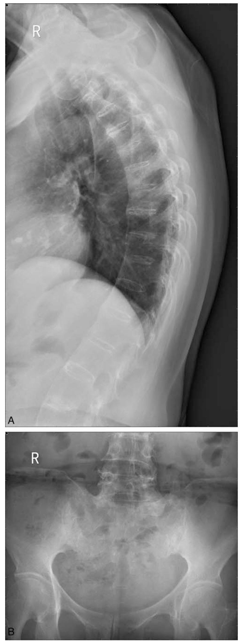
This composite of two X-ray images demonstrates classic radiographic signs of Ankylosing Spondylitis (AS). Panel A is a lateral chest/thoracic spine radiograph showing characteristic 'bamboo spine' appearance. This is characterized by extensive syndesmophyte formation, where bony bridges fuse the thoracic vertebral bodies together, creating a continuous, rigid vertical column. Notably, the intervertebral disc spaces remain relatively preserved in height despite the fusion. Panel B is an anteroposterior (AP) pelvic radiograph focusing on the sacroiliac joints. It shows Grade III bilateral sacroiliitis, evidenced by significant subchondral sclerosis (increased bone density) and narrowing with irregular joint margins. The sacrum and iliac wings exhibit focal areas of erosion and fusion. These findings are hallmarks of seronegative spondyloarthropathies and are used clinically to assess disease progression and staging in patients presenting with chronic inflammatory back pain and HLA-B27 positivity.
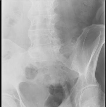
This diagnostic X-ray (radiograph) depicts the lumbar spine and pelvis, highlighting characteristic findings of late-stage Ankylosing Spondylitis (AS). The lumbar vertebrae exhibit extensive ossification of the outer fibers of the annulus fibrosus, forming marginal syndesmophytes. These bony bridges connect adjacent vertebral bodies, resulting in the classic 'bamboo spine' appearance. Furthermore, there is visible fusion of the posterior spinous processes and facet joints. In the pelvic region, the bilateral sacroiliac (SI) joints demonstrate significant pathology, ranging from cortical blurring and erosions to partial ankylosis (fusion), where the joint spaces are narrowed or obliterated by bone bridging. These radiographic features are hallmark indicators of axial spondyloarthritis, reflecting chronic inflammation leading to spinal immobility and structural remodeling. The image serves as a clinical reference for identifying advanced syndesmophytes and sacroiliitis.
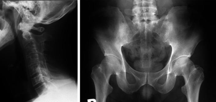
This composite diagnostic image features two X-ray radiographs illustrating the hallmark skeletal manifestations of advanced ankylosing spondylitis (SpA). The left panel is a lateral radiograph of the cervical spine showing 'bamboo spine' morphology. It demonstrates extensive marginal syndesmophytes and ankylosis leading to the fusion of vertebral bodies and complete ossification of the intervertebral disc spaces. The result is a continuous, rigid vertebral column with loss of the normal cervical lordosis. The right panel is an anteroposterior (AP) radiograph of the pelvis focusing on the sacroiliac (SI) joints. Both SI joints show Grade 4 sacroiliitis according to the Modified New York criteria, characterized by total ankylosis (fusion), significant subchondral sclerosis, and loss of visible joint space. These findings represent end-stage chronic inflammation in axial spondyloarthritis. The image is intended for medical education regarding the progression of rheumatological conditions and the interpretation of radiological signs of spinal fusion and sacroiliitis.
1. Definition
- Predominantly affects males (3:1 male-to-female ratio)
- Onset typically between ages 20-30 years
- HLA-B27 association is strong
- Hip joint is among the most commonly affected peripheral joints, often requiring THR
2. Anesthesia Goals
| Goal | Rationale |
|---|---|
| Protect the fragile, rigid spine | Prevent catastrophic cervical fractures during positioning/intubation |
| Secure airway safely | Difficult airway is highly probable due to cervical rigidity and TMJ disease |
| Maintain cardiovascular stability | Risk of aortic regurgitation, conduction defects, coronary artery disease |
| Optimize pulmonary function | Restrictive lung disease from thoracic cage immobility |
| Provide adequate analgesia | Chronic opioid tolerance is common (~36% of patients) |
| Prevent VTE | Immobility and hypercoagulable state |
| Avoid neurological injury | Risk of spinal cord injury from hyperextension and epidural hematoma |
3. Multisystem Considerations (Pre-Anesthetic Assessment Focus)
Airway
- Bamboo spine = difficult airway by default
- Reduced neck extension (cervical kyphosis/fusion) makes direct laryngoscopy potentially impossible
- Anterior atlantoaxial subluxation in ~14% (often asymptomatic)
- TMJ disease may limit mouth opening (reduced inter-incisor distance)
- Higher Mallampati scores in advanced disease
- Risk of devastating neurological injury from inadvertent hyperextension
- Cervical fracture most common at C5-C6 or C6-C7
Cardiovascular
- Inflammation and fibrosis of the aortic root → Aortic regurgitation (prevalence increases with disease duration)
- Extension to conduction system → Heart block / Supraventricular arrhythmias
- Aortic aneurysm, coronary artery disease
- Cardiovascular risk is mildly above general population (less than RA)
Pulmonary
- Restrictive lung disease from thoracic cage immobility and poor chest wall compliance
- Patients may rely entirely on diaphragmatic/abdominal breathing
- Reduced FVC, reduced TLC
- Scalene (interscalene) block is CONTRAINDICATED - reduces FVC by 30-40%, which these patients cannot compensate for
Renal
- Chronic kidney disease (from long-term NSAID use)
- Affects drug dosing, perioperative fluid management
Neurological
- Risk of cauda equina syndrome
- Atlantoaxial subluxation risk
- Preexisting neurological deficits must be documented
Medications
- NSAIDs (first-line for AS) - platelet dysfunction, renal impairment, GI bleeding risk
- DMARDs (non-biologic and biologic) - Biologic agents should be held for at least one dosage cycle prior to surgery; resume after wound healing. Methotrexate can be continued perioperatively.
- No oral glucocorticoids (unlike RA) - stress-dose steroids NOT routinely required
4. Preoperative Evaluation
History
- Duration and severity of disease (longer duration = more extra-articular manifestations)
- Neck range of motion and pain - any new neurological symptoms
- Jaw opening assessment
- Dyspnea on exertion, orthopnea (cardiac/pulmonary involvement)
- Chronic medication history, especially NSAIDs, DMARDs, biologics, opioids
- Previous anesthesia records - history of difficult intubation
Physical Examination
- Complete airway assessment: mouth opening, neck mobility, Mallampati score, thyromental distance
- Cardiovascular exam: murmurs (aortic regurgitation), arrhythmias
- Respiratory: chest expansion, breath sounds, SpO2
- Neurological: baseline deficits (weakness, sensory changes, bladder/bowel)
- Spinal deformity assessment: degree of kyphosis, hip flexion contractures
Investigations
| Investigation | Purpose |
|---|---|
| Cervical spine X-ray (AP/lateral + flexion-extension views) | Detect atlantoaxial subluxation, fusion extent, fracture risk |
| Pulmonary function tests (PFT) | Baseline spirometry - expected restrictive pattern |
| Chest X-ray | Pulmonary infiltrates, cardiomegaly |
| ECG | Conduction abnormalities, arrhythmias |
| Echocardiography | Aortic regurgitation, LV function, valvular disease |
| Complete blood count | Anemia of chronic disease |
| Renal function tests (S. Creatinine, BUN) | NSAID nephrotoxicity |
| Coagulation profile / bleeding time | NSAID-induced platelet dysfunction |
| HbA1c, blood sugar | If on long-term steroids |
| Blood cross-match/group & screen | Anticipated blood loss |
Anesthesia Planning
- Discuss airway management plan explicitly: awake fiberoptic intubation vs. video laryngoscopy
- Neuraxial vs. general anesthesia decision should be made preoperatively
- Discuss with rheumatologist regarding perioperative medication management
- Informed consent specifically discussing difficult airway risks
5. Intraoperative Management
A. Positioning
- THR is typically performed in the lateral decubitus position
- Use adequate padding for shoulders and neck - avoid any hyperextension
- A semi-sitting position can accommodate hip flexion deformities
- Careful log-roll positioning with all team members coordinated
- Document baseline neurological status before and assess after positioning
- Protect all pressure points; bony prominences are at higher fracture risk
B. Airway Management
| Situation | Preferred Technique |
|---|---|
| Severe cervical disease, predicted difficult airway | Awake Fiberoptic Intubation (AFOI) - gold standard; allows neurological monitoring during intubation with maintained spontaneous ventilation |
| Moderate disease | Video Laryngoscopy (GlideScope/C-MAC) - useful but failures reported |
| Cases not requiring ETT | LMA as primary airway or bridge to intubation (intubating LMA) |
| Cormack-Lehane III/IV on DL | Video laryngoscopy first; fiberoptic if fails |
- Soft cervical collar should remain on until patient is in the anaesthetic room
- Always have backup plans (plan A, B, C, D) documented
- Keep the neck in neutral position at all times
- Surgical airway (cricothyrotomy) must be immediately available
C. Choice of Anesthesia
- Suitable when neuraxial is contraindicated or fails
- RSI or modified RSI based on aspiration risk
- Maintain hypnosis with inhalational (sevoflurane, desflurane) or TIVA (propofol)
- Neuromuscular blockade + reversal
- Maintain normotension (avoid hypotension with AR)
- Preferred for THR when feasible - associated with:
- Decreased mortality
- Reduced VTE/DVT incidence
- Lower infection rates
- Less blood loss
- Reduced acute kidney injury
- Challenges in AS:
- Higher failure rate due to ossified ligaments and loss of interspinous spaces
- Ultrasound guidance or fluoroscopic guidance is recommended to improve success (PMID: 41318896 - RCT showing US-guided superior to landmark technique in AS patients)
- Paramedian approach may be easier than midline
- Smaller intrathecal doses may be needed - risk of high spinal block (reduced epidural space and altered CSF dynamics - documented in case reports including PMID: 42534176)
- Epidural hematoma risk is HIGHER in AS than general population - due to: traumatic needle placement, NSAID use, and narrowed epidural space making cord compression more symptomatic
- Post-procedure vigilance for epidural hematoma symptoms is mandatory
- Offers flexibility - spinal for immediate anesthesia, epidural catheter for postoperative analgesia extension
D. Monitoring
- Standard ASA monitoring (SpO2, NIBP, ECG, EtCO2, temperature)
- Invasive arterial line - justified for patients with significant AR, cardiac disease, or anticipated hemodynamic instability
- Central venous access - for those with poor peripheral access or hemodynamic instability
- Large-bore IV access (x2) - anticipated blood loss in THR
- Urine output monitoring (Foley catheter)
- Temperature monitoring and active warming (forced air warmer)
E. Intraoperative Pharmacology
- Tranexamic acid (TXA): Routinely recommended before incision to reduce blood loss - both IV and topical routes supported; single dose adequate
- Neuraxial opioids (if spinal/epidural used): morphine or hydromorphone extends postoperative analgesia
- Antiemetics: Ondansetron/dexamethasone (dexamethasone dose <15 mg to avoid adrenal suppression issues)
- Antibiotics: Cefazolin prophylaxis
- Vasopressors on standby: Phenylephrine/ephedrine for hypotension
F. Blood Management
- Cell salvage (intraoperative autologous transfusion)
- Permissive hypotension (controlled hypotension) to reduce blood loss - but CAUTION with AR (maintain adequate diastolic pressure)
- Trigger-based transfusion (Hb <7-8 g/dL)
6. Postoperative Management
Analgesia (Multimodal approach)
| Modality | Details |
|---|---|
| Neuraxial opioids | If spinal/epidural placed - extended analgesia |
| Epidural infusion | Bupivacaine ± fentanyl via epidural catheter |
| IV PCA | Morphine/hydromorphone - note chronic opioid users may need higher doses |
| NSAIDs | Use with caution - renal function monitoring required |
| Acetaminophen (paracetamol) | Scheduled around the clock |
| Peripheral nerve blocks | Femoral nerve block or fascia iliaca block for THR |
| Gabapentinoids | Pregabalin/gabapentin for opioid-sparing effect |
Respiratory
- Incentive spirometry and early physiotherapy
- Positioning: head-up position improves diaphragmatic excursion
- Continuous SpO2 monitoring - high risk of hypoxia due to restrictive lung disease
- Humidified oxygen supplementation
- Avoid excessive sedation (respiratory depression risk)
Cardiovascular
- Continuous ECG monitoring for arrhythmias (heart block risk)
- Maintain adequate intravascular volume
- Echocardiographic monitoring if significant AR (avoid tachycardia, maintain preload)
Thromboprophylaxis
- LMWH (enoxaparin) or factor Xa inhibitors (rivaroxaban, apixaban) - start 6-12 hours postoperatively
- Mechanical: compression stockings, intermittent pneumatic compression devices
- Early mobilization (when safe)
Wound Care
- If biologic DMARDs were held preoperatively, resume only after wound healing is confirmed
- Monitor for signs of infection (immunocompromised state on DMARDs)
Neurological Monitoring
- Immediate assessment of lower limb neurology post-neuraxial block
- Vigilance for epidural hematoma: back pain + bladder/bowel dysfunction + motor weakness = EMERGENCY - urgent MRI + neurosurgical review
- Assess for new neurological deficits following positioning
7. Complications
Airway/Respiratory
| Complication | Mechanism | Prevention |
|---|---|---|
| Failed/difficult intubation | Cervical fusion, TMJ disease, kyphosis | AFOI, video laryngoscopy, graded approach |
| Cervical spine fracture | Hyperextension during laryngoscopy | Neutral positioning, gentle technique |
| Quadriplegia/cord injury | Atlantoaxial subluxation + cord compression | Preop imaging, avoid extension |
| Respiratory failure | Restrictive lung disease + postop splinting | PFTs, incentive spirometry, adequate analgesia |
Neuraxial
| Complication | Notes |
|---|---|
| Failed spinal/epidural | Higher failure rate due to ossified ligaments |
| Epidural hematoma | Higher risk in AS - NSAID use + traumatic placement + narrow epidural space |
| High spinal block | Reduced epidural space alters CSF dynamics, drug spread unpredictable |
| Adhesive arachnoiditis | Rare - associated with AS itself and neuraxial techniques |
Cardiovascular
| Complication | Notes |
|---|---|
| Hemodynamic instability | Aortic regurgitation + vasodilation from neuraxial anesthesia |
| Arrhythmias | Heart block, SVT due to conduction system disease |
| Bone cement implantation syndrome (BCIS) | Fat/bone marrow/cement embolism at cementing - hypotension, hypoxia, cardiac arrest |
Surgical/THR-Specific
| Complication | Notes |
|---|---|
| DVT/Pulmonary embolism | High-risk surgery - mandatory prophylaxis |
| Significant blood loss/hemorrhage | Large raw surfaces during acetabular reaming |
| Periprosthetic fracture | Osteoporotic bone from AS + long-term disease |
| Dislocation of prosthesis | Positioning challenges during surgery |
| Wound infection | Immunosuppressed (DMARDs/biologics) |
Postoperative
- Residual neuromuscular blockade
- Postoperative nausea and vomiting (PONV)
- Chronic pain/opioid dependence
- Delayed wound healing (biologic agents)
- Renal impairment (NSAIDs, perioperative hypotension)
Summary Flow
AS Patient for THR
        ↓
Preop: Assess airway (cervical X-ray, Mallampati) + Echo + PFTs + Hold biologics
        ↓
Anaesthesia Plan: AFOI / VL for GA || US-guided spinal for neuraxial
        ↓
Intraop: Careful lateral positioning, TXA, invasive monitoring if needed
        ↓
Postop: Multimodal analgesia, SpO2 monitoring, early mobilization, LMWH, vigilance for epidural hematoma
- Miller's Anesthesia, 10e - Chapter 29 (Ankylosing Spondylitis, pp. 4291-4293) and Chapter 60 (pp. 9181-9182)
- Morgan and Mikhail's Clinical Anesthesiology, 7e - Chapter 38 (THA Intraoperative Management, p. 1513)
- Rheumatology, 2-Volume Set (Elsevier 2022) - Anesthesia in Rheumatic Disease (p. 500)
- Recent RCT: Mishra NK et al. (2026) - Ultrasound-assisted vs. landmark-guided subarachnoid block in AS for THR [PMID: 41318896]
- Case report of high spinal block with intrathecal mepivacaine in AS: Garcia-Mendez et al. (2026) [PMID: 42534176]
Hello, I’m an anesthesiology final year resident practicing anesthesiology.. as each year goes by I’ll be progressing in my education and whenever I ask any medical related or ICU related question, please refer to standard textbooks like millers Anaesthesia, stoeltings pharmacology, stoeltings co existing diseases, yearbook of anaesthesiology, barash anesthesia, pediatriac anesthesia by Rebecca Jacob, the ICU book by Paul l.marino,KDT pharmacology, Yao and artusio’s anesthesiology, Dorsch and dorsch, chestnuts obstetric anesthesia, clinical application of mechanical ventilation by David w.chang and standard journals,case reports, textbooks, guidelines and articles like British journal of anesthesia, pubmed and give me answers which are highly reliable and Evidence backed answers from guidelines, research papers, and journals. Give heavily researched, clinical-grade answers, use real-time search tools to hunt for data from reputable databases (like PubMed, NCBI, Cochrane Reviews) and institutional bodies (like the FDA, CDC, AHA, or WHO), drop conversational filler and lead directly with verified data, citing the specific studies, journals, or clinical guidelines where the evidence lives, use exact biomedical terminology and structured data (such as differential tables or mechanism breakdowns) without watering down the language, If the medical literature shows conflicting guidelines or insufficient data on a topic, try to explicitly map out that conflict rather than trying to smooth it over. Include Clinical Decision Support System (CDSS), Point-of-care clinical decision support, Rigidly cited, evidence-backed medical data, align with medical/healthcare standards, Save this format for future questions I ask. - Also Whenever I ask about a drug mention all these things in detail - group, chemical structure, formulation, dilution, available forms in market, routes of administration, dosage, mechanism of action, pharmacokinetics, pharmacodynamics, indications, contraindications, adverse effects and special points and a quick reference summary table.
Response Profile: Anesthesiology Final Year Resident
Primary Reference Stack (in order of priority)
| Domain | Primary Textbook(s) |
|---|---|
| Core Anesthesia | Miller's Anesthesia 10e, Barash Clinical Anesthesia 9e, Yao & Artusio's Anesthesiology |
| Pharmacology | Stoelting's Pharmacology & Physiology 6e, KDT (Tripathi) Pharmacology |
| Co-existing Disease | Stoelting's Anesthesia & Co-Existing Disease |
| Obstetric Anesthesia | Chestnut's Obstetric Anesthesia 6e |
| Pediatric Anesthesia | Pediatric Anesthesia - Rebecca Jacob |
| Airway/Equipment | Dorsch & Dorsch - Understanding Anesthesia Equipment |
| ICU/Critical Care | The ICU Book - Paul L. Marino |
| Mechanical Ventilation | Clinical Application of Mechanical Ventilation - David W. Chang |
| Journals | BJA, Anesthesiology (ASA), Anesthesia & Analgesia, JAMA, NEJM, Cochrane, PubMed/NCBI |
| Guidelines | ASA, ESAIC (ESA), SOAP, AAP, AHA/ACC, WHO, FDA, NICE, ACOG |
Answer Structure (Applied to Every Response)
- Definition / Classification - precise biomedical terminology
- Mechanism / Pathophysiology - molecular/cellular level where relevant
- Evidence-Based Content - graded evidence (RCT > Systematic Review > Cohort > Expert Opinion)
- Clinical Decision Support (CDSS) - structured decision trees or algorithms
- Differential / Comparison Tables - side-by-side structured data
- Conflicting Guidelines - explicitly mapped, not smoothed over
- Drug Questions - the 15-point pharmacology template (group, chemical structure, formulation, dilution, market forms, routes, dosage, MOA, PK, PD, indications, contraindications, adverse effects, special points, quick reference summary table)
- Citations - specific study names, PMIDs, journal names, guideline year and issuing body
- No filler language - direct, clinical-grade prose only
Evidence Grading I'll Use
| Grade | Source |
|---|---|
| IA | Meta-analysis / Systematic review of RCTs |
| IB | Single well-designed RCT |
| IIA | Well-designed controlled trial, non-randomized |
| IIB | Cohort or case-control study |
| III | Expert consensus / Guideline |
| IV | Case report / Case series |
Drug Template (Applied Every Time a Drug Is Asked)
- Group / Class
- Chemical Structure
- Formulation & Dilution
- Available Market Forms
- Routes of Administration
- Dosage (induction / maintenance / pediatric / special populations)
- Mechanism of Action
- Pharmacokinetics (ADME)
- Pharmacodynamics (system-by-system)
- Indications
- Contraindications
- Adverse Effects
- Drug Interactions
- Special Points (pregnancy, renal/hepatic impairment, reversal, monitoring)
- Quick Reference Summary Table