
Acid base balance full details for md exams 10 marks answer
Reading File
Now let me search the medical library in parallel for comprehensive acid-base balance content.
Finding Sources
Finding Sources
Excellent. Now let me read the most authoritative sources in parallel - Medical Physiology (Boron), Ganong's, and Harrison's for comprehensive content.
Reading File
Reading File
Reading File
Let me get the core acid-base physiology content from the major physiology textbooks and Harrison's detailed sections.
Finding Sources
Reading File
Let me now read the key acid-base sections from Harrison's (the most comprehensive for MD exams) and also from Ganong's for compensation formulas.
Reading File
Reading File
I now have excellent content from Harrison's and Ganong's. Let me get the compensation formulas and metabolic acidosis/alkalosis details from Harrison's more completely.
Reading File
I now have very rich content from Harrison's. Let me fetch the acid-base nomogram image from Harrison's and compile the full answer.
Loading Image
Loading Image
I now have all the key images and content needed. Let me compile the comprehensive MD exam answer.
ACID-BASE BALANCE - Comprehensive MD Exam Answer (10 Marks)
1. NORMAL ACID-BASE HOMEOSTASIS
Systemic arterial pH is maintained between 7.35 and 7.45 by three integrated mechanisms:
- Extracellular and intracellular chemical buffers (immediate, seconds)
- Respiratory regulation of PaCO2 (minutes to hours)
- Renal regulation of plasma HCO3- (hours to days)
The relationship between these components is expressed by the Henderson-Hasselbalch equation:
pH = pK' + log [HCO3-] / (0.03 x PaCO2)
Where pK' = 6.1, and 0.03 is the solubility coefficient of CO2. At steady state, pH = 7.4 when [HCO3-] = 24 mEq/L and PaCO2 = 40 mmHg.
Normal daily acid production: ~50-70 mEq of fixed (non-volatile) acid from metabolism.
2. BUFFER SYSTEMS
A. Bicarbonate Buffer (most important extracellular buffer)
CO2 + H2O ⇌ H2CO3 ⇌ H+ + HCO3-
- pKa = 6.1 (far from physiological pH, yet most effective because CO2 is an open system - lungs continuously regulate CO2)
- Buffers ~50% of acid load
B. Proteins (Hemoglobin - most important intracellular buffer)
- Hb is the major intracellular buffer in blood
- Deoxyhemoglobin is a better buffer than oxyhemoglobin (Haldane effect)
C. Phosphate Buffer
- HPO4²- / H2PO4- system (pKa = 6.8)
- Important in urine acidification and intracellular buffering
D. Bone Buffer
- In chronic acidosis, bone carbonate and phosphate are mobilized - contributes to osteomalacia/osteoporosis
3. RESPIRATORY REGULATION
The respiratory system controls PaCO2 by adjusting alveolar ventilation:
- Acidosis → stimulates peripheral (carotid body) and central chemoreceptors → hyperventilation → decreased PaCO2 → pH rises
- Alkalosis → decreased chemoreceptor drive → hypoventilation → increased PaCO2 → pH falls
Respiratory compensation is rapid (minutes to hours) but incomplete - it returns pH toward, but not to, normal.
4. RENAL REGULATION
The kidney regulates pH by:
- Reabsorption of filtered HCO3- (proximal tubule: 80-85%; TAL and distal: remainder)
- Titratable acid excretion (mainly as H2PO4- in distal tubule)
- Ammonium (NH4+) excretion - most important for net acid excretion
- Proximal tubule generates NH4+ from glutamine
- NH4+ excreted in urine carries H+ out of the body
Renal compensation is slow (days) but complete (fully restores pH in isolated respiratory disorders).
5. CLASSIFICATION OF ACID-BASE DISORDERS
| Disorder | pH | Primary Change | Compensation |
|---|---|---|---|
| Metabolic acidosis | ↓ | HCO3- ↓ | PaCO2 ↓ (hyperventilation) |
| Metabolic alkalosis | ↑ | HCO3- ↑ | PaCO2 ↑ (hypoventilation) |
| Respiratory acidosis | ↓ | PaCO2 ↑ | HCO3- ↑ (renal) |
| Respiratory alkalosis | ↑ | PaCO2 ↓ | HCO3- ↓ (renal) |
6. COMPENSATION FORMULAS (Critical for MD Exams)
| Disorder | Expected Compensation |
|---|---|
| Metabolic acidosis | PaCO2 = 1.5 x [HCO3-] + 8 ± 2 (Winter's formula) |
| Metabolic alkalosis | PaCO2 increases by 0.7 mmHg per 1 mEq/L rise in HCO3- |
| Acute resp. acidosis | HCO3- increases by 1 mEq/L per 10 mmHg rise in PaCO2 |
| Chronic resp. acidosis | HCO3- increases by 3.5 mEq/L per 10 mmHg rise in PaCO2 |
| Acute resp. alkalosis | HCO3- falls by 2 mEq/L per 10 mmHg fall in PaCO2 |
| Chronic resp. alkalosis | HCO3- falls by 5 mEq/L per 10 mmHg fall in PaCO2 (only disorder where pH fully normalizes) |
- Harrison's Principles of Internal Medicine 22E, p. 411
7. ACID-BASE NOMOGRAM
The Davenport/acid-base nomogram plots pH vs. HCO3- with PCO2 isobars, showing 95% confidence bands for each simple disorder:
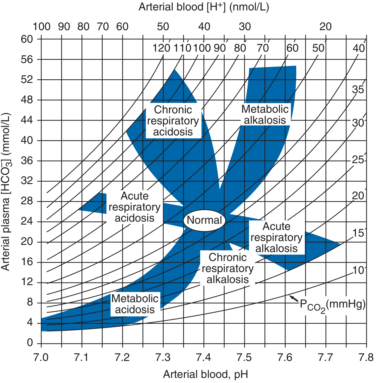
Acid-base nomogram (Harrison's Principles of Internal Medicine 22E) - shaded zones represent 95% confidence limits for simple disorders
8. METABOLIC ACIDOSIS
Definition
Primary fall in plasma HCO3- (< 22 mEq/L), with compensatory fall in PaCO2.
Causes - classified by Anion Gap
Anion Gap (AG) = Na+ - (Cl- + HCO3-)
- Normal AG = 6-12 mEq/L (average ~10 mEq/L)
- Corrected AG: add 2.5 mEq/L per 1 g/dL fall in serum albumin below 4.5 g/dL
High Anion Gap (HAGMA) - mnemonic MUDPILES:
- M - Methanol
- U - Uraemia (renal failure)
- D - DKA (diabetic ketoacidosis)
- P - Propylene glycol / Paraldehyde
- I - Isoniazid / Iron
- L - Lactic acidosis (type A: tissue hypoxia; type B: metformin, liver failure)
- E - Ethylene glycol
- S - Salicylates
Normal Anion Gap (hyperchloraemic) acidosis - mnemonic HARD-UP:
- Diarrhoea (most common - loss of HCO3-)
- Renal tubular acidosis (types 1, 2, 4)
- Ureterosigmoidostomy
- Addison's disease
- Post-hypocapnia
- Carbonic anhydrase inhibitors (acetazolamide)
Delta Ratio (for HAGMA) = (AG - 12) / (24 - HCO3-)
- <0.4: pure normal AG acidosis
- 0.4-0.8: mixed HAGMA + normal AG acidosis
- 1-2: pure HAGMA
-
2: HAGMA + concurrent metabolic alkalosis
Clinical Features
- Kussmaul breathing (deep, rapid - compensatory hyperventilation in severe metabolic acidosis)
- Decreased cardiac contractility, hypotension
- Decreased insulin sensitivity, hyperkalaemia
- Confusion, lethargy, coma
Treatment
- Treat underlying cause
- NaHCO3 rarely needed; use if pH < 7.1 or severe acidaemia with haemodynamic compromise
9. METABOLIC ALKALOSIS
Definition
Primary rise in plasma HCO3- (>26 mEq/L), with compensatory hypoventilation (PaCO2 rises).
Causes
Chloride-responsive (urine Cl- <10 mEq/L):
- Vomiting/nasogastric suction (loss of HCl)
- Diuretics (thiazides, loop)
- Post-hypercapnia
- Villous adenoma
Chloride-resistant (urine Cl- >20 mEq/L):
- Hyperaldosteronism (primary/secondary)
- Cushing's syndrome
- Bartter's/Gitelman's syndrome
- Severe hypokalaemia
Maintenance factors
- Volume depletion → increased aldosterone → renal HCO3- retention
- Hypokalaemia → increased renal H+ secretion
Treatment
- Chloride-responsive: IV saline (0.9%) + KCl; correct the underlying cause
- Chloride-resistant: treat underlying mineralocorticoid excess
10. RESPIRATORY ACIDOSIS
Definition
Primary rise in PaCO2 (>45 mmHg) with compensatory rise in HCO3-.
Causes (hypoventilation)
| CNS depression | Opiates, sedatives, anaesthesia, brainstem lesion |
|---|---|
| Neuromuscular | Myasthenia gravis, GBS, poliomyelitis, MND |
| Airway obstruction | COPD, severe asthma, foreign body |
| Chest wall | Kyphoscoliosis, obesity hypoventilation |
| Parenchymal | End-stage pulmonary fibrosis |
Acute vs Chronic
- Acute: HCO3- rises by 1 mEq/L per 10 mmHg rise in PaCO2; pH falls markedly
- Chronic: HCO3- rises by 3.5 mEq/L per 10 mmHg rise in PaCO2; pH nearly normal
Treatment
- Treat the cause
- Mechanical ventilation if severe
- Oxygen cautiously in COPD (avoid eliminating hypoxic drive)
11. RESPIRATORY ALKALOSIS
Definition
Primary fall in PaCO2 (<35 mmHg) with compensatory fall in HCO3-.
Causes (hyperventilation)
- Anxiety, psychogenic
- Hypoxaemia (pneumonia, PE, high altitude, severe anaemia)
- Sepsis (early) - most common cause of resp. alkalosis in ICU
- CNS stimulation (salicylates, fever, head injury)
- Pregnancy (progesterone effect)
- Liver failure (hepatic encephalopathy)
- Iatrogenic (mechanical ventilation)
Compensation
- Acute: HCO3- falls 2 mEq/L per 10 mmHg fall in PaCO2
- Chronic: HCO3- falls 5 mEq/L per 10 mmHg fall - only disorder where full pH compensation can occur
Clinical Features
- Paraesthesias (perioral, fingertips), carpopedal spasm
- Lightheadedness, syncope
- Decreased ionised calcium (alkalosis increases protein binding of Ca2+)
12. MIXED ACID-BASE DISORDERS
Defined as independently coexisting disorders (not just compensation). Common in critically ill patients.
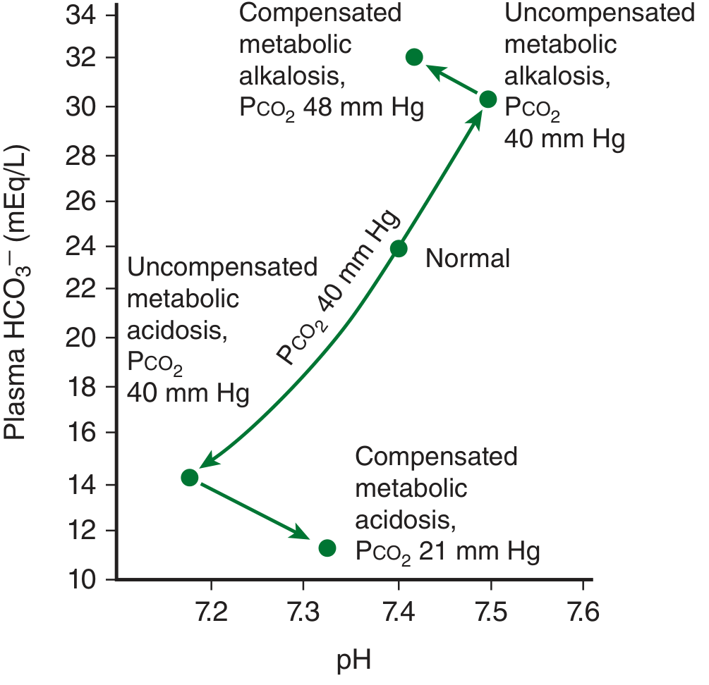
Acid-base paths during metabolic acidosis and alkalosis (Ganong's Review of Medical Physiology, 26th Ed.)
| Mixed Disorder | pH change | Example |
|---|---|---|
| Met. acidosis + Resp. acidosis | Severe ↓↓ | Cardiac arrest, severe COPD + DKA |
| Met. acidosis + Resp. alkalosis | May be near normal | Sepsis + shock, salicylate poisoning |
| Met. alkalosis + Resp. acidosis | May be near normal | COPD + diuretics/vomiting |
| Met. acidosis + Met. alkalosis | pH variable | DKA + vomiting |
Key: If PaCO2 deviates from the predicted compensation (using Winter's formula or compensation table), a mixed disorder is present.
13. SYSTEMATIC APPROACH TO ABG INTERPRETATION (Step-by-Step)
- Check pH - acidaemia (<7.35) or alkalaemia (>7.45)?
- Check PaCO2 - elevated, normal, or low?
- Check HCO3- - elevated, normal, or low?
- Identify the primary disorder - which change (CO2 or HCO3-) explains the pH direction?
- Calculate expected compensation - using formulas above
- Compare actual vs. expected compensation - deviation = mixed disorder
- Calculate Anion Gap - if metabolic acidosis present
- Calculate Delta Ratio - if HAGMA present
- Correlate with clinical context
14. KEY NORMAL VALUES (Reference)
| Parameter | Normal Range |
|---|---|
| Arterial pH | 7.35-7.45 |
| PaCO2 | 35-45 mmHg |
| HCO3- | 22-26 mEq/L |
| PaO2 | 80-100 mmHg |
| Base excess | -2 to +2 mEq/L |
| Anion Gap | 6-12 mEq/L |
15. SPECIAL TOPICS FOR MD EXAMS
Renal Tubular Acidosis (RTA) - Normal AG acidosis
| Type | Defect | Urine pH | K+ | Cause |
|---|---|---|---|---|
| Type 1 (Distal) | Failure to acidify urine | >5.5 | Low | SLE, Sjogren's, amphotericin |
| Type 2 (Proximal) | Failure to reabsorb HCO3- | <5.5 | Low | Multiple myeloma, Wilson's |
| Type 4 | Aldosterone deficiency/resistance | <5.5 | High | DM nephropathy, Addison's |
Lactic Acidosis
- Type A: Tissue hypoperfusion - shock, cardiac arrest, severe anaemia, CO poisoning
- Type B: No obvious hypoperfusion - metformin toxicity, liver failure, thiamine deficiency, malignancy
Salicylate Poisoning - Classic Mixed Disorder
- Early: Respiratory alkalosis (direct CNS stimulation)
- Late: Metabolic acidosis (HAGMA) + Respiratory alkalosis simultaneously
- Classic exam scenario
Sources: Harrison's Principles of Internal Medicine 22E (2025), Ganong's Review of Medical Physiology 26th Ed., Medical Physiology (Boron & Boulpaep)
This is a shared conversation. Sign in to Orris to start your own chat.