


Analyse these KNRUHS pyqs and sort them as priority wise Most repeated Most likely to be asked in 2026 Less likely to be asked
KNRUHS Physiology PYQ Analysis - Blood Chapter
Priority Classification
🔴 TIER 1 — MOST REPEATED (High Priority, Study First)
1. Erythropoiesis ⭐⭐⭐ (HIGHEST PRIORITY)
- March-21, July-19, Dec-17, July/Aug-14, July-12, July-2000, Aug-1980, April-86, Oct-98, Nov-91
- Asked as: Stages, factors, morphological changes, factors regulating erythropoiesis, effect of Vit B12/iron
- Must prepare: All stages + factors (EPO, iron, B12, folate, androgens) + exam-ready diagram
2. Blood Coagulation / Clotting Mechanism ⭐⭐⭐
- Mar-21, Nov-13, May-07, Oct-1995, Oct-1993, April-84, Oct-1983, Aug-1980, Oct/Nov-18 (intrinsic pathway)
- Asked as: Mechanism of coagulation, clotting factors, intrinsic pathway, cascade amplification, fibrinolytic system
- Must prepare: Intrinsic + Extrinsic + Common pathway + anticoagulants together
3. Anticoagulants ⭐⭐⭐
- Dec-17, July-11, April-03, Feb-01, July-87, Nov-91, April-95, Oct-1998 (heparin/dicoumarol/sodium citrate), Aug-15 (intrinsic + anticoagulants)
- Asked as: Name 3 anticoagulants + mode of action, heparin/dicoumarol/sodium citrate mechanism, Vitamin K antagonist
4. Mismatched / Incompatible Blood Transfusion ⭐⭐⭐
- July-21, Aug-18, Nov-14, Jan-10, April-94, Oct-1984, April-1983
- Asked as: Hazards, effects, consequences, precautions to avoid
5. Platelets (Structure, Count, Functions) ⭐⭐
- Jan-12, Feb-2000 (×2), Oct-1998, April-96, April-95, April-94, July-1991
- Asked as: Functions, morphology + count, enumerate functions + explain one, structure
6. Functions of Plasma Proteins ⭐⭐
- July-19, March/April-05, April-03 (×2), Aug-01, May-1988, Nov-1994, July-1991, April-1995
- Asked as: Functions, isolation, concentration, albumin function, plasma proteins and their functions
7. Rh Factor ⭐⭐
- July-09, May-1999, Oct-1996, July-2000, April-1997, July-1991
- Also embedded in Anticoagulants short Qs (April-03 old)
- Asked as: Rh factor + significance, importance in blood transfusion
8. Blood Groups (ABO + Landsteiner's Laws) ⭐⭐
- July-13, Sep/Oct-07, Feb-09, Oct-04, Oct-1999, May-1988, Oct/Nov-02, Oct-1998
- Asked as: Blood group systems, physiological basis, Landsteiner's laws, determine own blood group
9. Anaemia (Classification + Types) ⭐⭐
- Jan-12, Sep/Oct-07, Feb-2000, May-1999 (long Qs); Nov-20, Jan-11, July-09 (very short Qs)
- Asked as: Anaemia, nutritional deficiency anaemias, classify anaemias, macrocytic/megaloblastic, morphological classification
🟠 TIER 2 — MOST LIKELY TO BE ASKED IN 2026
| Topic | Last Asked | Likelihood Reason |
|---|---|---|
| Erythropoiesis | March-21 | Asked in every recent exam; always comes back |
| Erythropoietin | July-21, July-18, July-10 | 3 consecutive July exams - due again |
| WBC / Leucocytes (Functions, Types) | July-21 (VSQ), April-03 | Very short Qs trend upward post-2018 |
| Eosinophils / Role in allergy | March-21, Oct/Nov-18 | Repeated in last 2 exams |
| Blood coagulation (intrinsic) | Oct/Nov-18, Aug-16 | Due as a long question again |
| Erythroblastosis Fetalis | July-21, July-11, Sep-06 | Asked as short Q in July-21 |
| Haemophilia | July-21, Feb-01 | Returned in July-21 after long gap |
| Humoral immunity | July-21, Feb-2000 | Long gap - returned 2021 |
| T-lymphocytes / Immunity | Aug-17, July/Aug-14 | Regular 3-year cycle |
| Anti-Coagulants (VSQ) | Dec-17, Jan-10, Sep/Oct-07 | Steady short Q frequency |
| ESR (define + normal + factors) | Aug-16, Aug-15, Jan-11 | Short Q staple |
| Megaloblastic Anaemia | Oct/Nov-18 | Clinical relevance, due for repeat |
| Reticulo-endothelial system | Aug-18, April-03 | Recent reappearance |
| Haemostasis + Bleeding disorders | April-03 + Feb-01 | Due for a long Q revival |
| Mismatched transfusion | July-21 | Just appeared - may come as short Q |
| Plasma proteins | July-19 | Recent long Q appearance |
🟢 TIER 3 — LESS LIKELY TO BE ASKED IN 2026
| Topic | Last Asked | Reason Less Likely |
|---|---|---|
| Reticular formation | Nov-1994 | Very old, niche neurophysiology topic |
| Hypothalamus nuclei / integrative processes | Oct-1993 | Very old, rarely repeated |
| Body defense mechanisms (skin/mouth/airway) | Feb-2000 | Vague, non-specific - rarely revisited |
| Haemorrhage & compensatory mechanisms | Feb-2001 (old) | Old format question |
| Blood volume + estimation | Oct-1997, Nov-1994 | Rarely asked in recent years |
| Fick's principle | Aug-2001 | Cardio topic, appeared only once |
| Indicator dilution technique | April-03 | Appeared once |
| Physiological leucocytosis causes | March/April-05 | Appeared once |
| Packed cell volume in haemorrhagic shock | Sep-06 | Niche clinical application |
| Normal diameter of erythrocytes | Oct-1996, Nov-1992 | Very old |
| Normal life span of RBC + determination | Oct-1996 | Old and narrow |
| Oncotic pressure of plasma | Oct-1993 | Very old |
| Basophil and its functions | April-1995 | Single appearance |
| Formation of lymph / composition | Oct-1993 | Old, low recent frequency |
| Vitamin K dependent clotting factors | July/Aug-14 | Appears occasionally as VSQ |
| Secretory vesicles / Exocytosis / Mitochondria | Jan-12, Jan-11, Feb-09 | General physiology, not blood-specific |
| Erythroblastosis fetalis (as long Q) | Oct-1983 | Only as short Q recently |
| Blood transfusion rules (Apr-1983) | April-1983 | Very old long Q format |
| Protein functions of plasma (Apr-1985) | April-1985 | Extremely old |
📊 Quick Reference Summary Table
| Priority | Topic | Times Asked | Focus Area |
|---|---|---|---|
| 🔴 #1 | Erythropoiesis | 10-12× | Stages + Factors + Regulation |
| 🔴 #2 | Blood Coagulation | 8-10× | Intrinsic + Extrinsic + Cascade |
| 🔴 #3 | Anticoagulants | 8-9× | Heparin, Dicoumarol, Sodium Citrate |
| 🔴 #4 | Mismatched Transfusion | 6-7× | Effects + Hazards + Precautions |
| 🔴 #5 | Platelets | 8× | Structure + Count + Functions |
| 🔴 #6 | Plasma Proteins | 7-8× | Types + Functions + Albumin |
| 🔴 #7 | Rh Factor | 6-7× | Significance + Incompatibility |
| 🔴 #8 | Blood Groups / Landsteiner | 6× | ABO system + Clinical importance |
| 🔴 #9 | Anaemia | 5-6× | Classification + Nutritional types |
| 🟠 #10 | Erythropoietin | 3× (all July!) | EPO mechanism + renal role |
| 🟠 #11 | T & B lymphocytes / Immunity | 4× | Role in immunity |
| 🟠 #12 | ESR | 3× | Definition + Normal value + Factors |
| 🟠 #13 | Erythroblastosis Fetalis | 4× | Cause + Prevention + Rh basis |
| 🟠 #14 | Haemostasis + Haemophilia | 4× | Haemostasis process + bleeding disorders |
| 🟠 #15 | Leucocytes (WBC types + functions) | 4× | Classification + Defense role |
| 🟠 #16 | Jaundice / Haemolytic Jaundice | 3× | Types + Clinical distinction |
| 🟠 #17 | Haemoglobin (structure/catabolism) | 2× | Formation factors + breakdown |
| 🟠 #18 | Reticulo-endothelial System | 2× (recent) | Functions + Cell types |
| 🟠 #19 | Fate of Hb / RBC | 3× | Post-hemolysis pathway |
| 🟠 #20 | Megaloblastic Anaemia | 1× (recent) | Causes + features |
| 🟢 | Reticular formation | 1× (1994) | Very old |
| 🟢 | Hypothalamus nuclei | 1× (1993) | Very old |
| 🟢 | Fick's principle | 1× | Cardio/not blood |
| 🟢 | Blood volume estimation | 2× (old) | Old format |
| 🟢 | Oncotic pressure | 1× (1993) | Very old |
🎯 2026 Exam Strategy
- Erythropoiesis (with diagram)
- Blood coagulation - both intrinsic & extrinsic pathways
- Anticoagulants (heparin, warfarin, sodium citrate + mechanisms)
- Platelets - structure, count, functions
- Plasma proteins - types, concentrations, functions
- Blood groups + Rh factor
- Mismatched blood transfusion - hazards & effects
- Anaemia - classification (morphological + etiological)
- Erythropoietin
- T/B lymphocytes + immunity
- ESR, Eosinophils, Erythroblastosis fetalis, Haemophilia, Anti-coagulants, Lymph, Functions of spleen, Land Steiner's law, Cross matching, WBC functions
Hazards effects and consequences of mismatched blood transfusions
Hazards, Effects & Consequences of Mismatched Blood Transfusion
Definition
Mechanism (Pathophysiology)
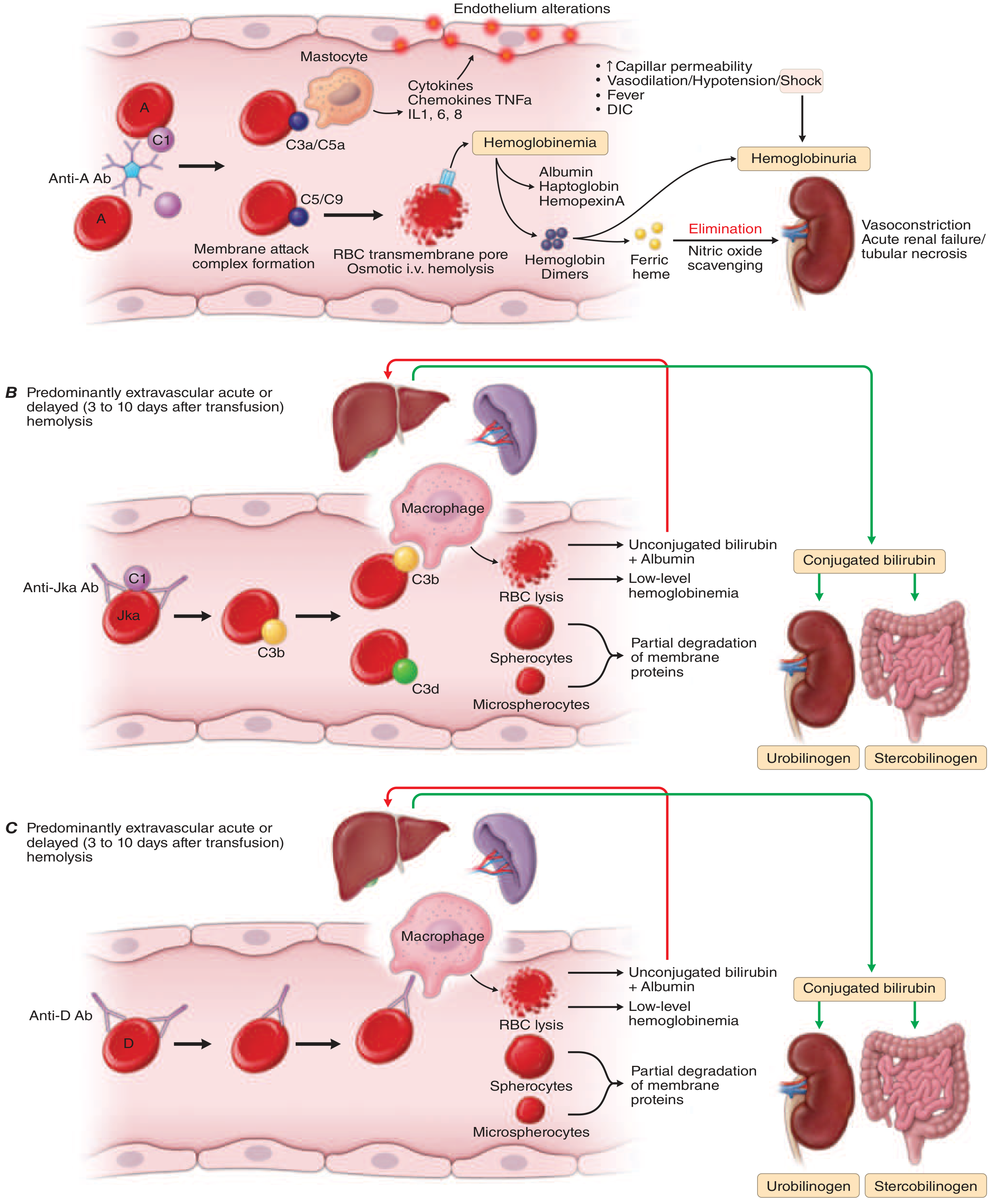
- Recipient anti-A/anti-B IgM antibodies bind to donor RBC surface antigens
- Antigen-antibody complex activates the complement cascade (C1 → C3 → C5 → C5b-C9)
- Membrane Attack Complex (MAC) forms pores in donor RBC membranes
- Intravascular hemolysis - massive destruction of transfused RBCs
- Free hemoglobin floods the plasma → hemoglobinemia
- Complement fragments C3a and C5a activate mast cells → release cytokines (IL-1, IL-6, IL-8, TNF-α)
- Kallikrein-kinin system activation → bradykinin release → hypotension
- RBC stroma releases erythrocytin → activates intrinsic coagulation → DIC
- Free Hb precipitates as acid hematin in distal tubules → acute renal failure
Classification of Hazards
A. Immunological Reactions
| Type | Onset | Mechanism |
|---|---|---|
| Acute Hemolytic Reaction | During/within 24 hrs | ABO/Rh incompatibility → intravascular hemolysis |
| Delayed Hemolytic Reaction | 3-10 days post transfusion | IgG antibodies → extravascular hemolysis in RES (spleen/liver) |
| Febrile Non-Hemolytic | During transfusion | Cytokines in blood products / recipient antibodies vs donor WBCs |
| Allergic/Anaphylactic | Minutes | IgE mediated; anti-IgA antibodies in IgA-deficient recipients |
| Transfusion-related Acute Lung Injury (TRALI) | Within 6 hrs | Donor anti-HLA antibodies activate recipient neutrophils in lungs |
B. Non-Immunological Reactions
- Circulatory overload (TACO)
- Bacterial contamination / septic reaction
- Air embolism
- Hypocalcemia (from citrate in stored blood)
- Hyperkalaemia
- Hypothermia from cold blood
Effects & Consequences of Acute Hemolytic Reaction
1. Immediate Symptoms (Clinical Features)
| Symptom | Significance |
|---|---|
| Fever + chills/rigors | Most common sign (complement + cytokine release) |
| Lumbar/flank pain | Renal ischemia from Hb precipitation |
| Chest pain + dyspnea | Agglutination in pulmonary vasculature |
| Hypotension / shock | Kallikrein-kinin activation → bradykinin → vasodilation |
| Facial flushing | Histamine release from mast cells |
| Nausea, vomiting | Systemic inflammatory response |
| Hemoglobinuria (dark red urine) | Free Hb exceeds haptoglobin binding capacity (>150 mg/dL plasma Hb) |
| Bleeding at IV site | Early sign of DIC |
Under general anesthesia, fever and pain are masked - the presenting signs are hemoglobinuria, unexplained hypotension, or bleeding diathesis - Miller's Anesthesia, 10e.
2. Acute Renal Failure (Most Serious Consequence)
- Free Hb saturates plasma haptoglobin (capacity ~100 mg Hb/100 mL plasma)
- Excess free Hb reaches kidneys → in acidic urine, precipitates as acid hematin in distal convoluted tubules
- Mechanical tubular obstruction → oliguria/anuria
- Additionally, renal vasoconstriction from nitric oxide scavenging by free Hb reduces renal blood flow → acute tubular necrosis (ATN)
- Result: Acute Kidney Injury (AKI)
3. Disseminated Intravascular Coagulation (DIC)
- Lysed RBC stroma releases erythrocytin (thromboplastin-like substance)
- Activates the intrinsic clotting pathway
- Consumption of clotting factors I (fibrinogen), II, V, VIII, and platelets
- Simultaneously, fibrinolysis is activated
- Net result: paradoxical bleeding despite widespread clotting
- Prolonged PT and PTT
- Decreased fibrinogen
- Decreased platelet count
- Elevated FDPs (Fibrin Degradation Products)
- Positive D-dimer
4. Shock (Circulatory Collapse)
- Complement activation → C3a, C5a → mast cell degranulation → histamine + serotonin release → vasodilation
- Kallikrein-kinin activation → bradykinin → marked hypotension
- Combined with haemorrhage from DIC → cardiogenic + distributive shock
- Death may result from anaphylaxis, renal failure, electrolyte imbalance, or circulatory overload
5. Jaundice (Prehepatic / Haemolytic)
- Massive intravascular haemolysis → large amounts of unconjugated bilirubin released
- Liver temporarily overwhelmed → unconjugated hyperbilirubinemia
- Scleral icterus, jaundice appear within hours
- Urine shows urobilinogen, urobilin, and red cell casts
6. Delayed Hemolytic Reaction (3-10 days later)
- Occurs with minor antigen incompatibilities (Kidd, Duffy, Kell systems)
- Recipient has low-titre IgG antibodies from prior sensitization (pregnancy/previous transfusion)
- Complement activation stops at C3b → opsonization → extravascular hemolysis in the RES (spleen, liver)
- Milder presentation: anaemia, jaundice, fever, positive DAT
- NO hemoglobinuria (as haemolysis is extravascular)
Investigations After Suspected Mismatched Transfusion
- Stop the transfusion immediately
- Haematological: Hb, PCV, haptoglobin (decreased), LDH (elevated), peripheral smear (spherocytes)
- Urine: Hemoglobinuria, urobilinogen, red cell casts
- Serology: Direct Antiglobulin Test / Coombs test (positive), cross-match repeat
- Coagulation: PT, aPTT, fibrinogen, platelet count (for DIC)
- Serum bilirubin: Unconjugated fraction raised
- Bacteriological: Culture of residual donor blood (to rule out bacterial contamination)
Management (Immediate Steps)
- Stop transfusion immediately
- Maintain urine output ≥75-100 mL/hour (IV fluids + mannitol → furosemide if needed)
- Alkalinize urine (IV sodium bicarbonate - prevents acid hematin precipitation)
- Treat hypotension with IV fluids/vasopressors
- Manage DIC - FFP, platelets, cryoprecipitate
- Send blood and urine samples to blood bank
- Return unused blood to blood bank for repeat crossmatch
- Monitor for ARDS, hepatic failure
Causes of Death in Incompatible Transfusion
- Anaphylactic shock (immediate)
- Acute renal failure (hours to days)
- DIC with uncontrollable haemorrhage
- Electrolyte imbalance (hyperkalaemia, hypocalcaemia)
- Circulatory overload
Precautions to Prevent Mismatched Transfusion
- ABO and Rh grouping of both donor and recipient
- Cross-matching before every transfusion (major + minor crossmatch)
- Check patient identity with two identifiers before administration
- Verify blood bag label against lab report
- Watch for similarly named patients in the ward (most common clerical error)
- Blood must be checked by two trained personnel
- Use barcode scanning systems in modern settings
- Start transfusion slowly (first 50 mL over 15 min under observation)
- Screen for irregular antibodies (indirect Coombs test) in recipients with previous transfusions or pregnancies
Anemia it's classifications in a flow chart form for university examination for 15 marks
A detailed medical flowchart for university physiology exam showing classification of anemia. The flowchart starts with "ANEMIA" at the top as the main box, then branches into three main pathways based on two classification systems. First system: "BASED ON ETIOLOGY/MECHANISM" splits into three branches: (1) "BLOOD LOSS" leading to Acute blood loss (Trauma, Surgery) and Chronic blood loss (GI lesions, Menorrhagia); (2) "INCREASED DESTRUCTION (HEMOLYTIC)" splitting into Intrinsic causes (Membrane defects: Hereditary spherocytosis, elliptocytosis; Enzyme defects: G6PD deficiency, PK deficiency; Hemoglobin defects: Sickle cell, Thalassemia) and Extrinsic causes (Immune: Autoimmune, Transfusion reactions, HDN; Non-immune: Malaria, Microangiopathy, Hypersplenism); (3) "DECREASED PRODUCTION" leading to Nutritional deficiency (Iron deficiency, B12 deficiency, Folate deficiency) and Bone marrow failure (Aplastic anemia, Myelodysplasia, Leukemia) and EPO deficiency (Chronic renal disease, Anemia of chronic disease). Second system: "BASED ON MORPHOLOGY (MCV/RBC size)" splits into three: (1) MICROCYTIC HYPOCHROMIC (MCV less than 80 fL) - Iron deficiency, Thalassemia, Sideroblastic, Anemia of chronic disease; (2) NORMOCYTIC NORMOCHROMIC (MCV 80-100 fL) - Acute blood loss, Hemolytic anemia, Aplastic anemia, Renal disease; (3) MACROCYTIC (MCV greater than 100 fL) - Megaloblastic: B12 deficiency, Folate deficiency; Non-megaloblastic: Liver disease, Alcoholism, Hypothyroidism. Use clean medical textbook style, white background, blue and dark navy color scheme for boxes, clear arrows connecting all nodes, professional font, well-organized layout suitable for MBBS university exam answer.
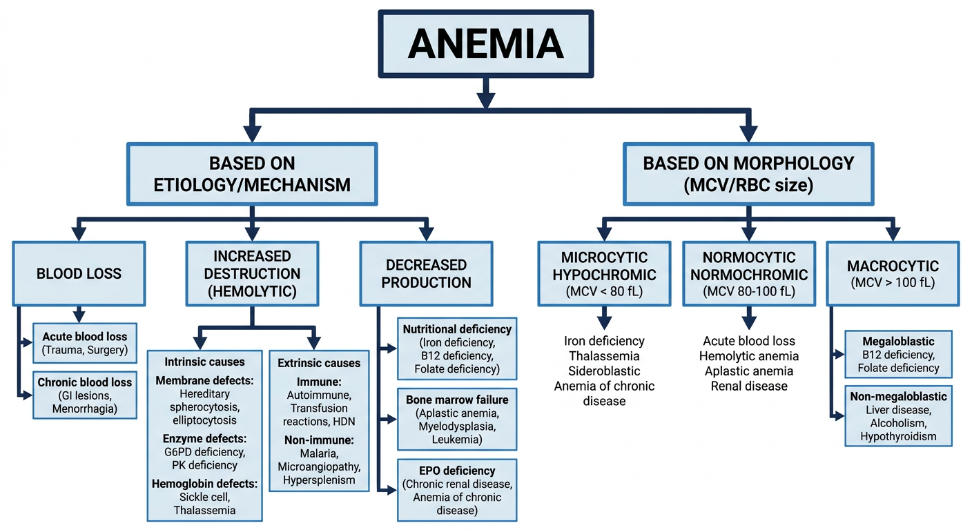
Anemia - Definition, Classification & Features (15 Marks)
Definition
| Group | Hb Threshold |
|---|---|
| Adult males | < 13 g/dL |
| Adult females | < 12 g/dL |
| Pregnant women | < 11 g/dL |
| Children (6-14 yrs) | < 12 g/dL |
Classification of Anemia
SYSTEM 1 - ETIOLOGICAL / PATHOPHYSIOLOGICAL Classification
┌─────────────────────┐
│ ANEMIA │
└──────────┬──────────┘
┌──────────────────┼───────────────────┐
▼ ▼ ▼
┌──────────────┐ ┌──────────────────┐ ┌──────────────────┐
│ BLOOD LOSS │ │ INCREASED RBC │ │ DECREASED RBC │
│ │ │ DESTRUCTION │ │ PRODUCTION │
│ (Hemorrhagic│ │ (Hemolytic │ │ (Hypoprolifera- │
│ Anemia) │ │ Anemia) │ │ tive Anemia) │
└──────┬───────┘ └────────┬─────────┘ └──────────┬───────┘
│ │ │
┌──────┴──────┐ ┌───────┴──────┐ ┌────────┴────────┐
│ ACUTE │ │ INTRINSIC │ │ EXTRINSIC │
│ Blood Loss │ │ (within RBC)│ │ (outside RBC) │
│ - Trauma │ └───────┬──────┘ └────────┬────────┘
│ - Surgery │ │ │
└─────────────┘ ┌───────┼──────────┐ ┌───────┼───────────┐
┌──────────────┐ ▼ ▼ ▼ ▼ ▼ ▼
│ CHRONIC │ Mem- Enzyme Hb Immune Non-immune
│ Blood Loss │ brane defects defects
│ - GI bleed │ defects
│ - Menorrhagia│
└──────────────┘
A. Blood Loss Anemia
- Trauma, surgery, ruptured aneurysm
- Normocytic normochromic initially
- Reticulocytes increase after 3-5 days
- GI tract lesions (ulcer, carcinoma)
- Gynecological: menorrhagia, fibroids
- Eventually leads to iron deficiency → microcytic hypochromic
B. Hemolytic Anemia (Increased RBC Destruction)
| Sub-type | Disorders |
|---|---|
| Membrane defects | Hereditary spherocytosis, Hereditary elliptocytosis |
| Enzyme defects | G6PD deficiency (Hexose monophosphate shunt), Pyruvate kinase deficiency (glycolytic) |
| Hemoglobin defects | Sickle cell anemia, Thalassemia syndromes, Unstable hemoglobins |
| Acquired genetic | Paroxysmal Nocturnal Hemoglobinuria (PNH) |
| Sub-type | Examples |
|---|---|
| Immune-mediated | Autoimmune hemolytic anemia, HDN (Rh disease), Transfusion reactions, Drug-induced |
| Microangiopathic | HUS, DIC, TTP (mechanical shearing) |
| Infections | Malaria, Babesiosis, Clostridium |
| Hypersplenism | Splenomegaly → increased RBC sequestration |
C. Decreased Production (Hypoproliferative / Dyserythropoietic)
| Cause | Disorders |
|---|---|
| Nutritional deficiency | Iron deficiency, B12 deficiency, Folate deficiency, Copper, Vitamin C |
| Bone marrow failure | Aplastic anemia, Pure red cell aplasia |
| Marrow replacement | Leukemia, lymphoma, metastatic carcinoma, myelofibrosis |
| Stem cell defects | Myelodysplastic syndrome (MDS), Fanconi anemia |
| EPO deficiency | Chronic renal disease, Anemia of chronic inflammation |
| Endocrine disorders | Hypothyroidism, hypopituitarism, Addison's disease |
SYSTEM 2 - MORPHOLOGICAL Classification (Based on RBC Size - MCV)
┌──────────────────────┐
│ ANEMIA │
└──────────┬───────────┘
┌────────────────────────┼────────────────────────┐
▼ ▼ ▼
┌──────────────────┐ ┌──────────────────┐ ┌──────────────────┐
│ MICROCYTIC │ │ NORMOCYTIC │ │ MACROCYTIC │
│ HYPOCHROMIC │ │ NORMOCHROMIC │ │ │
│ MCV < 80 fL │ │ MCV 80-100 fL │ │ MCV > 100 fL │
│ MCH < 27 pg │ │ MCH 27-33 pg │ │ │
└────────┬─────────┘ └────────┬─────────┘ └────────┬─────────┘
│ │ │
┌──────┴───────┐ ┌───────┴───────┐ ┌────────┴────────┐
│ TAILS │ │ │ │ MEGALOBLASTIC │
│ Iron def. │ Acute blood loss │ B12 deficiency │
│ Thalassemia │ Hemolytic anemia │ Folate deficiency │
│ Sideroblastic│ Aplastic anemia │ MDS │
│ Anemia of │ Renal disease │ Chemotherapy │
│ chronic dis. │ Hypothyroidism ├─────────────────────┤
└──────────────┘ Mixed deficiency │ NON-MEGALOBLASTIC │
└───────────────┘ │ Liver disease │
│ Alcoholism │
│ Hypothyroidism │
│ Reticulocytosis │
└─────────────────────┘
SYSTEM 3 - Based on Bone Marrow Response (Reticulocyte Count)
| Type | Reticulocyte Count | Interpretation | Examples |
|---|---|---|---|
| Hyperregenerative | > 2% (elevated) | Bone marrow responding | Hemolytic anemias, Acute blood loss |
| Hyporegenerative | < 1% (low/normal) | Bone marrow failing | Iron deficiency, Aplastic anemia, B12 deficiency, Renal anemia |
Quick Comparison Table - Key Anemias
| Type | MCV | MCH | Reticulocytes | Key Lab Finding |
|---|---|---|---|---|
| Iron deficiency | ↓ | ↓ | ↓ | ↓ Serum ferritin, ↑ TIBC |
| B12 deficiency | ↑ | Normal | ↓ | Hypersegmented neutrophils, ↓ serum B12 |
| Folate deficiency | ↑ | Normal | ↓ | ↓ RBC folate |
| Thalassemia | ↓ | ↓ | ↑ | Hb electrophoresis abnormal |
| Hemolytic | Normal | Normal | ↑↑ | ↑ LDH, ↓ haptoglobin, +ve Coombs |
| Aplastic | Normal | Normal | ↓↓ | Pancytopenia, empty marrow |
| Sickle cell | Normal | Normal | ↑ | HbS on electrophoresis |
| Anemia of chronic disease | Normal/↓ | Normal/↓ | ↓ | ↓ TIBC, ↑ ferritin (trapped iron) |
Clinical Features of Anemia (General)
- Fatigue, weakness, exertional dyspnea
- Palpitations, headache, dizziness
- Tinnitus, fainting
- Reduced exercise tolerance
- Pallor (conjunctival, palmar, sublingual)
- Tachycardia, wide pulse pressure
- Systolic flow murmur (hyperdynamic circulation)
- Ankle edema in severe cases
- Specific signs: koilonychia (iron def.), glossitis (B12), jaundice (hemolytic)
Summary Flowchart (Exam-Ready)
ANEMIA
│
├── BY ETIOLOGY
│ ├── Blood Loss → Acute / Chronic
│ ├── Hemolytic → Intrinsic (membrane, enzyme, Hb defects)
│ │ → Extrinsic (immune, microangiopathic, infection)
│ └── Decreased Production → Nutritional / Marrow failure / EPO deficiency
│
├── BY MORPHOLOGY (MCV)
│ ├── Microcytic (<80 fL) → Fe def., Thalassemia, Sideroblastic, ACD
│ ├── Normocytic (80-100) → Blood loss, Hemolytic, Aplastic, Renal
│ └── Macrocytic (>100) → Megaloblastic (B12, Folate) / Non-megaloblastic (Liver, Alcohol)
│
└── BY BONE MARROW RESPONSE
├── Hyperregenerative (Retics ↑) → Hemolysis, Blood loss
└── Hyporegenerative (Retics ↓) → Iron def., Aplastic, B12 def.