
Humerus bone
Reading File
The humerus is the long bone of the upper arm, articulating with the scapula at the shoulder and the radius/ulna at the elbow. Key landmarks include the head, greater/lesser tubercles, bicipital groove, deltoid tuberosity, medial/lateral epicondyles, and the trochlea and capitulum distally.
Searching sources...
Finding Sources
Finding Sources
Good results. I found an imaging anatomy atlas with humerus bone images and detailed anatomy sections in forensic anthropology and orthopaedics. Let me read the key anatomy content and fetch the image.
Reading File
Reading File
Reading File
Excellent content retrieved. Now let me fetch the humerus anatomy image and also search for more specific content on the proximal humerus anatomy.
Loading Image
Finding Sources
Finding Sources
Now let me fetch the proximal humerus anatomy content from Gray's Anatomy and also get the imaging atlas content on humerus.
Reading File
Reading File
I now have very rich content. Let me fetch the Gray's Anatomy proximal humerus diagram and then compile the full answer.
Loading Image
Reading File
I now have comprehensive, well-sourced content with two high-quality anatomical images. Here is the complete, detailed answer:
The Humerus
The humerus is the largest bone of the upper limb, forming the skeletal framework of the arm (the region between the shoulder and elbow). It has a triangular cross-section with anterior, lateral, and medial borders, and three surfaces: anterolateral, anteromedial, and posterior.
- Forensic Anthropology: A Comprehensive Introduction, p. 4333
- Imaging Anatomy: Bones, Joints, Vessels and Nerves, p. 2159
Proximal End
The proximal humerus consists of the head, anatomical neck, greater tubercle, lesser tubercle, surgical neck, and superior shaft.
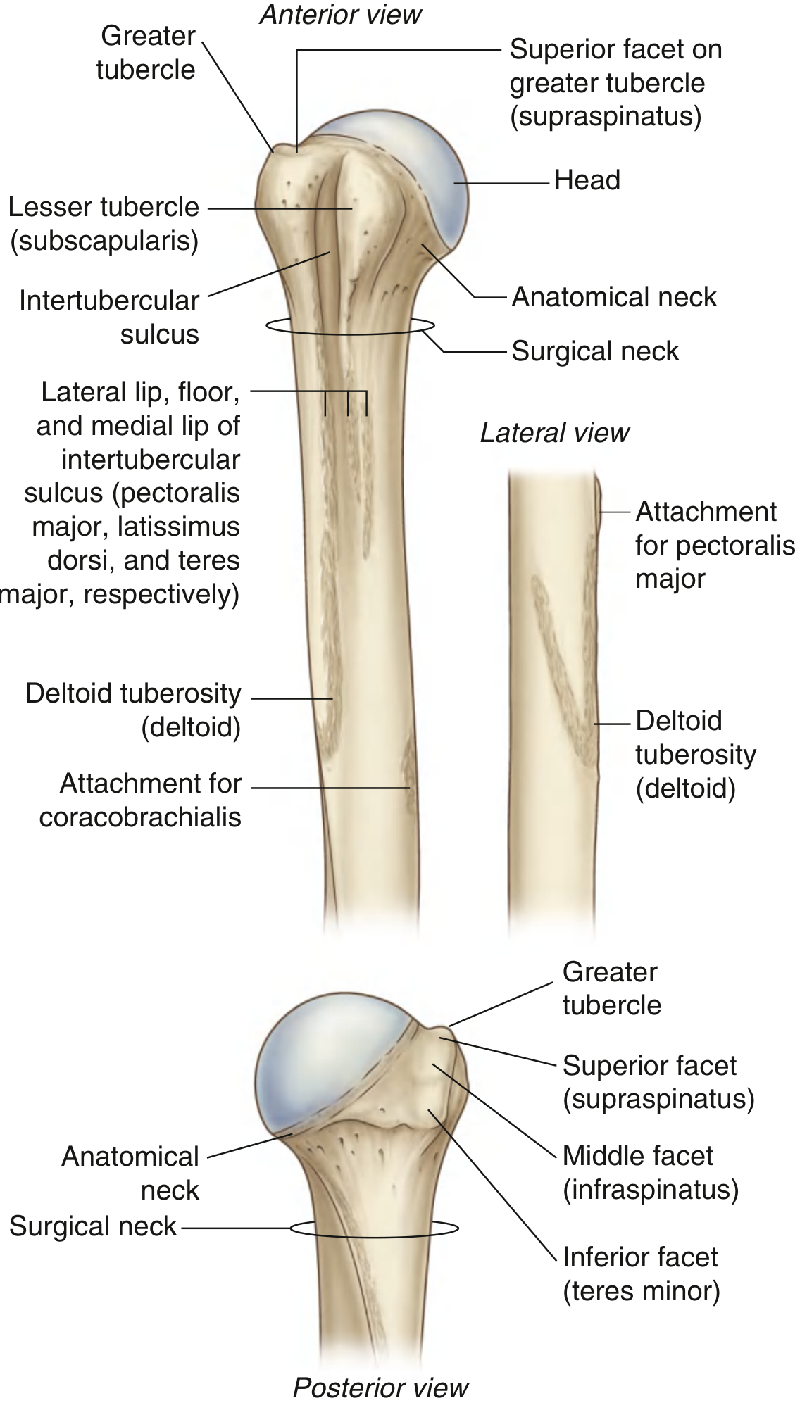
Head
- Half-spherical; projects medially and slightly superiorly.
- Articulates with the glenoid cavity of the scapula at the glenohumeral (shoulder) joint.
Anatomical Neck
- Narrow constriction immediately distal to the head.
- Lies between the head and the tubercles laterally, and between the head and shaft medially.
- Fractures here are rare because the bone is thickest at this level.
Greater Tubercle (lateral)
Three smooth facets for the rotator cuff muscles:
| Facet | Muscle |
|---|---|
| Superior | Supraspinatus |
| Middle | Infraspinatus |
| Inferior | Teres minor |
Lesser Tubercle (anterior)
- Has a large smooth impression for subscapularis attachment.
Intertubercular Sulcus (Bicipital Groove)
- Deep groove separating the greater and lesser tubercles; continues inferiorly on the proximal shaft.
- Carries the tendon of the long head of biceps brachii.
- The lateral lip gives attachment to pectoralis major; the floor to latissimus dorsi; the medial lip to teres major.
Surgical Neck
-
Horizontal constriction between the expanded proximal end and the narrower shaft.
-
The axillary nerve and posterior circumflex humeral artery pass immediately posterior to it.
-
The most common fracture site in the proximal humerus. Axillary nerve injury can occur.
-
Gray's Anatomy for Students, p. 823
Shaft
-
Cylindrical proximally, flattening distally.
-
Deltoid tuberosity: V-shaped roughening on the lateral surface at the mid-shaft; the deltoid muscle inserts here.
-
Coracobrachialis attachment: thin vertical roughening on the medial surface at a similar level.
-
Posterior spiral groove (radial groove): runs obliquely from proximal-medial to distal-lateral on the posterior shaft. The radial nerve and profunda brachii artery lie in this groove. The radial nerve enters it ~20 cm proximal to the medial epicondyle (approximately 74% of the total humeral length).
- Shaft fractures at this level commonly cause radial nerve palsy (wrist drop).
-
Nutrient foramen: opens distally, toward the elbow ("away from the knee, toward the elbow").
-
Miller's Review of Orthopaedics 9th Edition, p. 9682
-
Rockwood and Green's Fractures in Adults 10th ed, p. 1873
Distal End
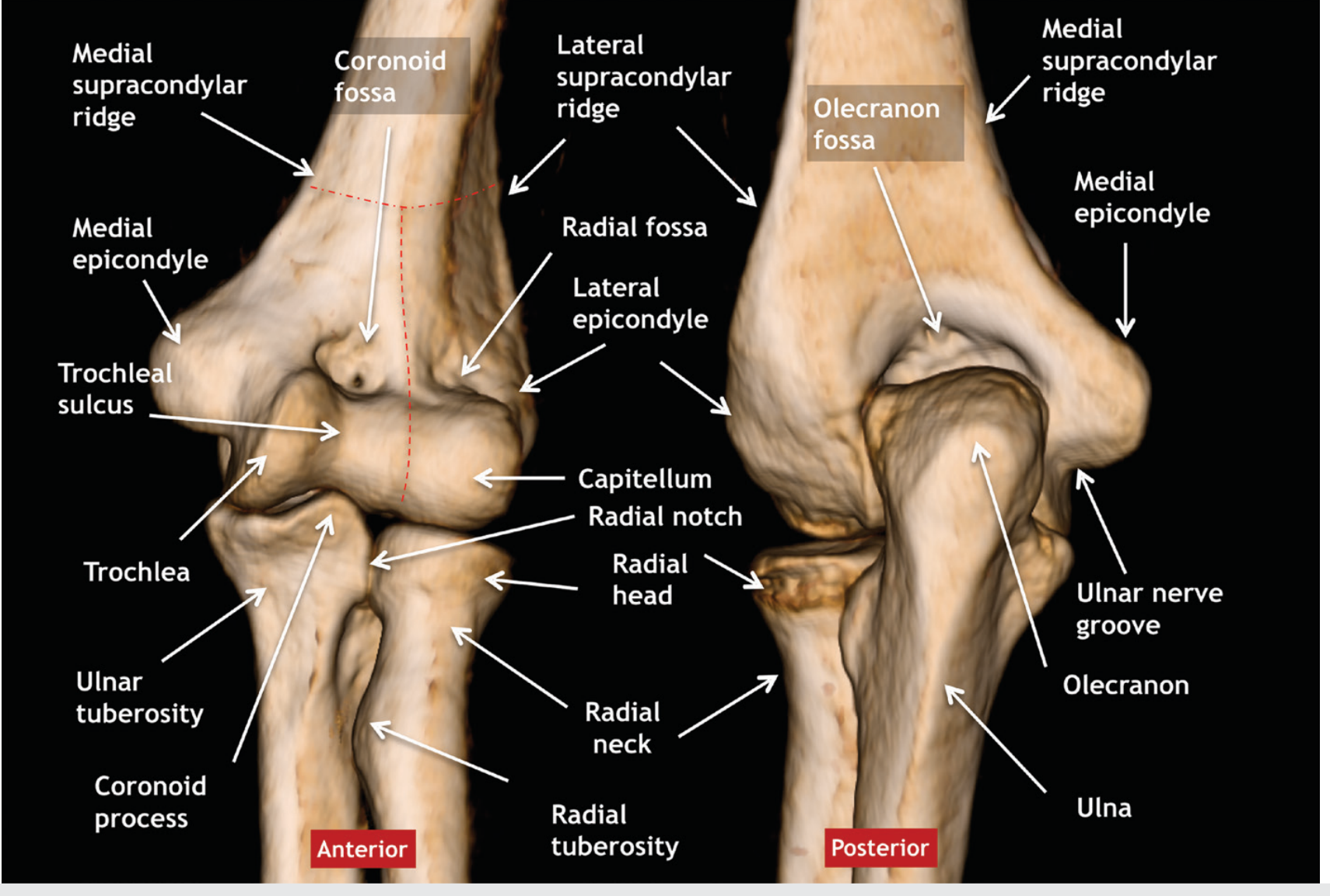
The distal humerus has two articular surfaces and several important landmarks:
| Structure | Position | Articulates with |
|---|---|---|
| Trochlea | Medial, spool/pulley shaped | Trochlear notch of ulna |
| Capitulum (Capitellum) | Anterolateral, rounded | Head of radius |
| Medial epicondyle | Medial bony prominence | - (common flexor origin) |
| Lateral epicondyle | Lateral bony prominence | - (common extensor origin) |
| Medial supracondylar ridge | Proximal to medial epicondyle | - |
| Lateral supracondylar ridge | Proximal to lateral epicondyle | - |
Three fossae accommodate adjacent bones during movement:
- Coronoid fossa (anterior): receives the coronoid process of the ulna during flexion.
- Radial fossa (anterolateral): accommodates the radial head during flexion.
- Olecranon fossa (posterior): receives the olecranon of the ulna during full extension. May be perforated, forming a septal aperture (normal variant - not to be confused with trauma).
The coronoid and olecranon fossae are separated by the thin supratrochlear septum.
The ulnar nerve groove runs posterior to the medial epicondyle.
- Imaging Anatomy: Bones, Joints, Vessels and Nerves, p. 3193
- Forensic Anthropology: A Comprehensive Introduction, p. 4342
Key Clinical Summary
| Structure | Clinical Relevance |
|---|---|
| Surgical neck | Most common proximal humerus fracture; risk to axillary nerve |
| Spiral groove | Humeral shaft fracture → radial nerve palsy (wrist drop) |
| Medial epicondyle | "Golfer's elbow" (medial epicondylitis); ulnar nerve runs posterior to it |
| Lateral epicondyle | "Tennis elbow" (lateral epicondylitis) |
| Greater tubercle | Avulsion in shoulder dislocation; rotator cuff attachment |
| Bicipital groove | Long head of biceps tendinopathy |
Siding the Humerus (Forensic/Anatomical Note)
-
The head is medial
-
The olecranon fossa is posterior
-
The deltoid tuberosity is lateral
-
The nutrient foramen opens distally (toward the elbow)
-
Forensic Anthropology: A Comprehensive Introduction, p. 4346
Swaras
Searching the Web
Swaras Indian classical music notes explained
Reading Web Page
https://en.wikipedia.org/wiki/Svara
Swaras (स्वर)
Swara (Sanskrit: स्वर) is a foundational concept in Indian classical music. The word literally means "to sound" and refers simultaneously to a breath, a vowel, a musical note, and a step of the octave. More than just a pitch, a swara is a deliberate musical note - naad (steady pitch sound) produced with intent and control for music-making.
The 7 Swaras - Saptaka (सप्तक)
The seven swaras are collectively called the Sargam (an acronym of Sa-Re-Ga-Ma). They are the Indian equivalent of Western solfège (Do-Re-Mi...).
| # | Short Name | Full Name | Meaning | Western Equivalent (when Sa = C) |
|---|---|---|---|---|
| 1 | Sa | Shadja (षड्ज) | "born of six" (six organs produce it) | C |
| 2 | Re | Rishabh (ऋषभ) | "the bull" | D |
| 3 | Ga | Gandhar (गान्धार) | "the fragrant one" | E |
| 4 | Ma | Madhyam (मध्यम) | "the middle one" | F |
| 5 | Pa | Pancham (पञ्चम) | "the fifth" | G |
| 6 | Dha | Dhaivat (धैवत) | "the bold one" | A |
| 7 | Ni | Nishad (निषाद) | "the one who sits" | B |
In Carnatic music, Re is called Ri and the system of variants differs slightly from Hindustani.
Achal vs Vikrit Swaras
| Type | Swaras | Meaning |
|---|---|---|
| Achal (Anchor) | Sa, Pa | Fixed, unalterable - same in all ragas |
| Vikrit (Variable) | Re, Ga, Ma, Dha, Ni | Can be komal or tivra |
The 12 Swaras (Full Chromatic Scale)
Beyond the 7 basic notes, Indian music recognizes 12 distinct pitches within an octave - formed by the komal (flat) and tivra (sharp) variants:
| Swara | Variants | Semitones from Sa | Western Note |
|---|---|---|---|
| Sa | Shuddha only | 0 | C |
| Re | Komal Re | 1 | D♭ |
| Re | Shuddha Re | 2 | D |
| Ga | Komal Ga | 3 | E♭ |
| Ga | Shuddha Ga | 4 | E |
| Ma | Shuddha Ma | 5 | F |
| Ma | Tivra Ma | 6 | F# |
| Pa | Shuddha only | 7 | G |
| Dha | Komal Dha | 8 | A♭ |
| Dha | Shuddha Dha | 9 | A |
| Ni | Komal Ni | 10 | B♭ |
| Ni | Shuddha Ni | 11 | B |
- Komal (कोमल) = soft/flat (lowered by a semitone)
- Tivra (तीव्र) = sharp (raised by a semitone; only applies to Ma)
- Shuddha (शुद्ध) = pure/natural
Types of Swaras
- Shuddha Swara - The pure, natural form of the note (e.g., Shuddha Re, Shuddha Ga)
- Komal Swara - Flattened form; applies to Re, Ga, Dha, Ni
- Tivra Swara - Sharpened form; applies only to Ma (Tivra Ma = F#)
- Achal Swara - Fixed notes that never change: Sa and Pa
Animal/Bird Origins (Traditional Belief)
Each swara is traditionally associated with the call of an animal or bird:
| Swara | Animal Sound |
|---|---|
| Sa | Peacock (मोर) |
| Re | Bull / Skylark |
| Ga | Goat |
| Ma | Heron / Crane (Krauncha) |
| Pa | Cuckoo (Koyal) |
| Dha | Horse / Frog |
| Ni | Elephant |
Swaras in Ragas
Ragas are built by selecting a subset of these 12 swaras and applying specific rules of ascent (aaroha) and descent (avaroha). The choice of shuddha, komal, or tivra variants gives each raga its unique emotional color (rasa). For example:
-
Komal Ga and Komal Ni give a raga a melancholic, devotional quality (e.g., Raga Bhairavi)
-
Tivra Ma gives a bright, mysterious quality (e.g., Raga Yaman)
-
-
This is a shared conversation. Sign in to Orris to start your own chat.