
Easily earn me chest x rays for respiratory disese in medicine ,examples with images,causes , differentiate points bw them
Reading File
Now let me search the medical library for chest X-ray findings in respiratory disease, running multiple parallel queries:
Finding Sources
Finding Sources
Finding Sources
Searching the Web
chest X-ray respiratory diseases patterns examples images differentiation medicine
Good, I found relevant textbook blocks. Let me now read the key sections and fetch relevant images in parallel:
Finding Sources
Finding Sources
Finding Sources
Reading File
Reading Web Page
https://radiologyassistant.nl/chest/chest-x-ray/lung-disease
Excellent! Now let me fetch key images from the textbook and read more sections in parallel:
Reading File
Finding Sources
Finding Sources
Loading Image
Loading Image
Great - I have excellent images and content. Let me fetch more key X-ray images for the other conditions now:
Loading Image
Loading Image
Reading File
Reading File
Excellent content! Let me fetch a few more key images - pleural effusion, pulmonary edema, and get TB/pneumothorax images:
Loading Image
Loading Image
Finding Sources
Finding Sources
I have all the content I need. Let me fetch the TB and pneumothorax images to complete the set:
Loading Image
Reading File
Reading File
Now let me fetch the TB cavitary X-ray and the pneumothorax image:
Loading Image
Loading Image
I now have all the images and textbook content I need. Let me compose the comprehensive guide:
🫁 Chest X-Ray Patterns in Respiratory Disease - The Complete Guide
Sources: Murray & Nadel's Respiratory Medicine, Goldman-Cecil Medicine, Harrison's Principles (22nd Ed.), Rosen's Emergency Medicine, Grainger & Allison's Diagnostic Radiology
Quick Memory Framework: The 5 Core CXR Patterns
| Pattern | Appearance | Think of |
|---|---|---|
| Consolidation | White/opaque area - airspace filled | Pneumonia, pulmonary oedema |
| Hyperinflation | Black/over-dark lungs, flat diaphragm | COPD/Emphysema |
| Pleural effusion | White lower zone, blunted angle | Heart failure, infection, cancer |
| Pneumothorax | Black rim + no lung markings | Trauma, spontaneous |
| Reticulation/Nodules | Fine mesh/lace or dots | TB, ILD, sarcoidosis |
1. LOBAR PNEUMONIA (Consolidation)
CXR Appearance:
- Homogeneous white opacity conforming to a lobe
- Air bronchograms (dark branching airways inside the white area - pathognomonic of airspace filling)
- Lobar or segmental distribution
- No volume loss (the lobe is FULL, not collapsed)
Causes:
- Streptococcus pneumoniae (most common, right upper lobe classic)
- Klebsiella pneumoniae (right upper lobe, "bulging fissure" sign)
- Community-acquired pneumonia (CAP)
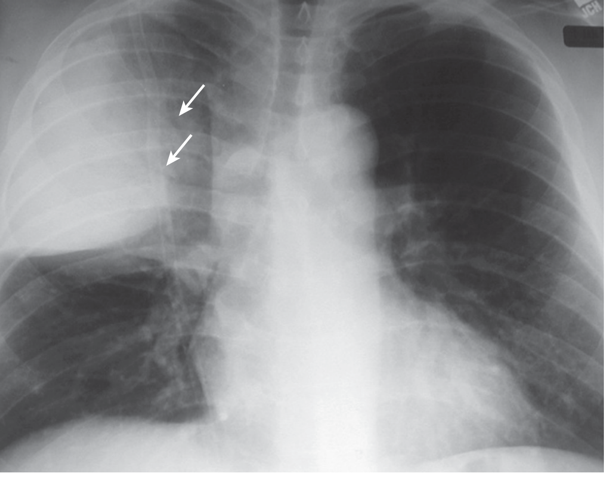
Key points: The arrow shows air bronchograms - dark air-filled airways visible through the white consolidated lobe. No volume loss = consolidation (not collapse).
2. BRONCHOPNEUMONIA (Patchy Consolidation)
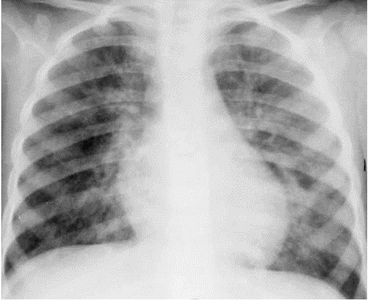
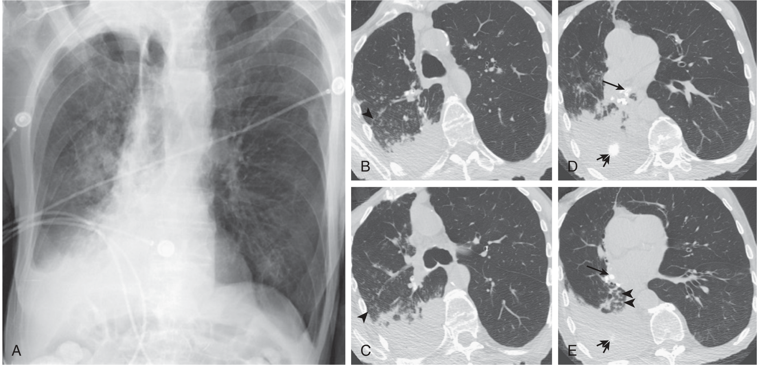
CXR Appearance:
- Patchy, multifocal, ill-defined opacities - scattered like "cotton balls"
- Bilateral or unilateral
- No single lobe boundary
- May have small centrilobular nodules
Causes:
- Staphylococcus aureus (MRSA - can cavitate)
- Mycoplasma pneumoniae (bilateral, basal)
- Haemophilus influenzae
- Aspiration pneumonia (RLL predominant, dependent regions)
- Measles, adenovirus (viral - peribronchial pattern)
3. PULMONARY OEDEMA (Perihilar Bat-Wing / Alveolar Flooding)
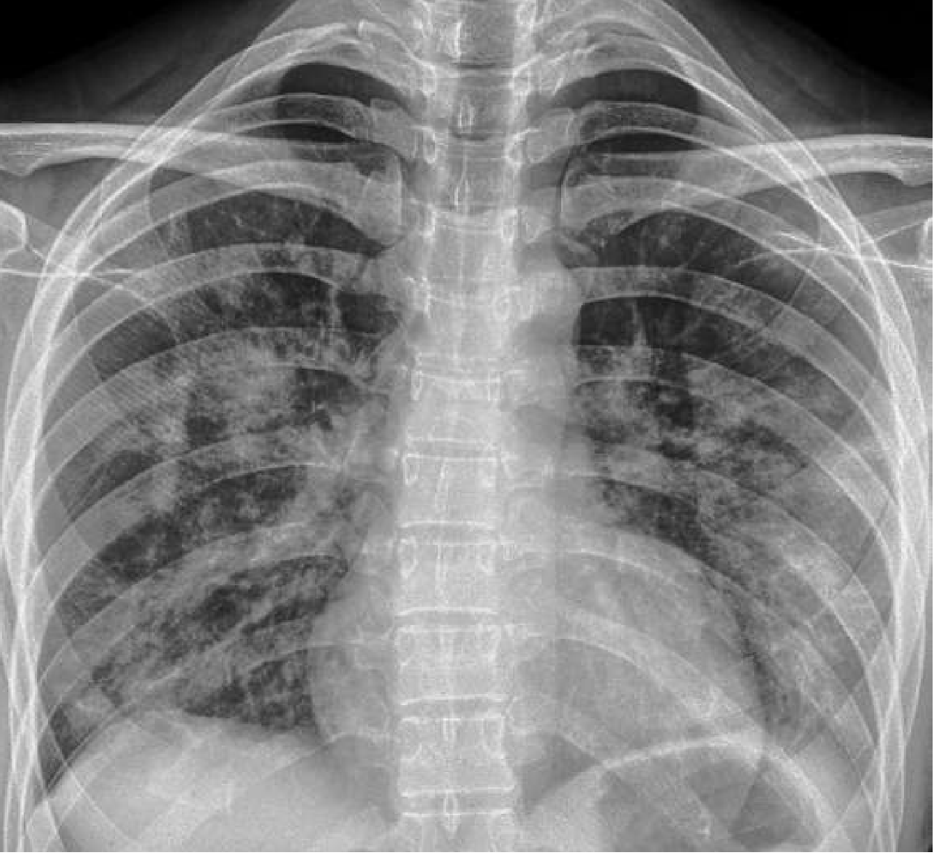
CXR Appearance:
- Bilateral perihilar ("bat-wing") infiltrates
- Kerley B lines - 1-2 cm horizontal lines at lateral lung bases (dilated lymphatics/fluid in interlobular septa)
- Upper lobe vascular diversion (cephalization of flow)
- Cardiomegaly (heart > 50% of thoracic diameter)
- Blunted costophrenic angles (effusions)
- Hazy/ground-glass shadowing bilateral, perihilar > peripheral
Causes:
- Left ventricular failure / heart failure (cardiogenic)
- ARDS / sepsis (non-cardiogenic - no cardiomegaly, no effusions)
- Renal failure, volume overload
- Mitral stenosis
4. PLEURAL EFFUSION
CXR Appearance:
- Blunting of the costophrenic angle (>200 mL visible on PA)
- Homogeneous basal opacity with a concave upper margin (meniscus sign)
- Fluid layers laterally higher than medially
- Massive effusion: complete opacification of a hemithorax
- On supine films: diffuse haziness of the hemithorax
Causes:
- Transudate: Heart failure (bilateral, R > L), hypoalbuminaemia, renal failure
- Exudate: Pneumonia (parapneumonic), TB, malignancy, pulmonary embolism
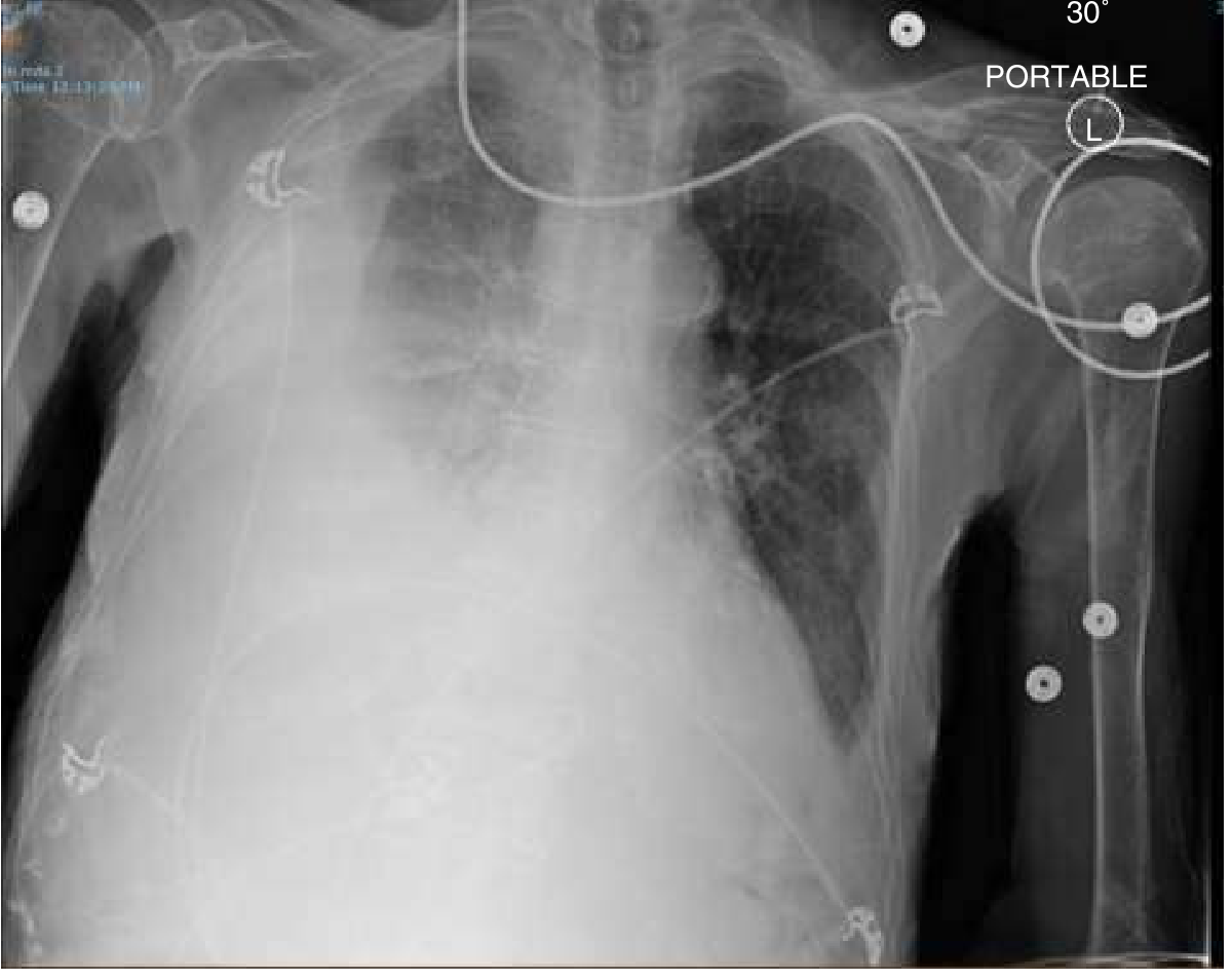
How to spot: Start at the costophrenic angles - they should be sharp and acute. Any blunting = suspect effusion.
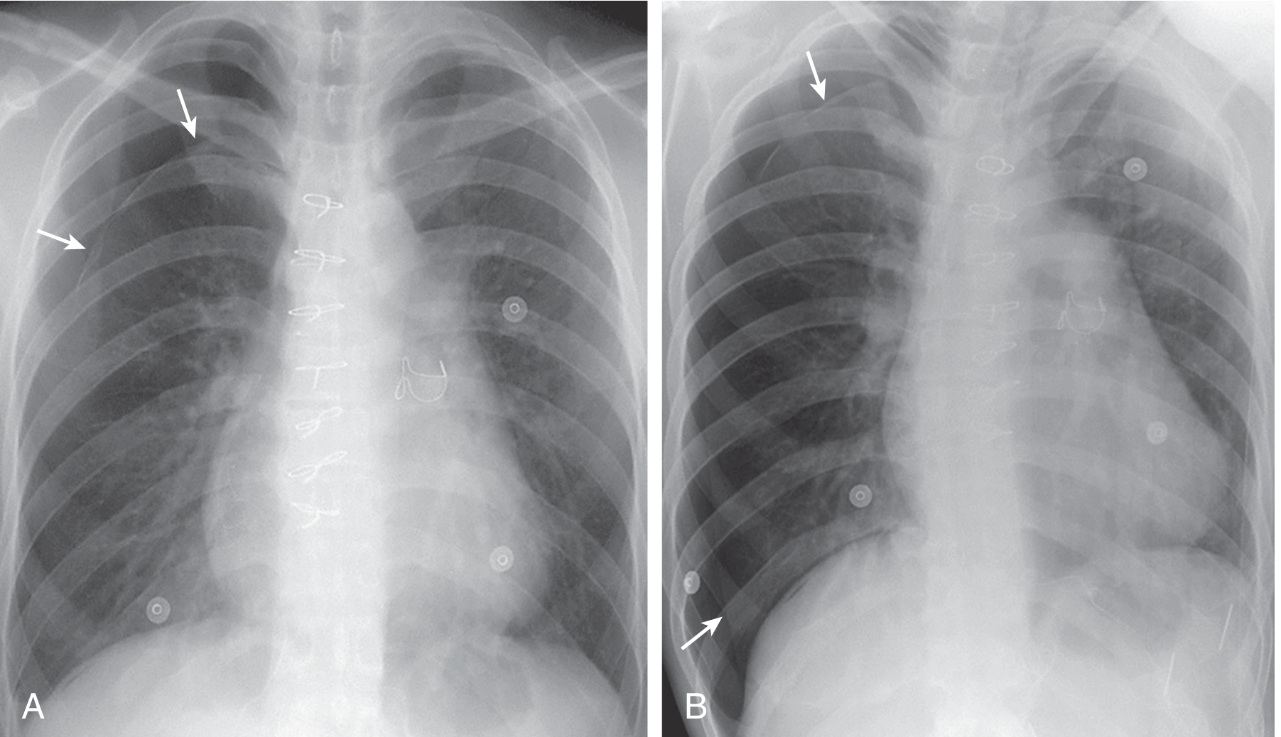
5. PNEUMOTHORAX
CXR Appearance:
- Visible pleural line - a thin white line parallel to the chest wall
- Complete absence of lung markings peripheral to that line (black space)
- Lung "falls away" from the chest wall
- Tension PTX: mediastinal shift AWAY from affected side + depressed hemidiaphragm - medical emergency!
Causes:
- Primary spontaneous: Tall, thin young males (ruptured apical bleb)
- Secondary spontaneous: COPD (emphysematous bullae), asthma, TB, cystic fibrosis
- Traumatic: Rib fractures, iatrogenic (central line, thoracentesis)
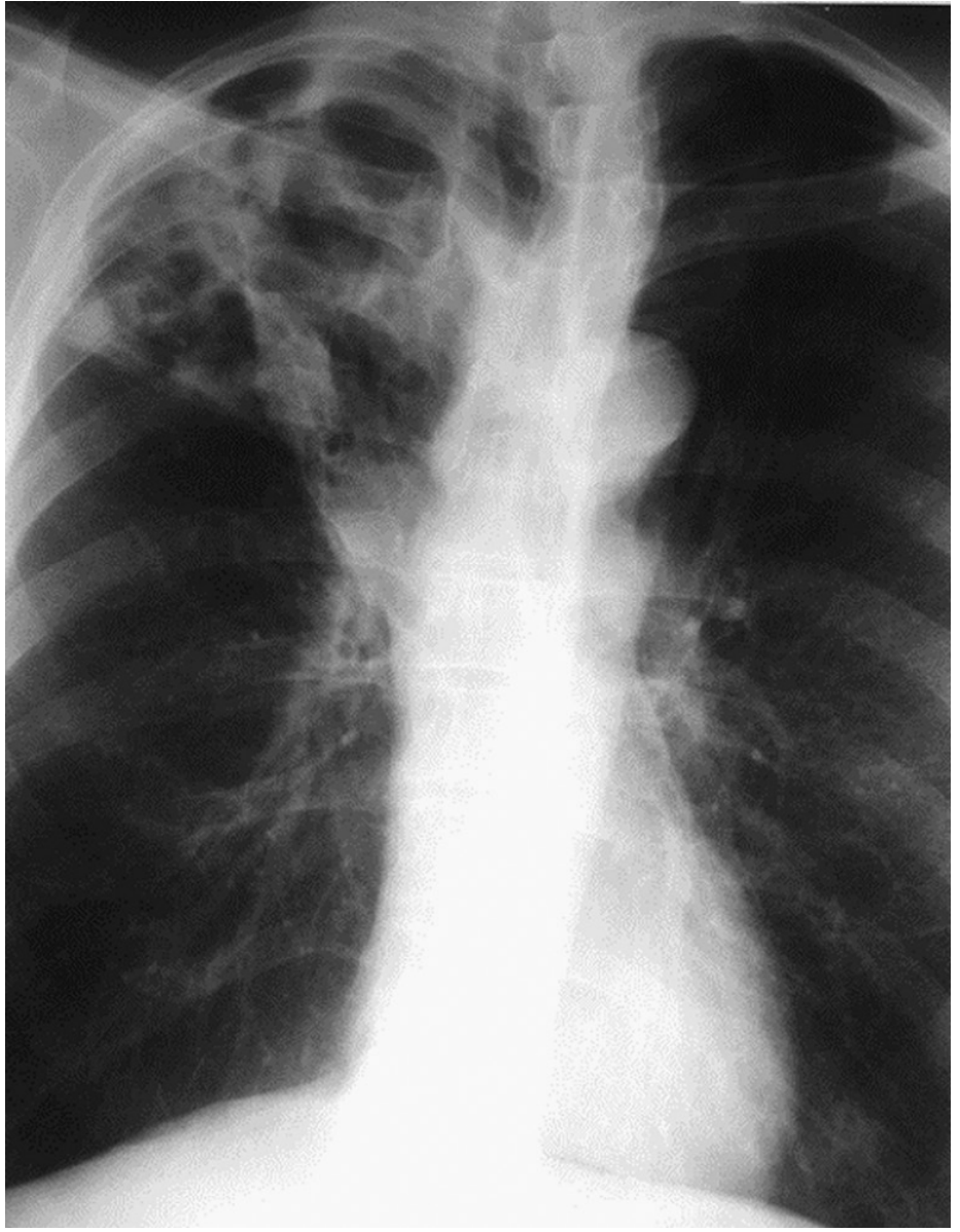
6. TUBERCULOSIS (TB)
CXR Appearance:
| Type | Location | Pattern |
|---|---|---|
| Primary TB | Middle/lower zone + ipsilateral hilar adenopathy | Consolidation + enlarged hilum |
| Reactivation TB | Apical/posterior segment upper lobes (R > L) | Cavitation, fibrosis, calcification |
| Miliary TB | Diffuse both lungs | 1-3mm nodules "millet seeds" throughout |
| HIV-associated TB | Lower zones, diffuse | Atypical - no cavity, adenopathy |
Causes: Mycobacterium tuberculosis - reactivation of latent infection
7. COPD / EMPHYSEMA
CXR Appearance:
- Hyperinflation - lungs look too dark, >6 anterior ribs visible above diaphragm on PA
- Flattened hemidiaphragms (most reliable sign)
- Increased AP diameter ("barrel chest")
- Increased retrosternal airspace on lateral view
- Prominent pulmonary arteries (cor pulmonale if right heart fails)
- Bullae (black avascular areas with thin walls) in emphysema
Causes: Smoking (>90%), alpha-1 antitrypsin deficiency, occupational dust
Memory trick: Flattened diaphragm + dark lungs + SMALL heart = COPD
8. COVID-19 PNEUMONIA (Organizing Pneumonia Pattern)
CXR Appearance:
- Bilateral, peripheral, lower-zone predominant consolidation
- "Ground glass" opacities
- Rounded/confluent consolidations
- Spares central/perihilar regions (unlike pulmonary oedema)
- Often accompanied by small pleural effusions
MASTER DIFFERENTIATION TABLE
| Feature | Pneumonia (Lobar) | Pulm. Oedema | Pleural Effusion | Pneumothorax | TB (Reactivation) | COPD |
|---|---|---|---|---|---|---|
| Location | Lobar/segmental | Bilateral perihilar | Basal | Apex (rim) | Upper lobe | Diffuse |
| Opacity | Dense white, lobar | Perihilar haze | Lower zone white | Black (avascular) | Patchy + cavities | Hyper-dark |
| Air bronchograms | YES (hallmark) | May be present | No | No | Variable | No |
| Meniscus sign | No | No | YES | No | No | No |
| Kerley B lines | No | YES | Sometimes | No | No | No |
| Cardiomegaly | No | YES (cardiac cause) | No | No | No | Late (cor pulm) |
| Cavitation | Possible (MRSA) | No | No | No | YES (classic) | Bullae |
| Trachea/mediastinum | Midline | Midline | Shifted AWAY | Shifted AWAY (tension) | Shifted TOWARD (fibrosis) | Midline |
| Diaphragm | Normal | Normal-elevated | Obscured | Depressed (tension) | Elevated (fibrosis) | Flat |
| Volume | Preserved | Increased | Decreased (compressed) | Increased | Decreased | Increased |
| Key memory hook | Air bronchograms | Bat wings + Kerley B | Costophrenic blunting | Pleural line + black | Upper lobe cavity | Flat diaphragm |
QUICK MNEMONICS
For PLEURAL EFFUSION vs CONSOLIDATION:
- Effusion: Concave upper border (meniscus), no air bronchograms, shift AWAY
- Consolidation: Air bronchograms, no shift (unless collapse), lobar shape
For PULMONARY OEDEMA (ABCDE):
Alveolar oedema (bat-wing), B-lines (Kerley), Cardiomegaly, Diversion of vessels (upper lobe), Effusions (bilateral)
For TB upper zone lesions:
"FACTS" = Fibrocavitary, Apical, Calcification, Top (upper) lobe, Scarring
Pneumothorax vs Bullae:
- Pneumothorax: rim of air wraps the ENTIRE edge of lung
- Bullae: confined, localized, thin-walled bubble, lung markings present beyond it
CXR INTERPRETATION APPROACH (ABCDE System)
- A - Airways: trachea midline? carina angle (<70°)?
- B - Bones/soft tissue: rib fractures, mastectomy?
- C - Cardiac: size, shape (>50% of thorax = cardiomegaly)
- D - Diaphragm: flat (COPD), high (effusion, phrenic paralysis), costophrenic angles sharp?
- E - Everything else: lung fields zone by zone, hilum, mediastinum
Sources: Murray & Nadel's Respiratory Medicine | Goldman-Cecil Medicine | Harrison's Principles of Internal Medicine 22nd Ed. | Rosen's Emergency Medicine | Grainger & Allison's Diagnostic Radiology
This is a shared conversation. Sign in to Orris to start your own chat.