
Dish vs opll
DISH vs OPLL diffuse idiopathic skeletal hyperostosis ossification posterior longitudinal ligament comparison
DISH diffuse idiopathic skeletal hyperostosis spine X-ray flowing ossification
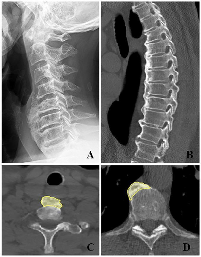
This composite of four radiological images demonstrates the characteristic features of Diffuse Idiopathic Skeletal Hyperostosis (DISH) in the cervical and thoracic spine. Image A (lateral X-ray) and Image B (sagittal CT) show 'flowing' ossification along the anterolateral aspect of the vertebral column, bridging at least four contiguous vertebral bodies while maintaining relatively preserved intervertebral disc spaces. Axial CT scans (C and D) highlight the regional variation in hyperostosis distribution. In the cervical spine (C), the new bone formation (outlined in yellow) is positioned symmetrically along the anterior midline of the vertebral body, resulting in the anterior displacement of the trachea and esophagus. Conversely, the axial view of the thoracic spine (D) reveals asymmetrical ossification predominantly on the right anterolateral side, likely due to the mechanical presence of the pulsating descending aorta on the left, which inhibits bone formation. These images serve as classic examples of Forestier's disease, emphasizing the diagnostic criteria of multisegmental bridging ossification and the influence of adjacent vascular structures on disease morphology.
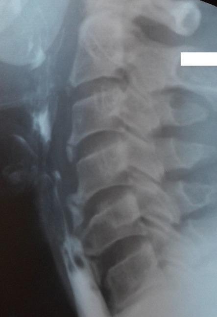
This diagnostic image is a lateral cervical X-ray combined with a barium swallow study. It depicts the cervical spine from C2 to C7 and the adjacent esophagus. Prominent anterior flowing osteophytes and ossification of the anterior longitudinal ligament are visible along the vertebral bodies, characteristic of Diffuse Idiopathic Skeletal Hyperostosis (DISH) or Forestier's disease. The barium contrast highlights the esophageal lumen, demonstrating a significant extrinsic compression and narrowing at the level of the C5-C6 vertebrae caused by these large anterior bony outgrowths. This finding provides a radiological explanation for mechanical dysphagia. The image serves as a clinical example of how degenerative spinal changes can impact adjacent soft tissue structures in the neck, specifically the upper digestive tract.
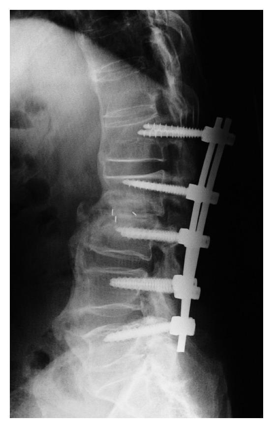
This lateral plain X-ray of the thoracolumbar spine demonstrates posterior spinal instrumentation and signs of Diffuse Idiopathic Skeletal Hyperostosis (DISH). The instrumentation consists of five pedicle screws connected by a vertical stabilizing rod. A critical finding is the significant 'back-out' and loosening of the most superior pedicle screw (at the L1 level), where the screw head is displaced posteriorly from the vertebral body compared to the other flush-mounted screws. The underlying anatomy shows characteristic 'flowing' anterior ossification of the vertebral bodies, consistent with DISH. There is noticeable narrowing and destructive change at the L2-L3 intervertebral disc space, suggesting pseudoarthrosis or instability. Small radiopaque markers are visible within the L2-L3 disc space, likely from a previous interbody fusion procedure. This diagnostic image illustrates a mechanical complication of spinal fusion in a patient with ankylosing spinal pathology, highlighting the risks of instrumentation failure and screw migration.
OPLL ossification posterior longitudinal ligament cervical spine CT
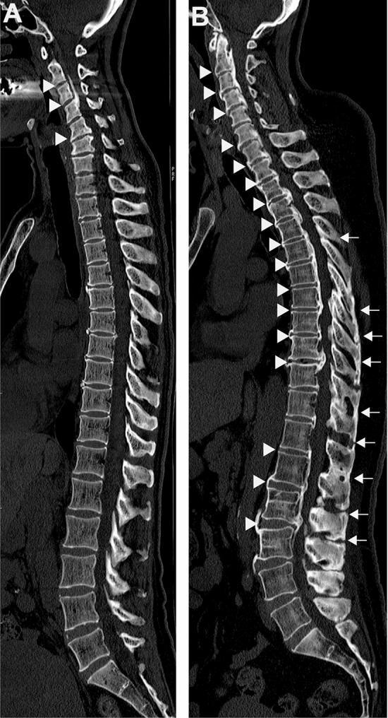
This diagnostic image displays sagittal CT reconstructions of the entire human spine, illustrating two distinct patterns of spinal ligament ossification. Image A demonstrates 'localized' pathology, primarily showing Ossification of the Posterior Longitudinal Ligament (OPLL) confined to the cervical spine region, indicated by white arrowheads. Image B represents a 'diffuse' or systemic involvement, showing multilevel ossification across the entire spinal column. In image B, white arrowheads highlight extensive OPLL and Ossification of the Anterior Longitudinal Ligament (OALL) spanning the cervical, thoracic, and lumbar segments. Additionally, white arrows in the thoracolumbar region of image B indicate concomitant Ossification of the Ligamentum Flavum (OLF) and Ossification of the Supra/Interspinous Ligaments (OSIL). The images serve to contrast localized cervical involvement with generalized spinal ligament ossification, which may be clinically associated with metabolic conditions such as non-alcoholic fatty liver disease (NAFLD) and diabetes mellitus. These radiographic features are critical for diagnosing progressive myelopathy or radiculopathy risks.
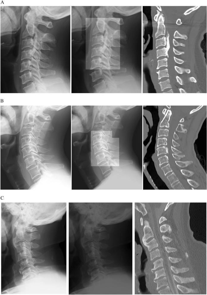
A multi-panel medical comparison chart demonstrating the detection of Ossification of the Posterior Longitudinal Ligament (OPLL) in the cervical spine using deep learning. The image is organized into three rows (A, B, C) and three columns. The left column displays lateral plain radiographs of the cervical spine. The middle column shows the same radiographs with deep learning algorithm-generated heatmaps or bounding regions highlighting suspected ossification. The right column presents sagittal CT scans, serving as the clinical ground truth. Row A illustrates continuous-type OPLL, visible as a thick, linear radiopaque structure spanning C2 to C4. Row B shows segmental-type OPLL, appearing as isolated ossified patches posterior to the C5 and C6 vertebral bodies. Row C serves as a negative control, displaying a normal cervical spine with no ligamentous ossification. This comparison illustrates how algorithmic analysis can identify subtle pathological bone formations in standard X-rays, confirmed by diagnostic CT imaging, supporting orthopaedic and radiological training in spinal pathology.
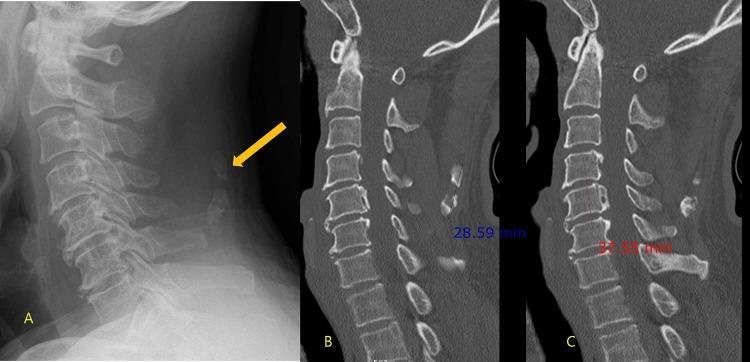
This composite diagnostic image displays a lateral cervical spine radiograph (A) alongside sagittal reconstructed CT scans (B, C) of a 60-year-old male. Image A demonstrates a focal radiopaque density within the posterior soft tissues (indicated by a yellow arrow), consistent with ossification of the nuchal ligament (ONL), while deeper structures like the posterior longitudinal ligament are obscured by overlying bone. CT image B provides a detailed sagittal view of this ONL, measuring its longitudinal extent from C4 to C5 at 2.9 cm. CT image C reveals a separate, clinically significant pathology: ossification of the posterior longitudinal ligament (OPLL), which presents as a continuous hyperdense strip along the posterior margins of the C4 to C6 vertebral bodies, measuring 3.8 cm. This comparison emphasizes the diagnostic superiority of CT over plain radiography for identifying OPLL, which may be masked on X-rays by the spinal column, and highlights the importance of further imaging when superficial ligamentous ossification is detected.
DISH vs OPLL - Comprehensive Comparison
At a Glance
| Feature | DISH | OPLL |
|---|---|---|
| Full name | Diffuse Idiopathic Skeletal Hyperostosis | Ossification of the Posterior Longitudinal Ligament |
| Ligament involved | Anterior longitudinal ligament (ALL) + entheses | Posterior longitudinal ligament (PLL) |
| Spinal canal | Usually NOT narrowed | Directly narrows spinal canal |
| Myelopathy risk | Low (unless concurrent OPLL) | High - primary cause of myelopathy |
| Location on vertebra | ANTERIOR to vertebral bodies | POSTERIOR to vertebral bodies (within canal) |
| Spinal level | Predominantly thoracic (right-sided) | Predominantly cervical |
| Prevalence | Common (>10% in >70 yrs in West) | Rare in West; up to 4% in Japanese |
| Radiology tool | Plain X-ray / CT | CT preferred (MRI underestimates) |
| Disc spaces | Preserved | Preserved |
| Sacroiliac joints | Normal | Normal |
DISH (Diffuse Idiopathic Skeletal Hyperostosis)
Diagnostic Criteria (Resnick & Niwayama)
- Flowing ossification along the anterolateral aspect of at least 4 contiguous vertebral bodies
- Relative preservation of intervertebral disc height in the involved areas
- Absence of apophyseal joint ankylosis and sacroiliac joint erosion/sclerosis (distinguishes from AS)
Epidemiology
- Prevalence: ~10% in persons over 70 years; up to 35% in men >70 years in some series; higher on CT than plain X-ray (mean 19.5%)
- Male > Female; middle age to elderly
- More common in Western and Jewish populations; lower in Korean and African populations
- Mild DISH found in human remains dating back 4,000 years
Pathogenesis
- Cause unknown - multifactorial
- Metabolic associations: hyperinsulinemia, metabolic syndrome, type 2 DM, dyslipidemia, hyperuricemia, elevated GH/IGF-1, obesity
- Insulin directly promotes differentiation of mesenchymal cells in ligaments into chondrocytes and subsequent endochondral ossification
- BMP-2 (bone morphogenetic protein) promotes mesenchymal stem cell differentiation and mineralization
- Aortic pulsation hypothesis: thoracic RIGHT-sided predominance because the descending aorta on the LEFT inhibits bone formation mechanically
- Possible inflammatory component: MRI corner lesions similar to ASAS axSpA criteria; peripheral entheseal inflammation on power Doppler ultrasound
- Genetic factors: COL6A1 (also the candidate gene for OPLL) is associated with DISH in Japanese patients
Radiographic Features
- "Flowing" ossification along the right anterolateral thoracic spine (due to the left-sided aorta)
- Ossification is ANTERIOR - on the front of vertebral bodies
- Lucent line between ossification and vertebral body (important distinguishing sign from osteophytes)
- Peripheral enthesopathy: "whiskering" at iliac crests, ischial tuberosities, patellar poles, calcaneus
- Does NOT cause significant spinal canal stenosis unless concurrent OPLL develops
Clinical Features
- Often asymptomatic or mild back stiffness with reduced range of motion
- Cervical spine complications include: dysphagia, hoarseness, stridor (anterior bony mass compressing esophagus), aspiration pneumonia, sleep apnea
- Atlantoaxial complications (pseudoarthrosis, subluxation)
- OPLL may develop concurrently, causing myelopathy
- Fracture risk: ankylosed rigid spine fractures easily with minor trauma ("chalk-stick" fractures), highly unstable
- No significant HLA associations
OPLL (Ossification of the Posterior Longitudinal Ligament)
Epidemiology
- Prevalence: up to 4% in Japanese people (80x the prevalence seen in Caucasians); also seen in non-Asian populations
- Male:Female ratio = 2:1
- Typical onset: 6th decade (range 19-85 years)
- 50% of OPLL patients have concurrent DISH (and ~48.7% of cervical DISH patients have OPLL)
Classification (Radiographic Types)
- Continuous - long lesion extending across several vertebrae
- Segmental - one or several separate skip lesions
- Mixed - combination of continuous and segmental
- Localized (circumscribed) - mainly at the intervertebral disc level
Pathogenesis
- Shares genetic factors with DISH (COL6A1 gene)
- Associated with diabetes mellitus, metabolic syndrome
- Endochondral ossification within the PLL progressively thickens and encroaches on the spinal canal
Imaging
- CT is the gold standard - shows dense ossification posterior to vertebral bodies inside the canal
- MRI underestimates OPLL - can be misidentified as multilevel disc herniation (important pitfall)
- Plain X-ray: dense linear calcification posterior to vertebral bodies (may be missed)
Clinical Features
- Majority of patients are asymptomatic - prophylactic surgery is NOT indicated
- Symptomatic cases present with cervical myelopathy and/or radiculopathy:
- Upper limb weakness, numbness, clumsiness
- Lower limb spasticity, gait disturbance
- Bladder dysfunction in advanced cases
- Direct compression of cord from behind - the opposite direction to disc herniation (which compresses from in front)
Treatment of OPLL
- Posterior approach (laminectomy + fusion, or laminoplasty):
- Indirect decompression - the cord drifts posteriorly away from the OPLL
- Requires lordotic or neutral alignment
- Laminoplasty is contraindicated in kyphotic spine
- Anterior approach (ACDF, ACCF, or hybrid):
- More directly addresses the pathology
- Higher risk of dural leak because OPLL is frequently associated with dural ossification (up to 70% in some series)
- "Anterior floating technique": OPLL is thinned but not removed; the cord is decompressed laterally and the OPLL becomes "free-floating"
Key Distinguishing Points
| DISH | OPLL | |
|---|---|---|
| Ligament | Anterior longitudinal + entheses | Posterior longitudinal |
| Position relative to cord | Anterior to vertebra, away from canal | Inside canal, directly behind cord |
| Canal compromise | No (usually) | Yes - primary mechanism |
| Myelopathy | Rare (unless OPLL coexists) | Common - classic cause |
| Radiculopathy | Rare | Common |
| Dysphagia | Yes (anterior cervical mass) | No |
| Thoracic distribution | Right-sided (aortic effect) | Less common thoracic |
| MRI reliability | Good | Poor (pitfall - mimics disc herniation) |
| CT preferred | Helpful for complications | Yes - diagnostic modality of choice |
| Association with each other | Yes, ~50% overlap | Yes, ~50% overlap |
| HLA-B27 | Negative (key vs AS) | Negative |
| Sacroiliac joints | Normal | Normal |
| Disc height | Preserved | Preserved |
| Surgery for stenosis | Rarely needed | Often needed for myelopathy |