
Ivf setup 10 marks answer obgy
IVF in vitro fertilization steps procedure diagram
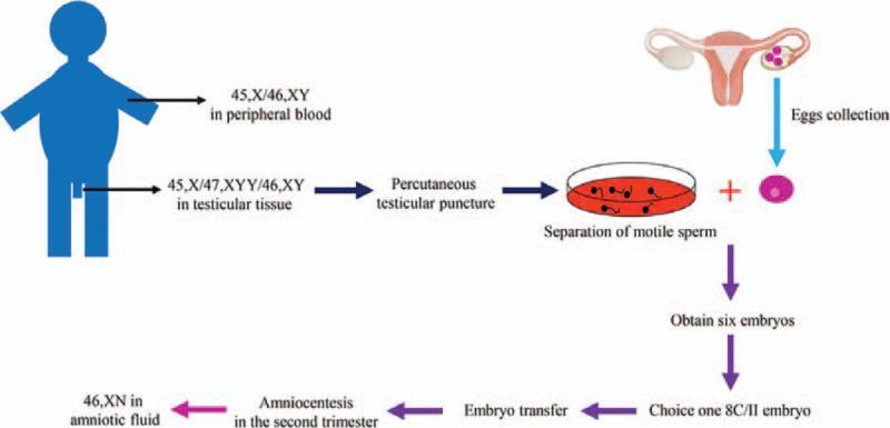
This medical flow diagram illustrates the clinical workflow of In Vitro Fertilization and Embryo Transfer (IVF-ET) in a case of male mosaicism. The process begins with a genetic evaluation of the male patient, showing a 45,X/46,XY karyotype in peripheral blood and a more complex 45,X/47,XYY/46,XY mosaicism in testicular tissue. The procedural steps follow: percutaneous testicular puncture for sperm retrieval, separation of motile sperm, and egg collection from the female reproductive system. These are combined to obtain six embryos, from which one '8C/II' embryo is selected for transfer. The final stages show the progression to pregnancy, including prenatal screening via amniocentesis in the second trimester, revealing a 46,XN karyotype in the amniotic fluid. This infographic serves as an educational tool for reproductive endocrinology and infertility (REI), demonstrating the management of gonadal dysgenesis and chromosomal mosaicism through assisted reproductive technology (ART) to achieve a successful pregnancy outcome.
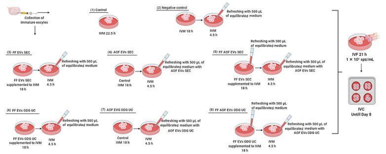
This medical schematic outlines an experimental workflow for bovine in vitro embryo production, focusing on the impact of extracellular vesicles (EVs) on oocyte maturation. The process begins with the collection of immature oocytes from cumulus-oocyte complexes. The diagram illustrates eight experimental groups for In Vitro Maturation (IVM). Group 1 (Control) undergoes 22.5 hours of IVM. Groups 2–8 utilize a two-phase maturation protocol (18h + 4.5h) involving different media refreshing steps and EV supplementations. These include follicular fluid-derived EVs (FF EVs) and ampullary oviductal fluid-derived EVs (AOF EVs), isolated via size exclusion chromatography (SEC) or OptiPrep density gradient ultracentrifugation (ODG UC). Visual highlights include pipette-mediated media refreshing with 500 ̀μL of equilibrated medium, with or without specific EV combinations. Following maturation, all groups proceed to In Vitro Fertilization (IVF) for 21 hours with a sperm concentration of 1 × 10⁶ spz/mL, and finally to In Vitro Culture (IVC) through Day 8. This flowchart demonstrates reproductive biotechnological research into cell-to-cell signaling and reproductive environment biomimicry.
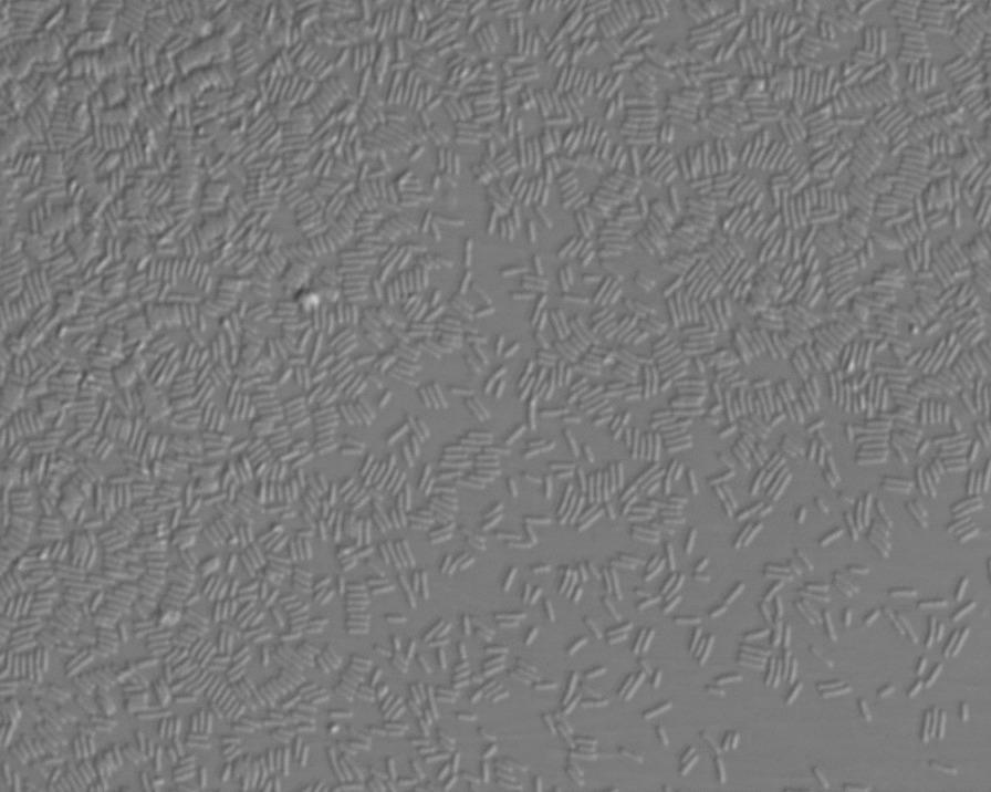
This diagnostic micrograph captures a significant bacterial contamination within an embryo culture medium during an in vitro fertilization (IVF) procedure. The image, likely obtained via phase-contrast or differential interference contrast (DIC) microscopy, shows a high-density proliferation of bacilli (rod-shaped bacteria) distributed throughout the liquid medium. The individual organisms exhibit a uniform, short-rod morphology with random orientation, suggesting rapid logarithmic growth. In several regions, the bacterial density is nearly confluent, creating a visually cluttered field against the hazy, grayish background of the culture environment. This visual evidence of contamination is clinically significant as it correlates with the degeneration of co-incubated oocytes, representing a major complication in assisted reproductive technology (ART). The image serves as an educational example of microbial iatrogenic contamination and its impact on embryological development and culture viability.
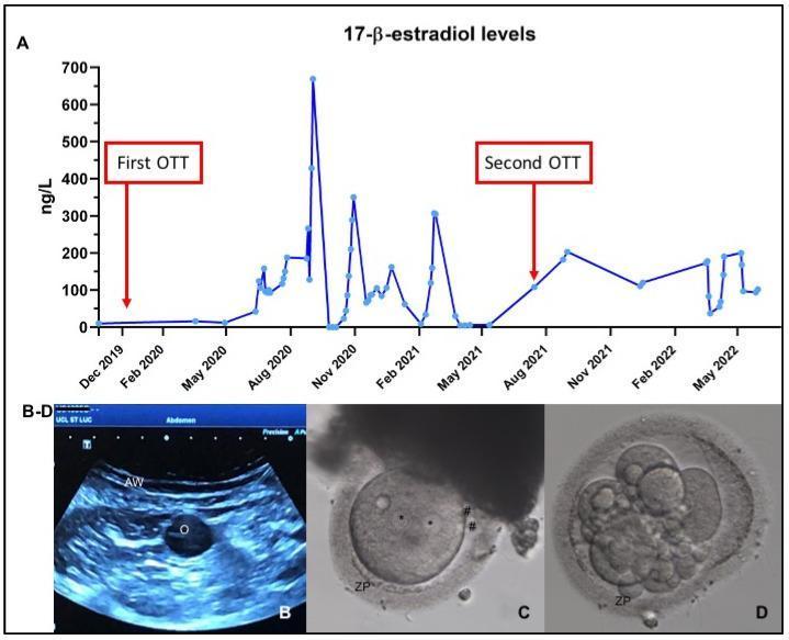
A composite medical illustration and imaging panel documenting ovarian function follow-up and in vitro fertilization (IVF) outcomes. Panel A is a line graph depicting 17-̢-estradiol levels (ng/L) over a two-year timeline, marking two separate ovarian tissue transplantation (OTT) events. Panel B is a transabdominal ultrasound image showing the abdominal wall (AW) and a hypoechoic follicle containing an oocyte (O) during a pick-up procedure. Panels C and D are microscopic diagnostic images of embryonic development. Panel C shows a Day 1 fertilized oocyte featuring two distinct pronuclei (*) and two extruded polar bodies (#) within the zona pellucida (ZP). Panel D demonstrates a Day 3 dividing embryo at the 6-cell stage, showing multiple blastomeres enclosed by the zona pellucida (ZP). This sequence illustrates the progression from clinical monitoring of hormone levels and follicle development to cellular-level fertilization and cleavage, relevant for reproductive endocrinology and infertility (REI) education.
In Vitro Fertilization (IVF) - Setup and Procedure
Definition
Indications
- Tubal factor infertility - bilateral tubal block, severe tubal damage (most common indication)
- Male factor infertility - oligospermia, asthenospermia, azoospermia (using ICSI)
- Unexplained infertility - failed IUI cycles
- Endometriosis - moderate to severe
- Ovulatory dysfunction - unresponsive to conventional treatment
- Diminished ovarian reserve
- Uterine factor (using surrogacy with own gametes)
- Genetic disease - where preimplantation genetic testing (PGT) is needed
Steps of IVF (Sequential Steps)
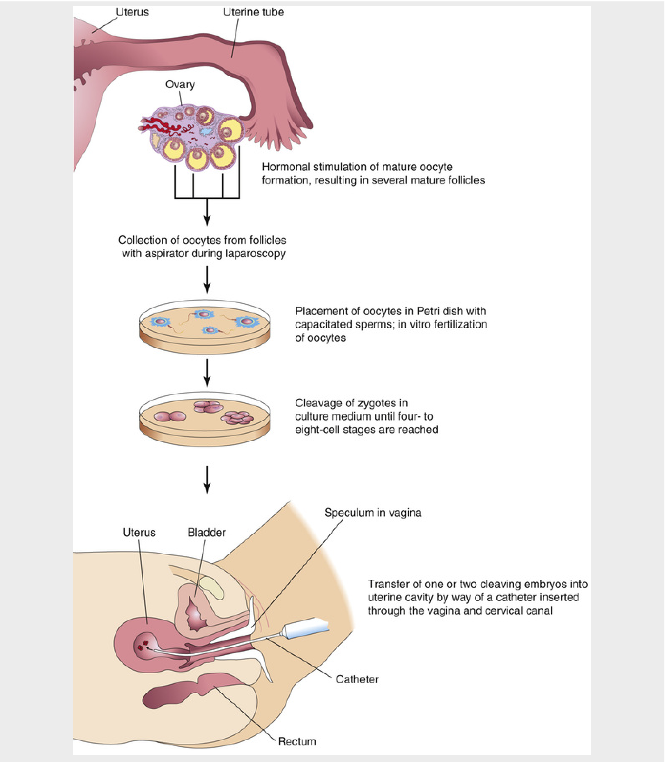
Step 1: Controlled Ovarian Hyperstimulation (COH)
- Clomiphene citrate (oral) or
- Exogenous gonadotropins (FSH +/- LH) by injection (superovulation)
- GnRH Agonist (Long protocol): Started on day 21 of the previous cycle (luteal phase). After 10-14 days of GnRH agonist administration, a pelvic ultrasound and E2 level confirm suppression, then gonadotropins are started. The GnRH agonist is continued until the hCG trigger. Provides better oocyte yields in normal responders.
- GnRH Antagonist (Short/Flexible protocol): Antagonist is added in the follicular phase when the lead follicle reaches 12-14 mm; has fewer side effects and shorter duration.
Step 2: Trigger Injection
- hCG (human chorionic gonadotropin) 5,000-10,000 IU is given to trigger final oocyte maturation (mimics the LH surge)
- Oocyte retrieval is scheduled 34-36 hours after the trigger
Step 3: Oocyte Retrieval (OPU - Oocyte Pick-Up)
- Performed under transvaginal ultrasound guidance using a needle passed through the vaginal wall into the ovarian follicles
- Alternatively, laparoscopic aspiration may be used
- Done under IV sedation/anesthesia
- The aspirated follicular fluid contains the cumulus-oocyte complex (COC)
Step 4: Sperm Collection and Preparation
- Semen sample collected by masturbation on the same day as oocyte retrieval
- In azoospermia: TESE/PESA/MESA (surgical sperm retrieval)
- Sperm is capacitated (processed by swim-up or density gradient centrifugation) to enhance fertilizing ability
Step 5: Insemination / Fertilization In Vitro
- Oocytes are placed in a Petri dish with special culture medium
- Capacitated spermatozoa are added to the dish (conventional IVF) OR
- ICSI (Intracytoplasmic Sperm Injection) - a single sperm is injected directly into the oocyte cytoplasm (used in male factor, failed fertilization, low oocyte count)
Step 6: Embryo Culture and Assessment
- Fertilization confirmed 15-20 hours post-insemination by presence of two pronuclei (2PN) and extrusion of the second polar body
- Embryo cleavage monitored microscopically:
- Day 2-3: 4-8 cell cleavage stage
- Day 4: Morula (16-cell compaction)
- Day 5-6: Blastocyst formation (inner cell mass + trophectoderm + blastocoel)
- Culture media: single media or sequential media (mimicking changing metabolic needs of the embryo)
- Extended culture to blastocyst (Day 5) is preferred as it allows selection of better-quality embryos (blastocyst transfer gives ~10% higher pregnancy rate than cleavage-stage transfer)
Step 7: Embryo Transfer (ET)
- 1-3 embryos (depending on maternal age and embryo quality) are loaded into a soft catheter
- Catheter is passed through the vagina → cervical canal → uterine cavity
- Embryos are deposited in the upper uterine cavity under ultrasound guidance
- Patient rests supine for a few hours post-transfer
- Remaining embryos are cryopreserved in liquid nitrogen for future use
Step 8: Luteal Phase Support
- Progesterone supplementation (vaginal pessaries, IM injections, or oral micronized progesterone) given to support endometrial receptivity and implantation
- Continued until 10-12 weeks if pregnancy is confirmed
Step 9: Pregnancy Testing
- Serum beta-hCG measured 14 days after embryo transfer
- If positive, TVS done at 5-6 weeks to confirm intrauterine pregnancy and fetal cardiac activity
Additional Techniques Under IVF Setup
| Technique | Indication |
|---|---|
| ICSI | Male factor, failed conventional IVF |
| PGT (Preimplantation Genetic Testing) | Known genetic disorders, recurrent miscarriage |
| Frozen Embryo Transfer (FET) | Use of cryopreserved embryos |
| Oocyte Donation | Premature ovarian insufficiency, poor responders |
| Gestational Surrogacy | Absent/non-functional uterus |
Complications
- Ovarian Hyperstimulation Syndrome (OHSS) - most serious complication
- Multiple pregnancy - higher incidence of twins/triplets
- Ectopic pregnancy - slightly higher risk
- Spontaneous abortion - increased incidence
- Birth defects - slightly increased risk of chromosomal abnormalities and low birth weight
- Cycle cancellation - inadequate (<3 follicles) or excessive response
Success Rates
- Live birth rate per cycle: approximately 30-40% in women under 35 years
- Success decreases with advancing maternal age and diminished ovarian reserve
- Blastocyst transfer shows higher implantation rates than cleavage-stage transfer