
What is hypopharynx
Reading File
Now let me search the medical library for hypopharynx content.
Finding Sources
Finding Sources
Good results. Now let me read the key sections from the most relevant sources.
Reading File
Reading File
Reading File
Let me also fetch the anatomy diagram from Murray & Nadel and read a bit more from Cummings for the lymphatics/blood supply.
Loading Image
Reading File
I now have comprehensive information to give a thorough answer. Let me compose the response.
The Hypopharynx
The hypopharynx (also called the "laryngopharynx") is the lowest subdivision of the pharynx. It is a subsite of the pharynx that connects the oropharynx above to the cervical esophagus below.
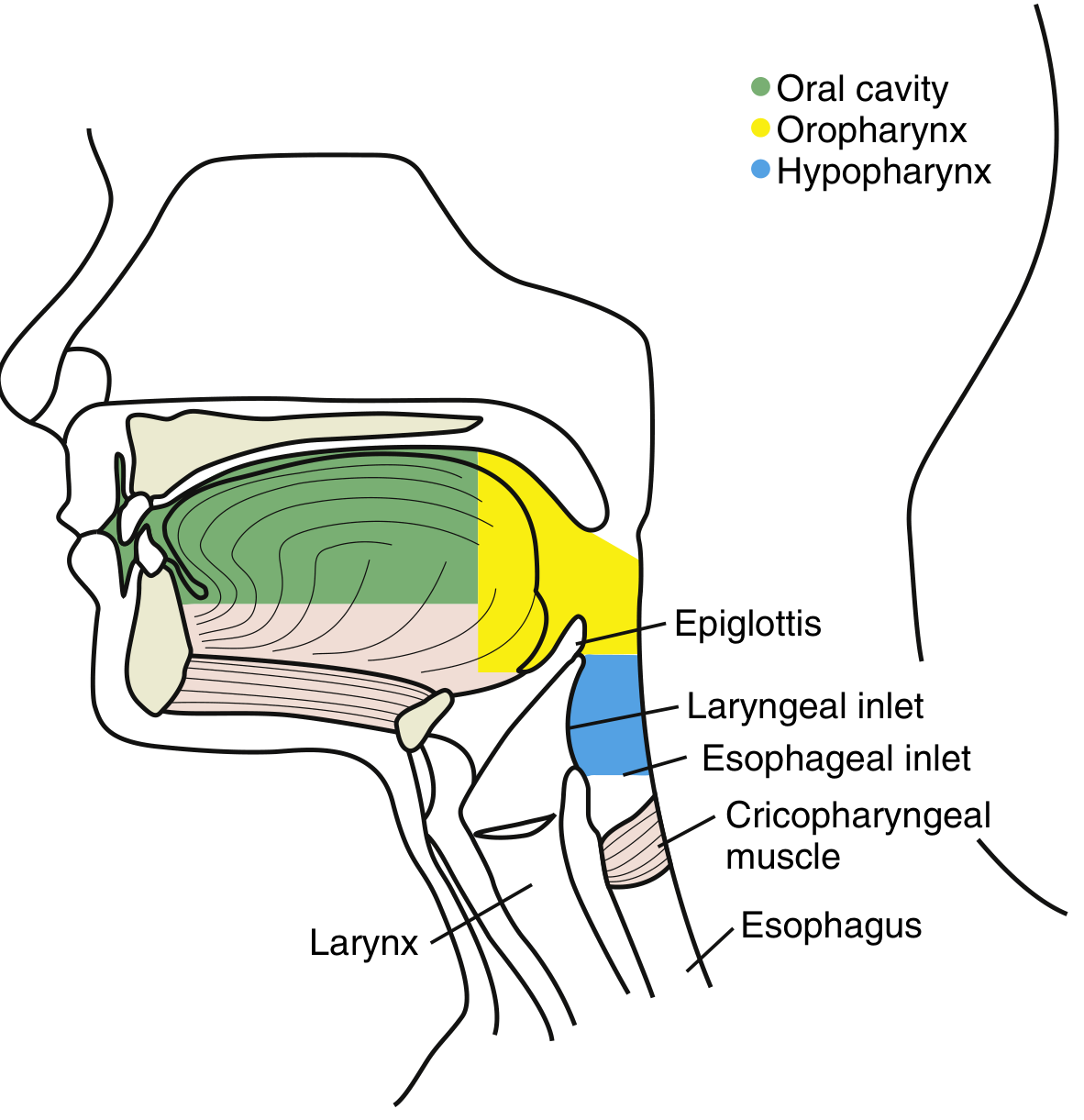
Boundaries
| Border | Landmark |
|---|---|
| Superior | Level of the pharyngoepiglottic folds / superior border of the vallecula / hyoid bone |
| Inferior | Lower border of the cricoid cartilage / cricopharyngeus muscle (esophageal introitus) |
| Anterior | Larynx (posterior surface of the epiglottis, aryepiglottic folds, cricoid cartilage) |
| Posterior | Retropharyngeal space / vertebral column |
(Cummings Otolaryngology, p. 1904; Goldman-Cecil Medicine)
Three Subsites
-
Piriform sinuses (fossae) - the most common site of hypopharyngeal malignancies
- Paired, inverted pyramid-shaped recesses flanking the laryngeal inlet
- Formed by anterior, medial, and lateral walls
- Base is at the pharyngoepiglottic fold; apex is just below the cricoid cartilage
- The medial piriform mucosa forms the posterolateral wall of the paraglottic space, separated from the endolarynx only by the aryepiglottic folds and lateral cricoarytenoid muscles
-
Postcricoid region
- Midline, inferiorly located subsite
- Extends from just below the posterior arytenoid cartilages to the esophageal introitus
- Lies medial to the tracheoesophageal groove, so tumors here can involve the recurrent laryngeal nerve, paratracheal nodes, and thyroid gland
-
Posterior pharyngeal wall
- Extends from the level of the hyoid bone to the superior aspect of the cricopharyngeus muscle
- Separated from vertebral/paravertebral structures by the retropharyngeal space
- Tumors can invade prevertebral tissues early
Histological Layers (Wall Structure)
From inside out, the hypopharynx has four layers:
- Mucosal lining - stratified squamous epithelium over loose stroma
- Fibrous layer - pharyngeal aponeurosis
- Muscular layer - posterior cricoarytenoid muscles (anterior) and middle/inferior constrictor muscles (posterior); the inferior constrictor condenses distally into the cricopharyngeus muscle
- Fascial layer - buccopharyngeal fascia
Key anatomical weak points:
- Killian's triangle - a potential defect at the superior junction of the inferior constrictor and cricopharyngeus, through which posterior pharyngeal wall tumors (and Zenker diverticula) can herniate
- Weakness at the thyrohyoid membrane (just inferior to the hyoid bone), where tumors may spread laterally along the vascular pedicle and superior laryngeal nerve
Blood Supply
- Arterial: Primarily the superior thyroid artery (branch of external carotid); collaterals from lingual and ascending pharyngeal arteries
- Venous: Mirrors the arterial supply
Innervation
- Sensory: Glossopharyngeal nerve (CN IX) and vagus nerve (CN X) - sensory signals pass to the nucleus solitarius in the brainstem, coordinating deglutition
- The internal branch of the superior laryngeal nerve passes through the superior lateral wall of the piriform sinus and through the thyrohyoid membrane
- This nerve synapses in the jugular ganglion alongside Arnold's nerve (from the external auditory canal) - this explains referred otalgia in piriform sinus tumors
Lymphatic Drainage
Drainage is subsite-specific:
- Piriform sinus - lymphatics pass through the thyrohyoid membrane to the jugulodigastric lymph nodes
- Postcricoid region - paratracheal and inferior cervical nodes
- Posterior wall - retropharyngeal and upper jugular nodes
Clinical Relevance
- The hypopharynx is a common site for squamous cell carcinoma (SCC), accounting for the majority of hypopharyngeal neoplasms
- Tumors here often present at an advanced stage because the region is "occult" (hidden) and symptoms are non-specific (dysphagia, otalgia, neck mass)
- Risk factors: tobacco, alcohol
- Piriform sinus tumors can invade the larynx due to close anatomical relationships
- Imaging with PET-CT is standard for staging; barium studies are particularly useful for piriform sinus apex and postcricoid assessment
- Treatment (surgery, radiotherapy, chemotherapy) carries a poor prognosis at advanced stages due to the challenging anatomy and reconstructive demands
(Cummings Otolaryngology Head and Neck Surgery; Goldman-Cecil Medicine; Schwartz's Principles of Surgery)
Ij my book it says location of larynx is in front of hypopharynx
Your book is correct, and so is what I said earlier - there is no contradiction here.
In my previous answer I wrote:
"bordered anteriorly by the larynx"
That means the larynx is in front of (anterior to) the hypopharynx. "Anterior border = the larynx" and "larynx is in front of the hypopharynx" are two ways of saying exactly the same thing.
Quick visual clarification
Think of a sagittal (side-view) cross-section of the neck:
FRONT (anterior)          BACK (posterior)
─────────────────────────────────────────────
   LARYNX    |   HYPOPHARYNX   |  VERTEBRAE
             ↑
        larynx is the anterior
        border of the hypopharynx
- The larynx sits in front of the hypopharynx
- The hypopharynx wraps around the back and sides of the larynx
- The piriform sinuses (part of the hypopharynx) are actually the two grooves running on either side of the larynx
- Behind the hypopharynx are the retropharyngeal space and vertebral column
So your book and my answer agree completely. The larynx is anterior to (= in front of) the hypopharynx.
This is a shared conversation. Sign in to Orris to start your own chat.