
Shoulder anatomy
shoulder anatomy diagram bones muscles rotator cuff
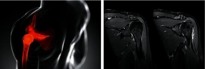
This composite educational graphic focuses on rotator cuff pathology and shoulder anatomy, featuring both an anatomical diagram and clinical diagnostic imaging. On the left, a schematic anatomical diagram of a human shoulder is shown in a coronal-style view; the humerus, scapula, and gleno-humeral joint are highlighted in red against a translucent body outline to indicate the anatomical region of interest, specifically the rotator cuff and subacromial space. On the right, two side-by-side diagnostic images are presented, consisting of Magnetic Resonance Imaging (MRI) scans in the coronal plane. These scans provide high-contrast visualization of the musculoskeletal structures, including the humeral head, acromion process, and the tendons of the rotator cuff (particularly the supraspinatus). The MRI images illustrate clinical findings such as tendon integrity, signal intensity changes suggestive of inflammation or fluid, and joint space morphology. This comparison serves to bridge basic anatomical concepts with real-world radiological diagnosis for orthopedic and sports medicine education.
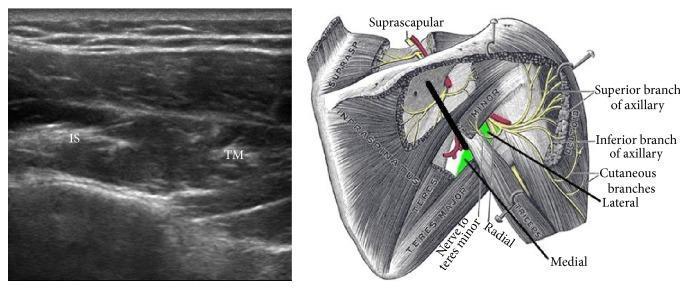
The visual content consists of two side-by-side images: a musculoskeletal diagnostic ultrasound (left) and an anatomical diagram of the posterior shoulder (right), illustrating the positioning for ultrasound-guided procedures. The ultrasound shows a short-axis view of the myotendinous junction within the infraspinatus fossa. Two distinct muscle groups are identified: the infraspinatus (IS) and the teres minor (TM), appearing as relatively hypoechoic structures with characteristic fibrillar echotexture separated by fascial planes. The anatomical diagram depicts the posterior aspect of the scapula and proximal humerus, highlighting the rotator cuff muscles, axillary nerve branches (superior and inferior), and the radial nerve. A thick black line on the diagram indicates the transducer's orientation, placed perpendicular to the axis of the spine of the scapula. This clinical setup is primarily used for identifying landmarks during ultrasound-guided axillary nerve blocks or diagnostic assessments of rotator cuff pathology. The educational focus is on correlating surface anatomy and transducer placement with real-time sonographic visualization of posterior shoulder musculature and neurovascular landmarks.
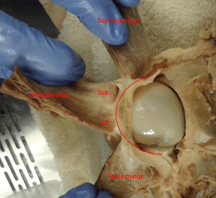
Anatomical Dissection: This clinical photograph provides a posterior view of a dissected left human shoulder, focusing on the rotator cuff complex. The supraspinatus, infraspinatus, and teres minor muscles are reflected laterally to reveal the underlying humeral head and its capsule. A curved red line overlay highlights the rotator cable, a thick fibrous band that spans the avascular zone of the rotator cuff. The infraspinatus muscle is further subdivided with labels 'Sup.' and 'Inf.', representing the superior and inferior portions of the muscle, respectively. The image demonstrates the varying degrees of adherence between the infraspinatus and the rotator cable, illustrating the functional anatomy of the shoulder's capsuloligamentous structures. This material is useful for studying musculoskeletal anatomy, specifically the insertions of the rotator cuff and the biomechanical role of the rotator cable in distributing forces across the shoulder joint.
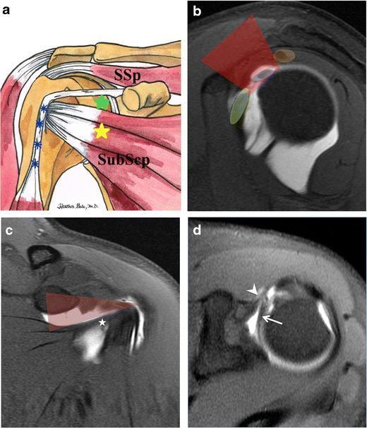
This composite educational resource illustrates the anatomy of the rotator interval of the shoulder. (a) An anatomical diagram of the left shoulder in external rotation highlights the rotator cuff interval (green star) located between the supraspinatus (SSp) and subscapularis (SubScp) tendons, with the long head of the biceps tendon (blue asterisks) traveling in the bicipital groove. (b-d) T1-weighted TSE fat-suppressed MR arthrography images in sagittal, coronal, and axial planes demonstrate the interval using contrast enhancement. In the sagittal view (b), the interval (red overlay) is flanked by the subscapularis (green) and supraspinatus (orange), with the biceps tendon (blue) centrally located. The coronal view (c) defines the triangular interval boundaries, bordered inferiorly by the subscapularis tendon (asterisk). The axial view (d) provides detailed visualization of the long head of the biceps brachii tendon (arrow) and the coracohumeral ligament (arrowhead) within the interval. This content demonstrates the anterior-rotator interval approach for intra-articular injection, emphasizing the preservation of the inferior glenohumeral ligament and subscapularis muscle integrity.
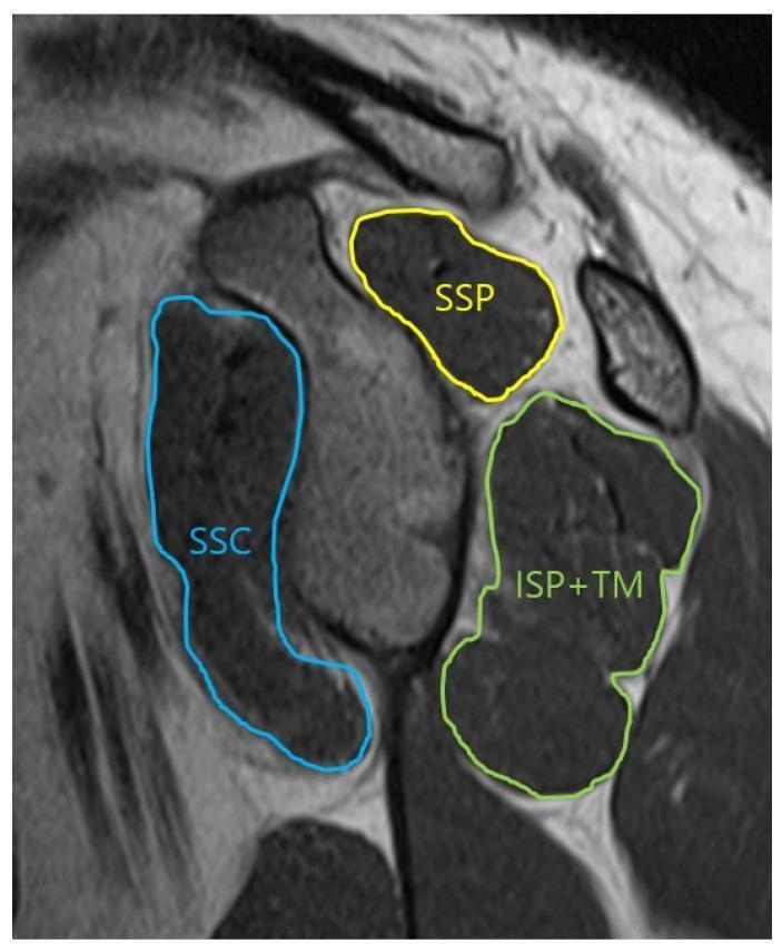
This oblique-sagittal T2 Turbo Spin Echo (TSE) MRI scan demonstrates the musculoskeletal anatomy of the shoulder in a glenoid face view, specifically for assessing the cross-sectional area of the rotator cuff muscles. The image features color-coded annotations outlining the muscular boundaries: the supraspinatus (SSP) is outlined in yellow in the superior position; the subscapularis (SSC) is traced in blue anteriorly; and the infraspinatus combined with the teres minor (ISP+TM) is outlined in green in the posterior-inferior quadrant. The musculature appears as intermediate to dark gray signal intensity, allowing for the evaluation of muscle volume and potential fatty infiltration or atrophy. This imaging view is clinically significant for orthopedic evaluation and preoperative planning in cases of rotator cuff tears or shoulder instability, providing a clear visualization of the muscular support surrounding the glenohumeral joint.
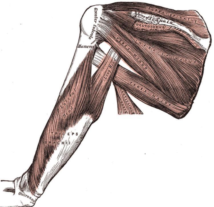
This medical anatomical diagram illustrates the musculature of the human shoulder and upper arm from a posterior perspective. The illustration highlights the rotator cuff complex and its relationship to the scapula and humerus. Key structures identified include the supraspinatus, originating above the spine of the scapula; the infraspinatus, occupying the infraspinous fossa; and the teres minor and teres major muscles positioned along the lateral border of the scapula. The deltoideus is shown partially overlying the posterior joint. On the humerus, the greater tubercle is clearly marked as an insertion point. The triceps brachii is depicted on the posterior arm, specifically identifying the long head—which passes between the teres minor and major—and the lateral head. The distal insertion of the triceps onto the olecranon of the ulna is visible. This diagram serves as a clinical education tool for understanding shoulder stability, biomechanics, and the spatial arrangement of the musculoskeletal system relevant to orthopedics and sports medicine.
Generate a study guide on rotator cuff anatomy