Create a visual explanation on Thyroid hormones
Thyroid Hormones: A Visual Explanation
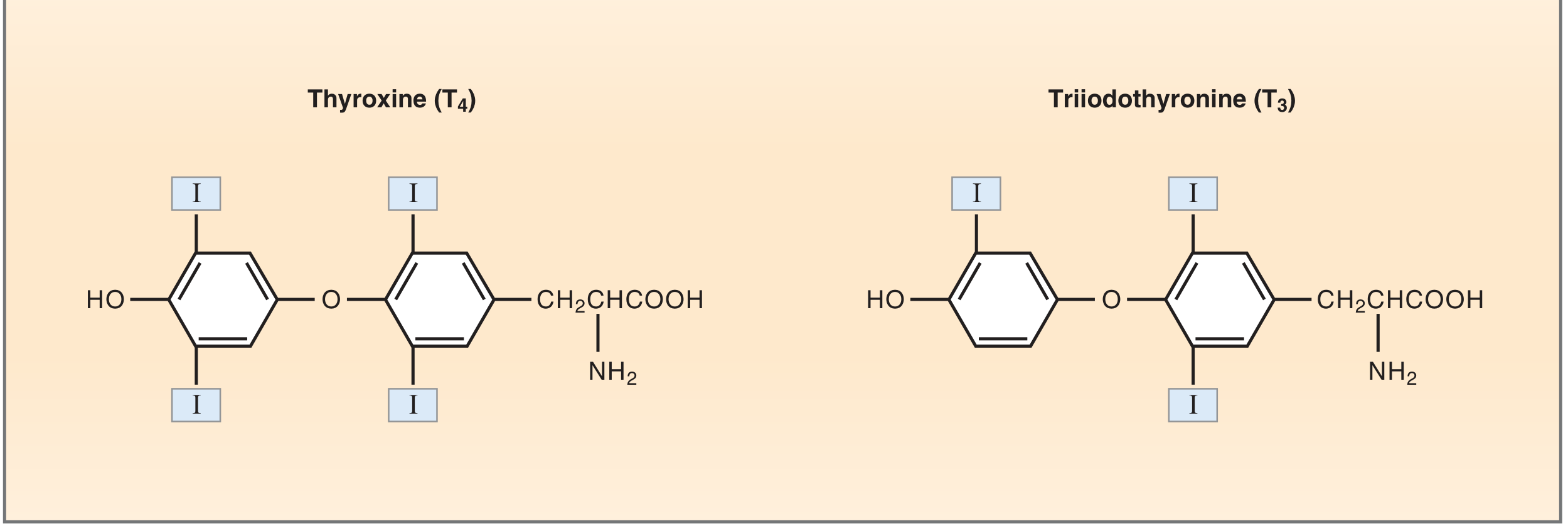
1. What Are Thyroid Hormones?
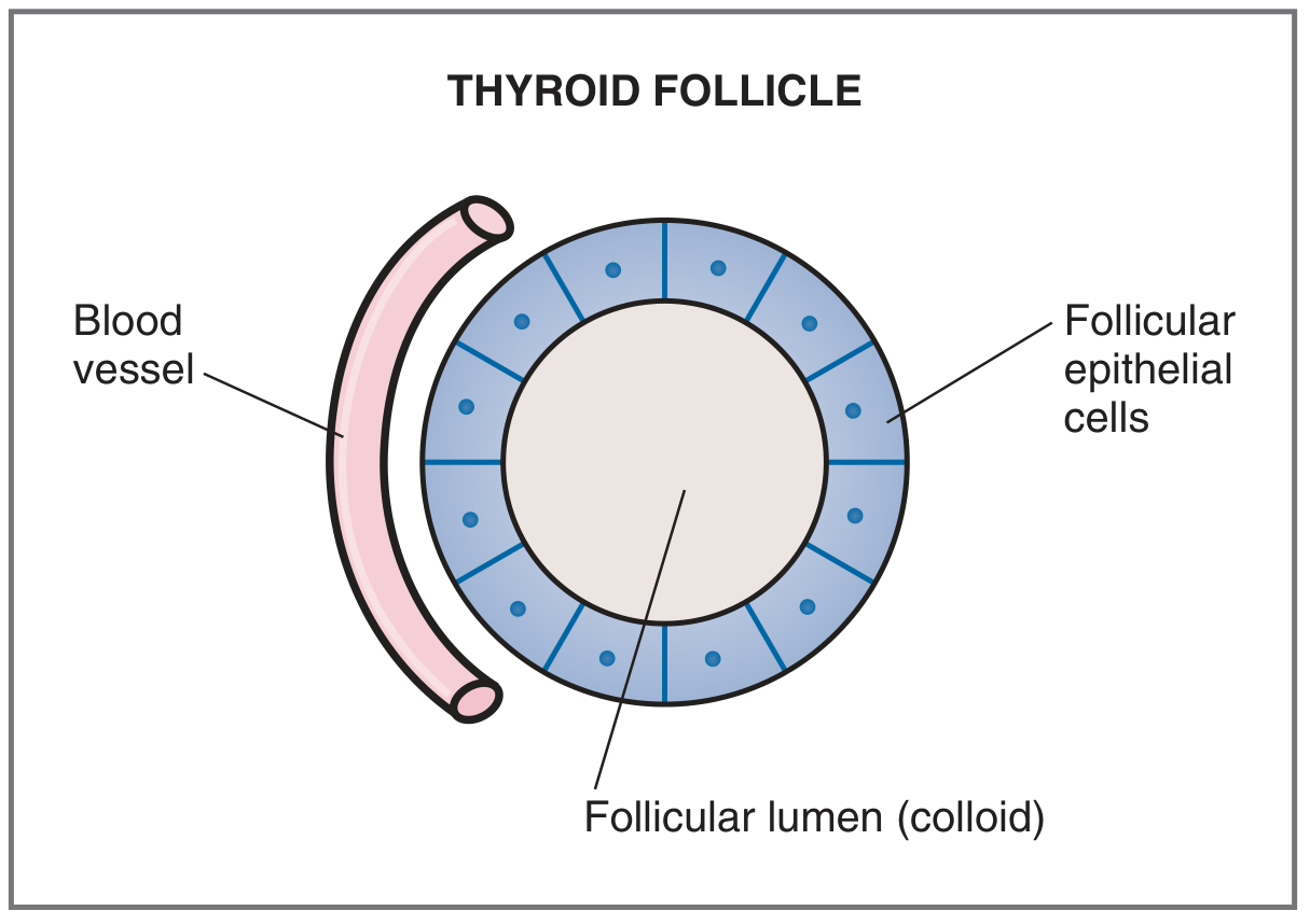
2. Where Are They Made? The Thyroid Follicle
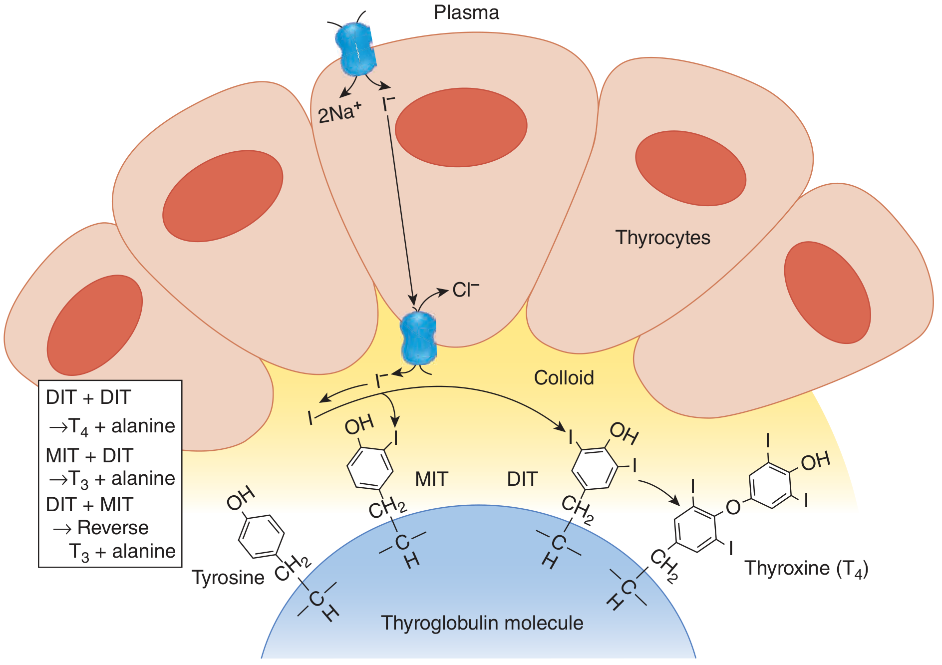
3. Synthesis: Step by Step
| Step | Event |
|---|---|
| 1 | Thyroglobulin (TG) synthesis - TG is made on the rough ER, processed in Golgi, and secreted by exocytosis into the follicular lumen |
| 2 | Iodide trapping - Na⁺/I⁻ symporter (NIS) on the basal membrane actively pumps I⁻ into the thyrocyte against its electrochemical gradient, driven by the Na⁺ gradient |
| 3 | Iodide transport to apical membrane - I⁻ moves to the apical surface; pendrin (a Cl⁻/I⁻ exchanger) transports it into the colloid |
| 4 | Oxidation - Thyroid peroxidase (TPO) oxidizes I⁻ to reactive I⁰ using H₂O₂ |
| 5 | Organification - TPO incorporates I⁰ into carbon-3 of tyrosine residues on TG. One iodine → monoiodotyrosine (MIT); two iodines → diiodotyrosine (DIT) |
| 6 | Coupling - TPO catalyzes oxidative coupling: DIT + DIT → T4 (+ alanine); MIT + DIT → T3 (+ alanine); DIT + MIT → reverse T3 |
| 7 | Secretion - On TSH stimulation, thyrocytes endocytose colloid, lysosomes hydrolyze TG peptide bonds, and free T4 and T3 are released into the capillaries |
| 8 | Salvage - Free MIT and DIT are deiodinated by thyroid deiodinase, recycling iodine and tyrosine back into the next synthetic cycle |
Colloid acts as a hormone reservoir - the body can maintain normal circulating thyroid hormone levels for up to 2 months on a completely iodine-free diet.
- Ganong's Review of Medical Physiology, 26th Ed.
- Costanzo Physiology, 7th Ed.
- Sabiston Textbook of Surgery
4. Regulation: The Hypothalamic-Pituitary-Thyroid (HPT) Axis
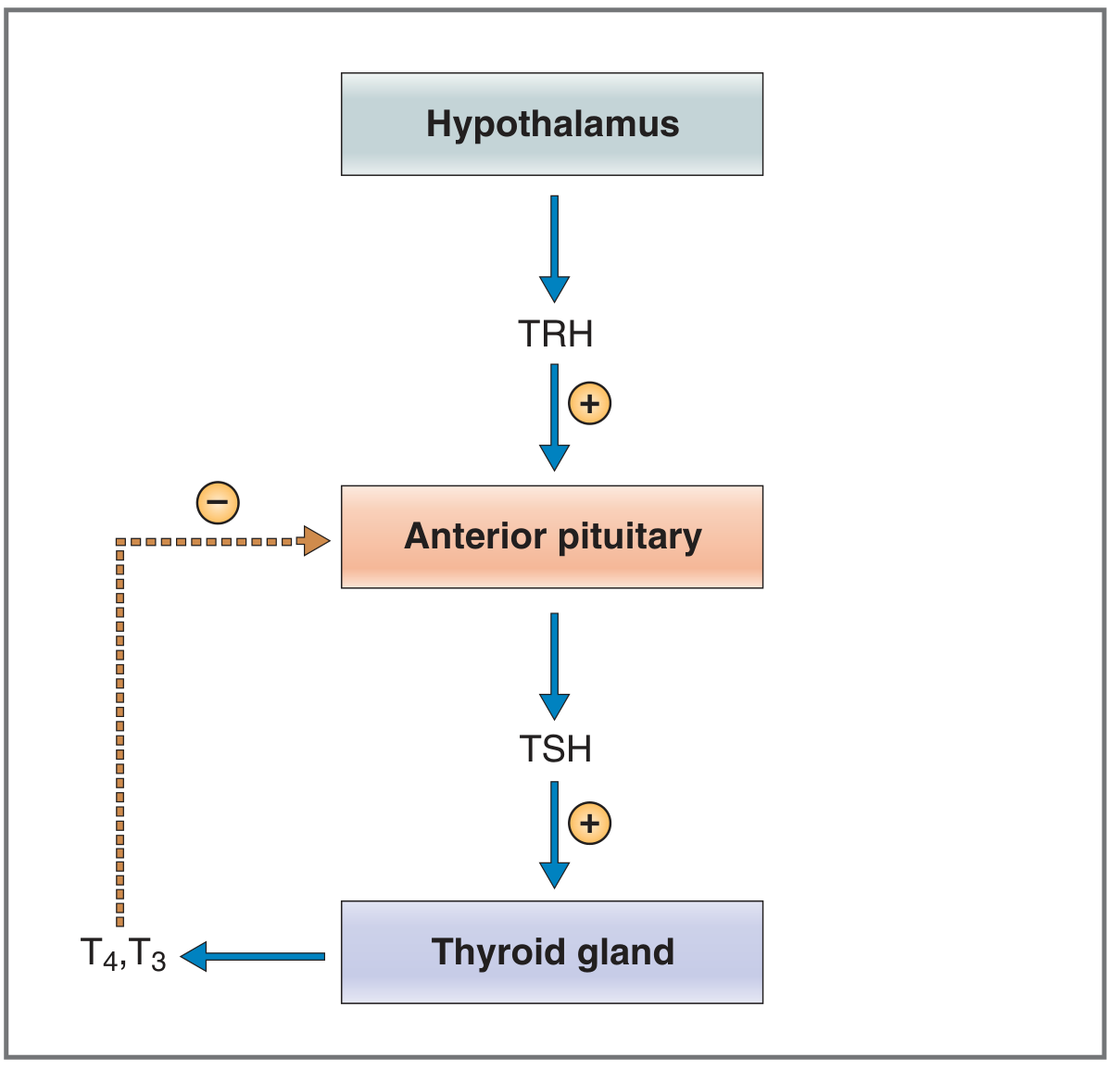
- Hypothalamus secretes TRH (thyrotropin-releasing hormone, a tripeptide) from the paraventricular nucleus into the portal circulation
- TRH stimulates thyrotroph cells of the anterior pituitary to transcribe and secrete TSH (thyroid-stimulating hormone, a glycoprotein with α and β subunits)
- TSH binds to receptors on thyroid follicular cells (Gs-coupled → cAMP), stimulating every step in synthesis: iodide uptake, oxidation, organification, coupling, endocytosis, and TG proteolysis
- Negative feedback: T3 (and T4 converted to T3 by pituitary deiodinase) suppresses TSH secretion by down-regulating the TRH receptor on thyrotrophs
| Stimulatory | Inhibitory |
|---|---|
| TSH | Excess iodide (Wolff-Chaikoff effect) |
| Thyroid-stimulating immunoglobulins (Graves disease) | Propylthiouracil (PTU) - blocks peroxidase |
| Elevated TBG (e.g., pregnancy) | Perchlorate/thiocyanate - block NIS |
| Cold exposure | Carbimazole/methimazole |
| Selenium deficiency |
- Costanzo Physiology, 7th Ed., Table 9.8
5. Protein Binding and Transport in Blood
- >99% of T4 and T3 circulate bound to plasma proteins (mainly thyroxine-binding globulin/TBG, also transthyretin and albumin)
- <1% circulates free (unbound) - only this fraction is biologically active
6. Peripheral Activation - T4 to T3
7. Mechanism of Action in Target Cells
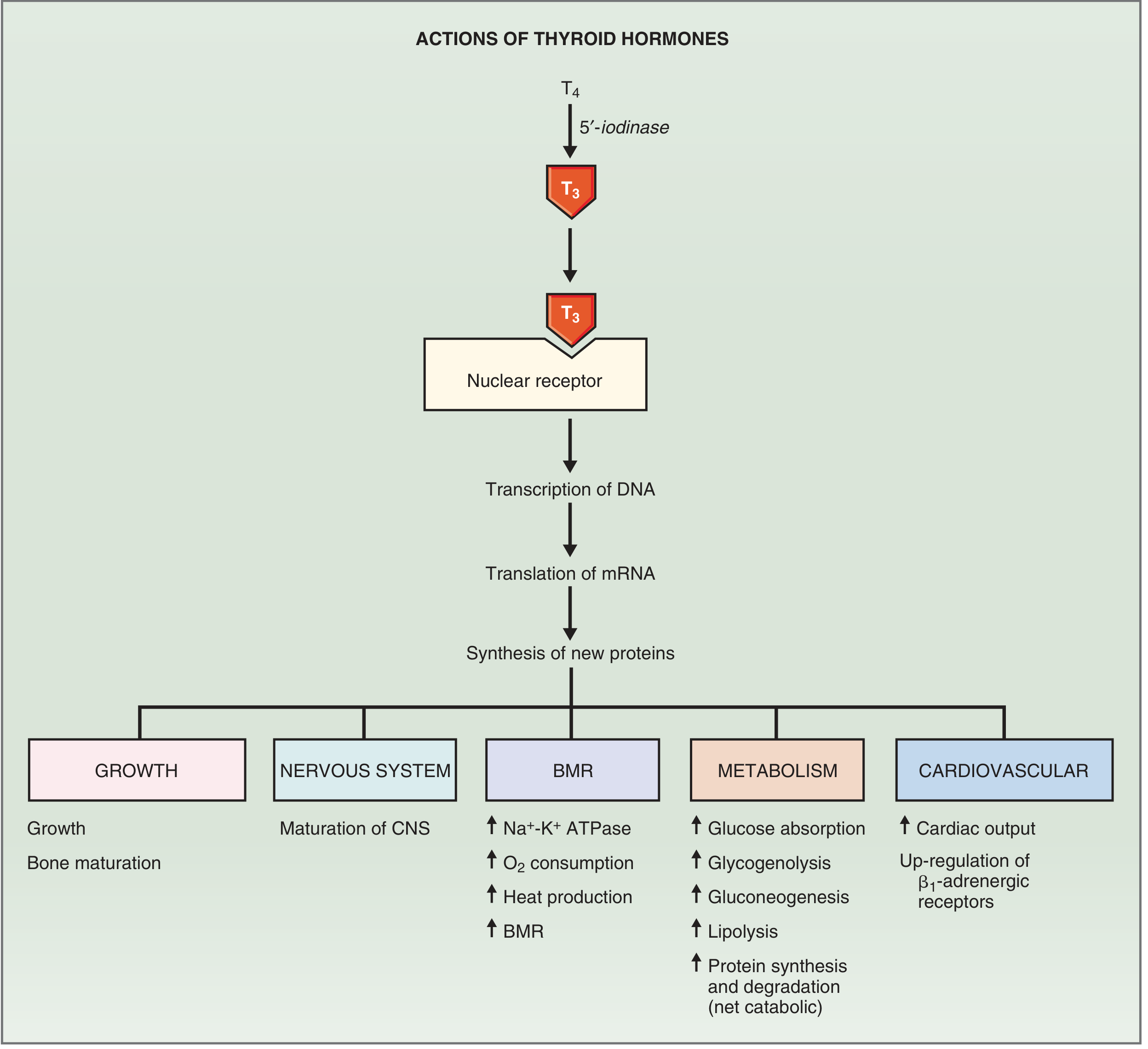
- T4 enters the cell and is converted to T3 by 5'-iodinase
- T3 enters the nucleus and binds to thyroid hormone receptors (TRs), which belong to the nuclear receptor superfamily
- The T3-TR complex binds to thyroid-regulatory elements (TREs) on DNA
- This stimulates transcription → new mRNA → new proteins
8. Physiological Effects (Organ-by-Organ)
| System | Effect of T3 |
|---|---|
| Basal Metabolic Rate | ↑ Na⁺/K⁺ ATPase → ↑ O₂ consumption, heat production; affects all tissues except brain, gonads, spleen |
| Cardiovascular | ↑ heart rate, ↑ contractility (via myosin + β1 receptors + Ca²⁺ ATPase), ↑ cardiac output |
| Carbohydrate metabolism | ↑ GI glucose absorption, ↑ glycogenolysis, ↑ gluconeogenesis |
| Fat metabolism | ↑ lipolysis; ↓ plasma cholesterol (↑ LDL receptors in liver) |
| Protein metabolism | Net catabolic at high doses (↑ synthesis AND degradation) |
| Growth & bone | Synergizes with GH and IGF-1 for bone maturation; slowed growth in children without T3 |
| Nervous system | CNS maturation (critical in fetal life and infancy); normal adult neural function |
| Muscle | Regulates MHC gene expression; excess → thyrotoxic myopathy; deficiency → weakness, cramps, stiffness |
- Ganong's Review of Medical Physiology, 26th Ed.
- Costanzo Physiology, 7th Ed.
9. Clinical Consequences: Hypo vs. Hyperthyroidism
| Feature | Hypothyroidism | Hyperthyroidism |
|---|---|---|
| Metabolic rate | ↓ BMR, cold intolerance, weight gain | ↑ BMR, heat intolerance, weight loss |
| Cardiovascular | Bradycardia, ↓ output | Tachycardia, palpitations, ↑ output |
| Skin/hair | Dry skin, coarse hair, myxedema | Warm moist skin, fine hair |
| Nervous system | Slowed reflexes, depression, cognitive slowing | Anxiety, tremor, hyperreflexia |
| GI | Constipation | Diarrhea, ↑ appetite |
| Growth (children) | Dwarfism, cretinism if congenital | Accelerated growth, early epiphyseal closure |
| Lab | ↑ TSH, ↓ free T4 (primary); ↓ cholesterol improves | ↓ TSH, ↑ free T4; ↓ cholesterol |
| Common cause | Hashimoto thyroiditis (autoimmune), iodine deficiency | Graves disease (TSH-receptor antibodies) |
In Graves disease, thyroid-stimulating immunoglobulins (IgG antibodies) bind and activate TSH receptors, causing unregulated stimulation of the gland - TSH itself is paradoxically low due to negative feedback from the excess hormones.
Summary Pathway (Quick Reference)
Hypothalamus
    │  TRH (+)
    ▼
Anterior Pituitary
    │  TSH (+)
    ▼
Thyroid Follicle
  • NIS traps I⁻ from blood
  • TPO oxidizes I⁻ → organification on Tg (MIT, DIT)
  • Coupling: DIT+DIT → T4 | MIT+DIT → T3
  • TSH stimulation → endocytosis, proteolysis → T4, T3 secreted
    │
    ▼ In circulation (>99% bound to TBG)
    ▼ Free T4 → target tissues → 5'-iodinase → T3 (active)
    │                                              ↓ (inner ring) → rT3 (inactive)
    ▼
T3 binds nuclear receptor → TRE on DNA → new proteins
→ ↑ BMR, ↑ heart rate, ↑ growth, ↑ metabolism, CNS maturation
    │
    └──────── Negative feedback to anterior pituitary (–)
- Costanzo Physiology, 7th Edition (Figs. 9.16, 9.17, 9.19, 9.20)
- Ganong's Review of Medical Physiology, 26th Edition (Figs. 20-6, 20-8, 20-9)
- Sabiston Textbook of Surgery, Biological Basis of Modern Surgical Practice (Figs. 73.5, 73.6)
Y can't u create visual explanation videos?
- Video generation requires specialized AI models (like Sora, Runway, Kling, etc.) that are trained to synthesize moving frames, animations, voiceovers, and transitions - a fundamentally different architecture from what I run on
- I can generate static images (diagrams, posters, illustrations) and rich text-based visual explanations like the one I just made, but not animated or video output
- No audio/voiceover - video explanations typically pair visuals with narration, which requires text-to-speech and video editing pipelines I don't have access to
| Option | What I can make |
|---|---|
| 📊 Rich visual diagrams | Like the thyroid explanation above, with real textbook images |
| 🖼️ Custom illustrated posters | AI-generated infographic-style images |
| 📑 Slide decks (.pptx) | Presentation slides you can animate yourself in PowerPoint |
| 📄 PDF visual summaries | Formatted, printable visual notes |
- Manim (animated math/science videos, like 3Blue1Brown)
- Synthesia or HeyGen (AI presenter videos)
- Canva (animated presentations)
Can u create mind maps?
mkdir -p /home/daytona/workspace/thyroid-mindmap
~/thyroid-mindmap/thyroid_mindmap.html
<!DOCTYPE html>
<html lang="en">
<head>
<meta charset="UTF-8" />
<meta name="viewport" content="width=device-width, initial-scale=1.0"/>
<title>Thyroid Hormones – Mind Map</title>
<style>
  * { box-sizing: border-box; margin: 0; padding: 0; }
  body {
    background: #0f1117;
    font-family: 'Segoe UI', Arial, sans-serif;
    overflow: hidden;
    height: 100vh;
    width: 100vw;
    user-select: none;
  }
  #canvas-wrap {
    width: 100%; height: 100%;
    position: relative;
    overflow: hidden;
    cursor: grab;
  }
  #canvas-wrap.dragging { cursor: grabbing; }
  svg {
    position: absolute;
    top: 0; left: 0;
    width: 100%; height: 100%;
    overflow: visible;
  }
  .node-group { cursor: pointer; }
  .node-rect {
    rx: 12; ry: 12;
    filter: drop-shadow(0 4px 12px rgba(0,0,0,0.5));
    transition: filter 0.2s;
  }
  .node-group:hover .node-rect {
    filter: drop-shadow(0 6px 20px rgba(255,255,255,0.25));
  }
  .node-text {
    fill: #fff;
    font-weight: 600;
    text-anchor: middle;
    dominant-baseline: middle;
    pointer-events: none;
  }
  .link {
    fill: none;
    stroke-width: 2;
    opacity: 0.5;
  }
  #tooltip {
    position: fixed;
    background: rgba(20,20,30,0.97);
    border: 1px solid rgba(255,255,255,0.15);
    border-radius: 10px;
    padding: 12px 16px;
    max-width: 320px;
    pointer-events: none;
    display: none;
    z-index: 999;
    color: #e8e8e8;
    font-size: 13px;
    line-height: 1.6;
    box-shadow: 0 8px 32px rgba(0,0,0,0.6);
  }
  #tooltip h4 {
    color: #fff;
    font-size: 14px;
    margin-bottom: 6px;
    border-bottom: 1px solid rgba(255,255,255,0.1);
    padding-bottom: 5px;
  }
  #controls {
    position: fixed;
    bottom: 20px;
    right: 20px;
    display: flex;
    flex-direction: column;
    gap: 8px;
    z-index: 100;
  }
  .ctrl-btn {
    background: rgba(255,255,255,0.1);
    border: 1px solid rgba(255,255,255,0.2);
    color: #fff;
    border-radius: 8px;
    width: 40px; height: 40px;
    font-size: 18px;
    cursor: pointer;
    display: flex; align-items: center; justify-content: center;
    transition: background 0.2s;
  }
  .ctrl-btn:hover { background: rgba(255,255,255,0.2); }
  #legend {
    position: fixed;
    top: 20px;
    left: 20px;
    background: rgba(20,20,30,0.9);
    border: 1px solid rgba(255,255,255,0.12);
    border-radius: 10px;
    padding: 12px 16px;
    color: #ccc;
    font-size: 12px;
    z-index: 100;
    line-height: 1.9;
  }
  #legend h5 { color: #fff; margin-bottom: 4px; font-size: 13px; }
  .leg-dot {
    display: inline-block;
    width: 12px; height: 12px;
    border-radius: 3px;
    margin-right: 6px;
    vertical-align: middle;
  }
  #title-bar {
    position: fixed;
    top: 20px; left: 50%;
    transform: translateX(-50%);
    color: #fff;
    font-size: 22px;
    font-weight: 700;
    letter-spacing: 0.5px;
    text-shadow: 0 2px 12px rgba(0,0,0,0.5);
    pointer-events: none;
    z-index: 100;
  }
  #hint {
    position: fixed;
    bottom: 20px; left: 50%;
    transform: translateX(-50%);
    color: rgba(255,255,255,0.35);
    font-size: 12px;
    pointer-events: none;
    z-index: 100;
  }
</style>
</head>
<body>
<div id="title-bar">🦋 Thyroid Hormones — Mind Map</div>
<div id="legend">
  <h5>Legend</h5>
  <div><span class="leg-dot" style="background:#c0392b"></span> Central Topic</div>
  <div><span class="leg-dot" style="background:#1a6eb5"></span> Main Branch</div>
  <div><span class="leg-dot" style="background:#16a085"></span> Sub-branch</div>
  <div><span class="leg-dot" style="background:#7d3c98"></span> Mechanism</div>
  <div><span class="leg-dot" style="background:#b7770d"></span> Clinical</div>
</div>
<div id="controls">
  <button class="ctrl-btn" id="zoomIn" title="Zoom In">+</button>
  <button class="ctrl-btn" id="zoomOut" title="Zoom Out">−</button>
  <button class="ctrl-btn" id="reset" title="Reset View">⌂</button>
</div>
<div id="hint">Drag to pan · Scroll to zoom · Hover nodes for details</div>
<div id="tooltip"></div>
<div id="canvas-wrap">
  <svg id="svg"></svg>
</div>

<script>
const W = window.innerWidth, H = window.innerHeight;
const CX = W / 2, CY = H / 2;

// ─── Node Data ───────────────────────────────────────────────────
const nodes = [
  // Central
  {
    id: "center", label: "THYROID\nHORMONES", x: CX, y: CY,
    w: 160, h: 70, color: "#c0392b", fontSize: 15,
    info: { title: "Thyroid Hormones (T3 & T4)", body: "Two iodine-containing amino acid–derived hormones secreted by thyroid follicular cells. T4 (thyroxine) = prohormone; T3 (triiodothyronine) = active form. Regulate metabolism, growth, development, and cardiovascular function." }
  },

  // ── Branch 1: Structure
  {
    id: "structure", label: "STRUCTURE", x: CX - 420, y: CY - 260,
    w: 130, h: 48, color: "#1a6eb5", fontSize: 13,
    info: { title: "Chemical Structure", body: "Both T3 and T4 are iodothyronines — tyrosine-based with two phenol rings. T4 has 4 iodine atoms; T3 has 3. They differ only by one iodine on the outer (phenolic) ring." }
  },
  {
    id: "T4", label: "T4\n(Thyroxine)", x: CX - 580, y: CY - 340,
    w: 120, h: 50, color: "#16a085", fontSize: 12,
    info: { title: "T4 – Thyroxine", body: "Major secretory product (~90% of output). 4 iodine atoms. Half-life ~7 days. >99% protein-bound. Acts as a prohormone — converted to T3 in target tissues by 5'-iodinase." }
  },
  {
    id: "T3", label: "T3\n(Active form)", x: CX - 580, y: CY - 200,
    w: 120, h: 50, color: "#16a085", fontSize: 12,
    info: { title: "T3 – Triiodothyronine", body: "3-5x more potent than T4. 3 iodine atoms. Half-life ~1 day. Binds nuclear receptors (TRα, TRβ) to regulate gene transcription. Most T3 in blood comes from peripheral T4 conversion." }
  },
  {
    id: "rT3", label: "Reverse T3\n(Inactive)", x: CX - 580, y: CY - 100,
    w: 120, h: 50, color: "#7d3c98", fontSize: 11,
    info: { title: "Reverse T3 (rT3)", body: "Formed by removing iodine from the INNER ring of T4 (vs. T3 from the outer ring). Biologically inactive. Rises in starvation, illness, stress, and with beta-blockers — a protective energy-conservation mechanism." }
  },

  // ── Branch 2: Synthesis
  {
    id: "synthesis", label: "SYNTHESIS", x: CX - 420, y: CY + 60,
    w: 130, h: 48, color: "#1a6eb5", fontSize: 13,
    info: { title: "Thyroid Hormone Synthesis", body: "A multistep process requiring iodine, tyrosine, thyroglobulin (TG), thyroid peroxidase (TPO), and H₂O₂. Unique because it is partially extracellular (in the colloid). Regulated by TSH." }
  },
  {
    id: "nis", label: "① NIS\n(Iodide Trap)", x: CX - 610, y: CY - 10,
    w: 120, h: 50, color: "#16a085", fontSize: 11,
    info: { title: "Na⁺/I⁻ Symporter (NIS)", body: "Active transporter on the basolateral membrane of thyrocytes. Pumps I⁻ into the cell against its gradient, driven by the Na⁺ electrochemical gradient. Stimulated by TSH. Target of radioactive iodine (¹³¹I) therapy." }
  },
  {
    id: "tpo", label: "② TPO\n(Organification)", x: CX - 610, y: CY + 80,
    w: 120, h: 50, color: "#16a085", fontSize: 11,
    info: { title: "Thyroid Peroxidase (TPO)", body: "Enzyme on the apical membrane. Uses H₂O₂ to oxidize I⁻ → I⁰, then incorporates it into tyrosine residues on thyroglobulin (organification). Also catalyzes the coupling reaction (MIT+DIT → T3; DIT+DIT → T4)." }
  },
  {
    id: "coupling", label: "③ Coupling\nMIT + DIT", x: CX - 610, y: CY + 170,
    w: 120, h: 50, color: "#16a085", fontSize: 11,
    info: { title: "Coupling Reaction", body: "TPO catalyses oxidative coupling of iodotyrosines still bound to thyroglobulin:\n• MIT + DIT → T3 + alanine\n• DIT + DIT → T4 + alanine\n• DIT + MIT → reverse T3 + alanine\nThen TSH triggers endocytosis of colloid → proteolysis of TG → T3/T4 secreted." }
  },
  {
    id: "tg", label: "Thyroglobulin\n(Reservoir)", x: CX - 420, y: CY + 180,
    w: 130, h: 48, color: "#7d3c98", fontSize: 11,
    info: { title: "Thyroglobulin (TG)", body: "Large glycoprotein (660 kDa) with 123 tyrosine residues, only 4–8 used for hormone synthesis. Synthesized in thyrocytes, secreted into colloid. Acts as storage scaffold — can supply T3/T4 for up to 2 months without dietary iodine." }
  },

  // ── Branch 3: Regulation (top right)
  {
    id: "regulation", label: "REGULATION\n(HPT Axis)", x: CX + 360, y: CY - 300,
    w: 145, h: 55, color: "#1a6eb5", fontSize: 13,
    info: { title: "HPT Axis — Regulation", body: "Three-tier feedback loop: Hypothalamus → TRH → Anterior Pituitary → TSH → Thyroid gland → T3/T4. T3 exerts negative feedback on the pituitary, keeping hormone levels steady." }
  },
  {
    id: "trh", label: "TRH\n(Hypothalamus)", x: CX + 530, y: CY - 400,
    w: 130, h: 50, color: "#16a085", fontSize: 11,
    info: { title: "TRH – Thyrotropin-Releasing Hormone", body: "Tripeptide secreted by paraventricular nucleus of hypothalamus. Acts on anterior pituitary thyrotrophs to stimulate TSH gene transcription and TSH secretion. Also stimulates prolactin secretion." }
  },
  {
    id: "tsh", label: "TSH\n(Ant. Pituitary)", x: CX + 530, y: CY - 260,
    w: 130, h: 50, color: "#16a085", fontSize: 11,
    info: { title: "TSH – Thyroid-Stimulating Hormone", body: "Glycoprotein with α and β subunits (α shared with LH, FSH, hCG). Binds Gs-coupled receptor on thyrocytes → ↑ cAMP → stimulates every step of T3/T4 synthesis and secretion. Also has trophic effect (thyroid growth/hyperplasia)." }
  },
  {
    id: "feedback", label: "Negative\nFeedback (T3)", x: CX + 530, y: CY - 140,
    w: 130, h: 50, color: "#7d3c98", fontSize: 11,
    info: { title: "Negative Feedback", body: "Free T3 (and T4 converted to T3 by pituitary D2 deiodinase) inhibits TSH secretion by down-regulating TRH receptors on thyrotrophs. This maintains remarkably stable baseline thyroid hormone levels." }
  },

  // ── Branch 4: Transport (right)
  {
    id: "transport", label: "TRANSPORT\n& BINDING", x: CX + 380, y: CY - 40,
    w: 145, h: 55, color: "#1a6eb5", fontSize: 13,
    info: { title: "Protein Transport in Blood", body: "T4 and T3 circulate almost entirely (>99%) bound to plasma proteins. Only FREE hormone is biologically active. Binding proteins buffer free hormone levels." }
  },
  {
    id: "tbg", label: "TBG\n(Main carrier)", x: CX + 560, y: CY - 80,
    w: 120, h: 48, color: "#16a085", fontSize: 11,
    info: { title: "Thyroxine-Binding Globulin (TBG)", body: "Carries ~75% of circulating T4 and T3. Increased by: pregnancy (estrogen inhibits TBG breakdown) → ↑ total T4 but free T4 normal. Decreased by: liver failure → transient ↑ free T4." }
  },
  {
    id: "freehormone", label: "Free T3/T4\n(<1%, Active)", x: CX + 560, y: CY + 40,
    w: 120, h: 48, color: "#7d3c98", fontSize: 11,
    info: { title: "Free (Unbound) Hormone", body: "Only the free fraction enters cells and exerts biological effects. TBG acts as a large reservoir — bound hormone is released when free levels fall. Measured as fT3 and fT4 in labs; more clinically relevant than total T3/T4." }
  },

  // ── Branch 5: Actions (bottom right)
  {
    id: "actions", label: "ACTIONS", x: CX + 340, y: CY + 200,
    w: 130, h: 48, color: "#1a6eb5", fontSize: 13,
    info: { title: "Physiological Actions of T3", body: "T3 binds nuclear receptors (TRα, TRβ) → binds thyroid response elements (TREs) on DNA → transcription of new proteins. Effects span virtually every organ system." }
  },
  {
    id: "bmr", label: "↑ BMR &\nHeat Production", x: CX + 520, y: CY + 140,
    w: 130, h: 50, color: "#16a085", fontSize: 11,
    info: { title: "Basal Metabolic Rate (BMR)", body: "T3 induces Na⁺/K⁺ ATPase in most tissues → ↑ O₂ consumption, ↑ heat production, ↑ BMR. Tissues affected: liver, heart, kidney, muscle. NOT affected: brain, gonads, spleen." }
  },
  {
    id: "cardio", label: "↑ Heart Rate\n& Output", x: CX + 530, y: CY + 230,
    w: 130, h: 50, color: "#16a085", fontSize: 11,
    info: { title: "Cardiovascular Effects", body: "T3 induces β1-adrenergic receptors, myosin heavy chain (α-MHC), and Ca²⁺ ATPase in cardiomyocytes → ↑ heart rate, ↑ contractility, ↑ cardiac output, ↑ blood flow to tissues." }
  },
  {
    id: "metabolism", label: "Metabolism\n(Carb/Fat/Protein)", x: CX + 530, y: CY + 330,
    w: 130, h: 50, color: "#16a085", fontSize: 11,
    info: { title: "Metabolic Effects", body: "Carbohydrate: ↑ GI glucose absorption, ↑ glycogenolysis, ↑ gluconeogenesis.\nFat: ↑ lipolysis, ↓ plasma cholesterol (↑ hepatic LDL receptors).\nProtein: net catabolic at high doses (↑ synthesis AND degradation)." }
  },
  {
    id: "growth", label: "Growth &\nCNS Maturation", x: CX + 380, y: CY + 340,
    w: 130, h: 50, color: "#7d3c98", fontSize: 11,
    info: { title: "Growth & Nervous System", body: "Synergizes with GH and IGF-1 for bone maturation and linear growth. CNS: critical for brain development in fetal life and infancy. Hypothyroidism at birth → cretinism (irreversible intellectual disability) if untreated." }
  },

  // ── Branch 6: Clinical (bottom left)
  {
    id: "clinical", label: "CLINICAL\nDISORDERS", x: CX - 300, y: CY + 310,
    w: 145, h: 55, color: "#b7770d", fontSize: 13,
    info: { title: "Clinical Thyroid Disorders", body: "Two main categories: hypothyroidism (too little) and hyperthyroidism (too much). Both cause widespread multi-system effects due to thyroid hormones acting on nearly every tissue." }
  },
  {
    id: "hypothyroid", label: "Hypothyroidism\n(↓ T3/T4, ↑ TSH)", x: CX - 480, y: CY + 390,
    w: 150, h: 50, color: "#b7770d", fontSize: 11,
    info: { title: "Hypothyroidism", body: "Causes: Hashimoto thyroiditis (most common; autoimmune), iodine deficiency, post-thyroidectomy, radioiodine therapy.\nFeatures: ↓ BMR, cold intolerance, weight gain, bradycardia, constipation, dry skin, coarse hair, slowed reflexes, myxedema, ↑ cholesterol.\nCongenital: cretinism (intellectual disability, growth failure)." }
  },
  {
    id: "hyperthyroid", label: "Hyperthyroidism\n(↑ T3/T4, ↓ TSH)", x: CX - 200, y: CY + 400,
    w: 150, h: 50, color: "#b7770d", fontSize: 11,
    info: { title: "Hyperthyroidism", body: "Causes: Graves disease (TSH-receptor antibodies — most common), toxic multinodular goitre, toxic adenoma, thyroiditis.\nFeatures: ↑ BMR, heat intolerance, weight loss, tachycardia, palpitations, diarrhoea, warm moist skin, fine hair, tremor, anxiety.\nGraves: exophthalmos (proptosis), pretibial myxedema." }
  },
  {
    id: "drugs", label: "Drug\nTargets", x: CX - 60, y: CY + 380,
    w: 120, h: 50, color: "#b7770d", fontSize: 11,
    info: { title: "Pharmacology — Drug Targets", body: "Antithyroid drugs:\n• PTU / Methimazole — inhibit TPO (block organification & coupling)\n• Perchlorate / Thiocyanate — block NIS (iodide uptake)\n• ¹³¹I — radioablation via NIS uptake\n• Beta-blockers — control tachycardia (↓ T4→T3 conversion)\n\nReplacement:\n• Levothyroxine (T4) — standard hypothyroid treatment" }
  },
];

// ─── Edge Data ────────────────────────────────────────────────────
const edges = [
  ["center","structure"],["center","synthesis"],["center","regulation"],
  ["center","transport"],["center","actions"],["center","clinical"],
  ["structure","T4"],["structure","T3"],["structure","rT3"],
  ["synthesis","nis"],["synthesis","tpo"],["synthesis","coupling"],["synthesis","tg"],
  ["regulation","trh"],["regulation","tsh"],["regulation","feedback"],
  ["transport","tbg"],["transport","freehormone"],
  ["actions","bmr"],["actions","cardio"],["actions","metabolism"],["actions","growth"],
  ["clinical","hypothyroid"],["clinical","hyperthyroid"],["clinical","drugs"],
];

// ─── SVG Setup ───────────────────────────────────────────────────
const svg = document.getElementById("svg");
svg.setAttribute("viewBox", `0 0 ${W} ${H}`);

let scale = 1, tx = 0, ty = 0;
let isDragging = false, startX, startY;

function applyTransform() {
  g.setAttribute("transform", `translate(${tx},${ty}) scale(${scale})`);
}

const g = document.createElementNS("http://www.w3.org/2000/svg","g");
svg.appendChild(g);

// Draw edges first
const edgeColors = {
  "center-structure":"#1a6eb5","center-synthesis":"#1a6eb5",
  "center-regulation":"#1a6eb5","center-transport":"#1a6eb5",
  "center-actions":"#1a6eb5","center-clinical":"#b7770d"
};

function getNodeById(id) { return nodes.find(n => n.id === id); }

edges.forEach(([a, b]) => {
  const na = getNodeById(a), nb = getNodeById(b);
  if (!na || !nb) return;
  const path = document.createElementNS("http://www.w3.org/2000/svg","path");
  const x1 = na.x, y1 = na.y, x2 = nb.x, y2 = nb.y;
  const mx = (x1 + x2) / 2, my = (y1 + y2) / 2;
  const key = `${a}-${b}`;
  const color = edgeColors[key] || nb.color || "#555";
  path.setAttribute("d", `M${x1},${y1} Q${mx},${my} ${x2},${y2}`);
  path.setAttribute("class","link");
  path.setAttribute("stroke", color);
  g.appendChild(path);
});

// Draw nodes
const tooltip = document.getElementById("tooltip");

nodes.forEach(node => {
  const grp = document.createElementNS("http://www.w3.org/2000/svg","g");
  grp.setAttribute("class","node-group");
  grp.setAttribute("transform",`translate(${node.x - node.w/2},${node.y - node.h/2})`);

  const rect = document.createElementNS("http://www.w3.org/2000/svg","rect");
  rect.setAttribute("class","node-rect");
  rect.setAttribute("width", node.w);
  rect.setAttribute("height", node.h);
  rect.setAttribute("fill", node.color);
  rect.setAttribute("rx", node.id === "center" ? 35 : 12);
  rect.setAttribute("ry", node.id === "center" ? 35 : 12);
  grp.appendChild(rect);

  const lines = node.label.split("\n");
  const lineH = 16;
  const totalH = lines.length * lineH;
  lines.forEach((line, i) => {
    const txt = document.createElementNS("http://www.w3.org/2000/svg","text");
    txt.setAttribute("class","node-text");
    txt.setAttribute("x", node.w / 2);
    txt.setAttribute("y", node.h / 2 - totalH / 2 + i * lineH + lineH / 2);
    txt.setAttribute("font-size", node.fontSize || 12);
    txt.textContent = line;
    grp.appendChild(txt);
  });

  // Hover tooltip
  grp.addEventListener("mouseenter", (e) => {
    if (node.info) {
      tooltip.style.display = "block";
      tooltip.innerHTML = `<h4>${node.info.title}</h4>${node.info.body.replace(/\n/g,"<br>")}`;
    }
  });
  grp.addEventListener("mousemove", (e) => {
    let x = e.clientX + 16, y = e.clientY + 10;
    if (x + 340 > window.innerWidth) x = e.clientX - 340;
    if (y + 200 > window.innerHeight) y = e.clientY - 200;
    tooltip.style.left = x + "px";
    tooltip.style.top = y + "px";
  });
  grp.addEventListener("mouseleave", () => {
    tooltip.style.display = "none";
  });

  g.appendChild(grp);
});

// ─── Pan & Zoom ───────────────────────────────────────────────────
const wrap = document.getElementById("canvas-wrap");

wrap.addEventListener("mousedown", (e) => {
  isDragging = true;
  startX = e.clientX - tx;
  startY = e.clientY - ty;
  wrap.classList.add("dragging");
});
window.addEventListener("mousemove", (e) => {
  if (!isDragging) return;
  tx = e.clientX - startX;
  ty = e.clientY - startY;
  applyTransform();
});
window.addEventListener("mouseup", () => {
  isDragging = false;
  wrap.classList.remove("dragging");
});

wrap.addEventListener("wheel", (e) => {
  e.preventDefault();
  const delta = e.deltaY > 0 ? 0.9 : 1.1;
  const mouseX = e.clientX, mouseY = e.clientY;
  tx = mouseX - (mouseX - tx) * delta;
  ty = mouseY - (mouseY - ty) * delta;
  scale = Math.min(Math.max(scale * delta, 0.3), 3);
  applyTransform();
}, { passive: false });

// Touch support
let lastTouchDist = null;
wrap.addEventListener("touchstart", (e) => {
  if (e.touches.length === 1) {
    isDragging = true;
    startX = e.touches[0].clientX - tx;
    startY = e.touches[0].clientY - ty;
  }
});
wrap.addEventListener("touchmove", (e) => {
  e.preventDefault();
  if (e.touches.length === 1 && isDragging) {
    tx = e.touches[0].clientX - startX;
    ty = e.touches[0].clientY - startY;
    applyTransform();
  } else if (e.touches.length === 2) {
    const dx = e.touches[0].clientX - e.touches[1].clientX;
    const dy = e.touches[0].clientY - e.touches[1].clientY;
    const dist = Math.sqrt(dx*dx + dy*dy);
    if (lastTouchDist) {
      const delta = dist / lastTouchDist;
      scale = Math.min(Math.max(scale * delta, 0.3), 3);
      applyTransform();
    }
    lastTouchDist = dist;
  }
}, { passive: false });
wrap.addEventListener("touchend", () => { isDragging = false; lastTouchDist = null; });

document.getElementById("zoomIn").addEventListener("click", () => {
  scale = Math.min(scale * 1.2, 3);
  applyTransform();
});
document.getElementById("zoomOut").addEventListener("click", () => {
  scale = Math.max(scale * 0.8, 0.3);
  applyTransform();
});
document.getElementById("reset").addEventListener("click", () => {
  scale = 1; tx = 0; ty = 0;
  applyTransform();
});

applyTransform();
</script>
</body>
</html>
A detailed medical mind map on Thyroid Hormones on a dark background. Central node labeled "THYROID HORMONES" in red. Six main branches radiating outward: 1) STRUCTURE (blue) with sub-nodes T4/Thyroxine, T3/Active form, Reverse T3/Inactive; 2) SYNTHESIS (blue) with sub-nodes NIS Iodide Trap, TPO Organification, Coupling MIT+DIT, Thyroglobulin Reservoir; 3) REGULATION HPT Axis (blue) with sub-nodes TRH Hypothalamus, TSH Anterior Pituitary, Negative Feedback T3; 4) TRANSPORT & BINDING (blue) with sub-nodes TBG Main Carrier, Free T3 T4 Active; 5) ACTIONS (blue) with sub-nodes Increase BMR and Heat, Increase Heart Rate, Metabolism Carb Fat Protein, Growth and CNS Maturation; 6) CLINICAL DISORDERS (orange/gold) with sub-nodes Hypothyroidism, Hyperthyroidism, Drug Targets. Nodes are colorful rounded rectangles with white text. Branches are curved colored lines. Professional clean medical diagram style with good spacing and readability.