
Osteomyelitis detailed explanation
Reading File
Now let me search the medical library for osteomyelitis content in parallel.
Finding Sources
Searching PubMed
"osteomyelitis"[MeSH Terms] AND treatment
Excellent. Now let me read the most comprehensive sources in parallel - Bailey & Love (surgery), Robbins pathology, Goldman-Cecil medicine, and Rosen's Emergency Medicine.
Reading File
Reading File
Reading File
Reading File
I have very rich material. Let me read the remaining critical sections - Bailey & Love (continued), Rosen's (treatment) and Robbins (morphology continued) to complete the picture.
Reading File
Reading File
Reading File
Now let me fetch the key textbook images and read the Rosen's treatment section.
Reading File
Loading Image
I now have comprehensive material from multiple authoritative sources. Let me compile the detailed explanation.
Osteomyelitis - Detailed Explanation
Definition
Osteomyelitis is inflammation of bone and marrow, virtually always secondary to infection. It may complicate any systemic infection but frequently presents as a solitary primary focus. All types of organisms - bacteria, viruses, parasites, and fungi - can cause it, but pyogenic bacteria and mycobacteria are by far the most common.
- Robbins, Cotran & Kumar Pathologic Basis of Disease
Routes of Infection
Three pathways allow organisms to reach bone:
| Route | Description | Typical Setting |
|---|---|---|
| Hematogenous | Bacteremia seeds bone via the bloodstream | Children (long bone metaphyses), adults (vertebrae) |
| Contiguous spread | Extension from adjacent soft tissues/joints | Diabetic foot, pressure sores |
| Direct implantation | Penetrating trauma, surgery, open fractures | Post-operative, compound fractures |
In children, hematogenous spread is most common. The metaphyses of long bones are preferentially involved because the slow blood flow through looped capillaries, combined with microtrauma, encourages bacterial seeding during a bacteremia. In adults, osteomyelitis is most often a complication of open fractures, surgical procedures, or diabetes (feet).
- Bailey and Love's Short Practice of Surgery, 28th Ed.
Microbiology / Causative Organisms
| Setting | Typical Organism(s) |
|---|---|
| General (all ages) | Staphylococcus aureus (80-90% of culture-positive cases) |
| Neonates | Group B Streptococcus, E. coli |
| Older children | S. aureus, Streptococcus pyogenes, Haemophilus influenzae |
| Sickle cell disease | Salmonella spp., gram-negative organisms |
| IV drug users / elderly | Gram-negatives, polymicrobial |
| Immunocompromised | Fungi (Coccidioides, Blastomyces, Cryptococcus), Mycobacteria |
| Post-surgical / trauma | Mixed bacterial infections |
Why S. aureus? Staphylococcal cell-wall proteins bind bone matrix components such as collagen, facilitating bacterial adherence to bone. MRSA is now the dominant organism in acute hematogenous osteomyelitis (AHO) in many regions.
- Robbins, Cotran & Kumar
Specific organisms are only identified in ~50% of patients on culture; PCR-based assays are expected to improve this rate.
Age-Related Vascular Anatomy (Key Concept)
The site of infection is determined by the osseous vascular anatomy, which changes with age:
- Neonates: Metaphyseal vessels penetrate the growth plate, so infection can spread to both the epiphysis and joint - septic arthritis frequently co-exists.
- Older children (>18 months): The growth plate acts as a barrier; metaphyseal involvement is typical, epiphysis is spared.
- Adults: After growth plate closure, anastomoses form between metaphyseal and epiphyseal vessels - infection tends to involve the epiphyses and subchondral regions. Vertebral osteomyelitis becomes more common.
- Grainger & Allison's Diagnostic Radiology
Pathology: Stages of Disease
Acute Phase (within 48 hours)
- Bacteria proliferate → neutrophilic infiltration
- Bone cell and marrow necrosis within 48 hours
- Bacteria + inflammatory cells spread via Haversian canals to reach the periosteum
- Periosteum (loosely attached in children) is elevated → subperiosteal abscess forms
- Periosteal lifting further impairs cortical blood supply → more necrosis
Subacute / Progressive Phase
- Increased pressure from abscess → pus perforates cortical bone
- Pus elevates the strong periosteum, rendering cortical bone avascular
- Periosteal elevation is a potent stimulus for new bone formation (involucrum)
- Soft tissue abscesses form; may channel to skin as draining sinuses
Chronic Phase
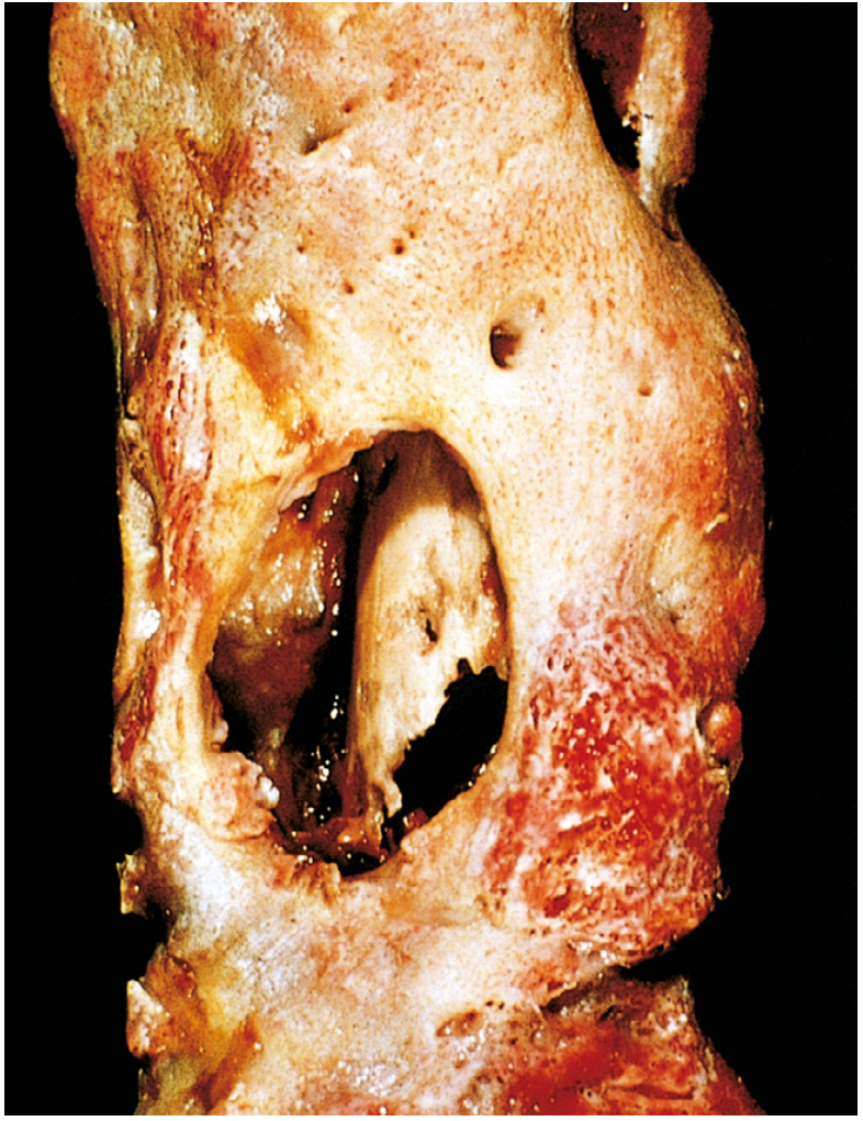
- Dead avascular bone = sequestrum
- New reactive bone forming a shell around the sequestrum = involucrum
- This creates the classic "bone-within-a-bone" appearance on imaging
- Marrow shows fibrosis and a chronic infiltrate of lymphocytes and plasma cells
- Pus may track through gaps (cloacae) in the involucrum to the surface
Brodie Abscess: A walled-off chronic abscess, appearing as a well-defined lytic lesion with a sclerotic rim - characteristic of subacute osteomyelitis.
Below is a gross pathology specimen showing chronic osteomyelitis - the outer involucrum (new viable bone shell) has been opened to reveal the inner white sequestrum (necrotic cortex):
Clinical Features
Symptoms
- Fever, rigors, may appear toxic
- Systemic: headache, fatigue, malaise, anorexia (less consistent in chronic disease)
- Children: sudden limp or inability to bear weight, localized warmth, swelling, erythema
Signs
- Point tenderness over the infected segment (most consistent finding)
- Palpable warmth and soft-tissue swelling with erythema
- In chronic disease: palpable involucrum/sequestrum, sinus tracts to skin
- A sympathetic effusion in the adjacent joint may develop even without joint infection
Acute Hematogenous Osteomyelitis (AHO) in Children
- Male preponderance (2:1 to 3:1)
- Involves long bones ~80% of cases
- Distal metaphysis most common site (increased vascularity)
- Blood cultures positive in ~40% of cases
- Rosen's Emergency Medicine
Vertebral Osteomyelitis (Adults)
- Increasingly common as population ages
- Risk factors: IV access devices, indwelling lines, urinary infections, IV drug use
- Presents with back pain, low-grade fever; neurological signs if epidural extension
Investigations
Laboratory
- CBC: Leukocytosis (variable)
- ESR and CRP: Elevated - useful for monitoring response to treatment
- Blood cultures: Positive in ~40% of pediatric AHO; lower in adults
- Bone biopsy + culture: Gold standard for organism identification
- PCR assays: Emerging to improve yield above culture
Imaging
| Modality | Key Points |
|---|---|
| Plain X-ray | Lags behind disease by 10-14 days; shows soft tissue swelling, cortical irregularity, periosteal reaction, lytic destruction |
| Ultrasound | Detects subperiosteal fluid/abscess early; guides drainage; especially useful in children |
| Bone scan (scintigraphy) | Sensitive early; useful for multifocal disease screening; less specific |
| CT | Best for defining extent of cortical destruction, detecting sequestra; higher radiation |
| MRI | Highest sensitivity and specificity; best for early disease, soft tissue involvement, spinal complications, epidural abscess |
MRI features: Bone marrow edema on T1 (low signal) and T2 (high signal); periosteal reaction; soft tissue edema. The penumbra sign in subacute infection = a peripheral high-signal ring of granulation tissue surrounding a low-signal abscess cavity.
Even on appropriate therapy, radiographic signs of improvement lag behind clinical recovery.
- Grainger & Allison's Diagnostic Radiology
Classification
By Duration
- Acute: < 2-4 weeks symptoms; hematogenous; responds to antibiotics alone (often)
- Subacute: Weeks to months; Brodie abscess typical; culture often negative
- Chronic: >4-6 weeks; sequestrum/involucrum/sinus tracts; requires surgery + prolonged antibiotics
Cierny-Mader Classification (Chronic Osteomyelitis)
Used clinically to guide surgical planning:
- Stage 1 (Medullary): Endosteal infection only
- Stage 2 (Superficial): Cortical surface involvement from contiguous source
- Stage 3 (Localized): Full-thickness cortical involvement, stable bone
- Stage 4 (Diffuse): Through-and-through infection, mechanically unstable bone
Host class: A (normal), B (compromised local or systemic), C (treatment worse than disease)
Special Subsets
Diabetic Foot Osteomyelitis
- Contiguous spread from soft tissue ulcers
- Polymicrobial; often includes gram-negatives and anaerobes
- Probe-to-bone test: positive = high specificity for osteomyelitis
- MRI is imaging modality of choice
Vertebral Osteomyelitis (Spondylodiscitis)
- Usually involves two adjacent vertebral bodies + intervening disc
- Most common site: lumbar spine
- Complications: epidural abscess, spinal cord compression, vertebral collapse
- MRI is mandatory to assess intraspinal involvement
Neonatal Osteomyelitis
- Multiple sites in ~50% of cases
- Septic arthritis frequently co-exists (shared vasculature)
- Flat bones (facial bones) more commonly involved than at other ages
- Whole-body MRI or skeletal scintigraphy for multifocal screening
CRMO (Chronic Recurrent Multifocal Osteomyelitis)
- Autoinflammatory (non-infectious) form
- Multiple foci, culture-negative
- Associated with psoriasis subtypes
- Diagnosed by imaging pattern; treated with NSAIDs/bisphosphonates
Treatment
General Principles
- Identify the organism (blood cultures, bone biopsy)
- Start empirical antibiotics, then narrow based on culture
- Drain any pus surgically
- Splint/rest the affected limb
- Treat underlying conditions (sickle cell, nutritional deficiency, diabetes)
Antibiotic Therapy
| Setting | Empirical Choice |
|---|---|
| Children (MSSA suspected) | Anti-staphylococcal penicillin (flucloxacillin) or 1st-gen cephalosporin |
| MRSA suspected (most AHO) | Vancomycin IV; or clindamycin if susceptible |
| Neonates | Vancomycin + cefotaxime (cover GBS + gram-negatives) |
| Adults (vertebral/diabetic) | Vancomycin + broad gram-negative cover until cultures return |
- IV-to-oral switch: Once clinical and laboratory improvement is documented, oral step-down is appropriate in uncomplicated cases. Debate continues on exact duration.
- Total duration typically 4-6 weeks for acute; 3-6 months for vertebral or chronic.
- A 2025 meta-analysis on oritavancin (PMID 41224240) found it effective for osteomyelitis, offering the advantage of once-weekly dosing - relevant for long-course outpatient treatment.
Surgical Treatment
- Indication: Abscess formation, failure of medical treatment, chronic osteomyelitis with sequestrum, sinus tracts, unstable bone
- Procedures: Debridement, sequestrectomy, saucerization, dead space management (antibiotic-impregnated beads, bone grafting, muscle flaps), stabilization if unstable
- Shortened IV and oral regimens apply only to uncomplicated cases in patients who are clinically and hematologically improving.
- Bailey and Love's Short Practice of Surgery
Complications
| Complication | Notes |
|---|---|
| Chronic osteomyelitis | Occurs in 5-25% of acute cases; delayed diagnosis, extensive necrosis are risk factors |
| Septic arthritis | Especially neonates and when metaphysis is intracapsular |
| Bacteremia / Sepsis | Systemic spread of infection |
| Growth disturbance | Physeal damage in children → limb shortening or deformity |
| Pathologic fracture | Through sites of bone destruction |
| Secondary amyloidosis | Complication of long-standing chronic infection |
| Squamous cell carcinoma | Malignant transformation in chronic draining sinus tracts (rare, late) |
| Sarcoma | Rare malignant transformation in infected bone |
| Brain abscess / meningitis | Hematogenous spread from vertebral/skull osteomyelitis |
| Spinal cord compression | Epidural extension in vertebral osteomyelitis |
Mycobacterial Osteomyelitis (Skeletal TB)
- More destructive and resistant than pyogenic osteomyelitis
- Occurs in ~1-3% of individuals with pulmonary or extrapulmonary TB
- Typically solitary, insidious onset; years may pass before diagnosis
- Classic site: spine (Pott's disease) - involves two adjacent vertebrae + disc
- Spina ventosa: TB dactylitis - cyst-like cavities with diaphyseal expansion, predominantly in hand/foot bones
- Treatment: standard anti-TB regimen (minimum 9-12 months)
- Robbins, Cotran & Kumar
Key Terms Summary
| Term | Meaning |
|---|---|
| Sequestrum | Fragment of dead, avascular necrotic bone |
| Involucrum | Shell of new periosteal bone surrounding the sequestrum |
| Cloaca | Gap/opening in the involucrum through which pus drains |
| Brodie abscess | Walled-off subacute abscess with sclerotic rim on imaging |
| Sinus tract | Channel from the infected bone to the skin surface |
| Penumbra sign | MRI finding in subacute osteomyelitis - high-signal granulation tissue rim around abscess |
Sources:
- Robbins, Cotran & Kumar - Pathologic Basis of Disease, Ch. 26
- Bailey and Love's Short Practice of Surgery, 28th Ed., Ch. 44
- Rosen's Emergency Medicine, Ch. 125
- Grainger & Allison's Diagnostic Radiology, Ch. 73
- Recent evidence: Systematic review on chronic pediatric osteomyelitis (PMID 41329650, 2025); Meta-analysis on oritavancin for osteomyelitis (PMID 41224240, 2025)
This is a shared conversation. Sign in to Orris to start your own chat.