

tell me about the x ray of patient having co shortness of breath having age 56 and ni sign of pyrexia with history of 16 chemotherapy and one is pending having breast carcinoma grade 3
bilateral pleural effusion chest X-ray
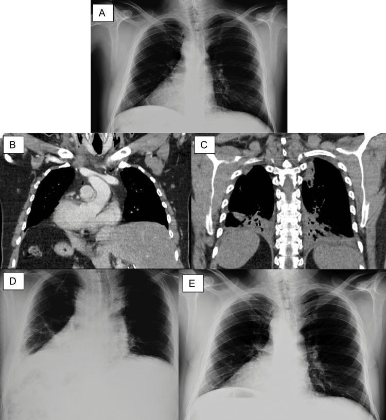
This composite figure displays a series of thoracic and abdominal imaging modalities illustrating Situs Inversus Totalis (SIT) and postoperative pulmonary complications. Image A is a preoperative posterior-anterior chest X-ray clearly demonstrating dextrocardia. Images B and C are coronal sections of a contrast-enhanced thoracoabdominal CT scan. Image B shows the heart apex pointing to the right (dextrocardia), with the liver in the left hypochondrium and the stomach in the right hypochondrium, diagnostic of SIT. Image C, a posterior coronal section, reveals bilateral posterobasal consolidations, basal atelectasis, and incipient bilateral pleural effusion. Images D and E are postoperative serial chest X-rays. Image D, taken 48 hours post-conservative management, shows significant but incomplete improvement with a persistent right-sided pleural effusion causing basal opacity. Image E is a follow-up X-ray showing full resolution of the atelectasis and pleural effusion, with clear lung fields and sharp costophrenic angles. The series provides an educational overview of the radiological presentation of complete organ transposition and the progression/resolution of secondary pulmonary atelectasis and effusion.
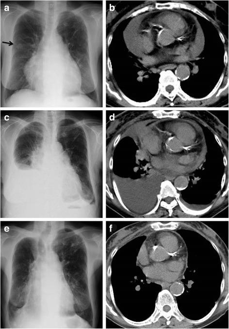
A multi-panel medical comparison chart featuring longitudinal chest X-rays (a, c, e) and corresponding axial chest computed tomography (CT) scans (b, d, f) illustrating the clinical progression and resolution of polyserositis associated with Meigs’ syndrome. Panel (a) shows a posteroanterior chest X-ray with cardiac enlargement and a right-sided pulmonary nodule (arrow). The adjacent CT scan (b) confirms a significant, large-volume pericardial effusion. Panel (c) demonstrates clinical progression on X-ray with a large right-sided pleural effusion and blunting of the left costophrenic angle, indicating bilateral involvement. The corresponding CT scan (d) shows a partial decrease in the pericardial effusion but reveals accompanying pleural effusions. Panels (e) and (f) display follow-up imaging eight months after surgical removal of an ovarian fibroma, showing complete resolution of the cardiomegaly, pleural effusions, and pericardial fluid. This clinical series highlights the diagnostic hallmarks of Meigs’ syndrome, characterized by the triad of benign ovarian tumor, ascites, and pleural/pericardial effusions that resolve following tumor excision.
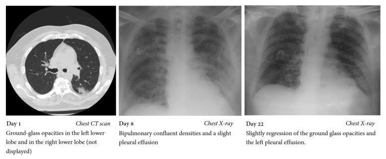
This composite educational image illustrates the progression and partial resolution of pulmonary pathology in a patient with COVID-19 ARDS and secondary fungal infection. Panel 1 (Day 1) shows an axial chest CT scan section demonstrating localized ground-glass opacities (GGOs) primarily in the posterior aspect of the left lower lobe. Panel 2 (Day 8) is an anteroposterior (AP) chest X-ray revealing significant disease progression, characterized by extensive bipulmonary confluent densities and signs of a slight bilateral pleural effusion, consistent with peak Acute Respiratory Distress Syndrome (ARDS) manifestation. Panel 3 (Day 22) is a follow-up AP chest X-ray showing slight regression of the bilateral confluent opacities and a reduction in the volume of the left pleural effusion. This temporal comparison highlights common radiological patterns in viral pneumonia, including the transition from focal ground-glass findings on CT to diffuse consolidation on X-ray, and subsequent monitoring of treatment response or secondary complications.
Chest X-Ray Analysis - Kavita, 56Y | Breast Carcinoma Grade 3 | 16/17 Chemotherapy Cycles
Radiological Findings
- More confluent and complete on the right side (left side on the film as labeled "R" for patient's right)
- Both costophrenic angles are completely obliterated and not visible
- The diaphragmatic outlines are entirely obscured
- The meniscus sign cannot be assessed clearly due to the density
- The upper lung fields appear relatively preserved with visible lung markings
- The lung parenchyma in the upper zones shows no obvious focal consolidation or mass lesion in the visible portions
- Hilar regions appear somewhat prominent but are partially obscured
- The cardiac silhouette is partially visible in the mid-chest region
- Mediastinal widening is difficult to exclude given the bilateral effusions
- There is no obvious pneumomediastinum
- Visible upper ribs, clavicles, and upper thoracic spine appear intact
- No obvious lytic lesions visible in the imaged bony structures (though full assessment is limited)
- No subcutaneous emphysema noted
- Image quality is adequate for the clinical findings
Clinical Correlation and Differential Diagnosis
1. Malignant Pleural Effusion (Most Likely)
- Direct pleural metastatic seeding
- Lymphatic obstruction
- Tumor invasion of the pleura
2. Paramalignant Effusion
- Hypoalbuminemia/cachexia (common after multiple chemotherapy cycles) causing transudates
- Pulmonary embolism (cancer-related hypercoagulability) causing exudate
- Superior vena cava obstruction or lymphatic obstruction
- Fishman's Pulmonary Diseases, p. 1026
3. Chemotherapy-Related Pulmonary Toxicity
4. Congestive Heart Failure (Cardiotoxicity)
5. Pulmonary/Pleural Metastases
Why Absence of Fever is Clinically Significant
- Infective parapneumonic effusion / empyema
- Bacterial or viral pneumonia causing reactive effusion
- Septic pulmonary emboli
Recommended Next Steps
| Investigation | Rationale |
|---|---|
| Thoracentesis + pleural fluid analysis | Cytology, LDH/protein (Light's criteria), pH, glucose - to confirm malignant vs transudative vs infective |
| Pleural fluid cytology | May identify malignant cells from breast carcinoma |
| Echocardiography | Assess for anthracycline-induced cardiomyopathy and pericardial effusion |
| CT Chest (contrast) | Full assessment of lung parenchyma, pleura, mediastinum, and bony metastases once effusions are drained |
| Serum albumin + LFTs | Nutritional status and hepatic metastases |
| D-dimer / CTPA | Rule out pulmonary embolism |
| BNP/NT-proBNP | Cardiac biomarker for heart failure |
Summary Statement
Disclaimer: This is an AI-assisted radiological interpretation for educational support. All findings must be formally reported and acted upon by a licensed radiologist and treating oncologist.