
Pericardium layers
The pericardium has two main layers: the fibrous pericardium (outer) and the serous pericardium (inner). The serous pericardium is further divided into the parietal layer (lines the fibrous pericardium) and the visceral layer (epicardium, adheres to the heart surface). Between the two serous layers is the pericardial cavity, which contains a small amount of fluid (15-50 mL) that reduces friction.
Searching sources...
Reading File
Finding Sources
Reading File
Reading File
Loading Image
Here is a full breakdown of the pericardium layers, sourced from Gray's Anatomy for Students and the Color Atlas of Human Anatomy:
Layers of the Pericardium
The pericardium is a fibroserous sac surrounding the heart and the roots of the great vessels. It has two main components:
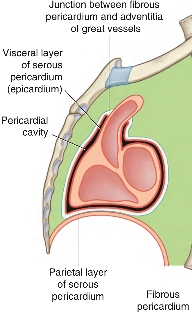
1. Fibrous Pericardium (Outer Layer)
- Tough, dense collagenous connective tissue
- Cone-shaped sac with its base on the diaphragm and apex continuous with the adventitia of the great vessels
- Attachments:
- Inferiorly: fused to the central tendon of the diaphragm (and a small left-side muscular area)
- Anteriorly: attached to the posterior sternum via sternopericardial ligaments
- Posteriorly: bands to the trachea and vertebral column
- Functions: anchors the heart in the thorax, limits cardiac distention, and prevents over-dilation
2. Serous Pericardium (Inner Layer)
A thin, double-layered closed sac sitting inside the fibrous pericardium. Like all serous membranes, it has two parts:
| Layer | Location | Also called |
|---|---|---|
| Parietal layer | Lines the inner surface of the fibrous pericardium | - |
| Visceral layer | Adheres directly to the heart surface and roots of great vessels | Epicardium |
The two layers are continuous with each other at the roots of the great vessels (at the sites of reflection).
3. Pericardial Cavity
- The potential space between the parietal and visceral serous layers
- Contains a small amount of serous fluid (normally ~15-50 mL) that acts as a lubricant
- Allows relatively uninhibited movement of the heart during contraction
Pericardial Sinuses (clinically important)
- Transverse pericardial sinus: a groove between the arterial (aorta + pulmonary trunk) and venous (pulmonary veins + venae cavae) reflections; surgeons use it to place clamps on the aorta during cardiac surgery
- Oblique pericardial sinus: a blind recess behind the left atrium, between the inferior pulmonary veins and inferior vena cava; a fluid collection here may not be detected on physical exam
Innervation & Blood Supply
- Innervation: phrenic nerve (C3-C5), vagus nerve, sympathetic trunk - the phrenic nerve travels within the fibrous pericardium
- Arterial supply: pericardiacophrenic artery (from internal thoracic artery)
- Venous drainage: pericardiacophrenic vein → brachiocephalic vein
Clinical Notes
- Pericarditis: inflammation can cause fibrinous adhesions between serous layers
- Pericardial effusion: excess fluid accumulates in the pericardial cavity (e.g., in pericarditis, malignancy, trauma)
- Cardiac tamponade: large effusion compresses the heart, impeding ventricular filling - a medical emergency
Sources: Gray's Anatomy for Students; Color Atlas of Human Anatomy Vol. 2, Internal Organs
This is a shared conversation. Sign in to Orris to start your own chat.