
Perineal membrane
Reading File
Searching sources...
Finding Sources
Finding Sources
Reading File
Reading File
Reading File
Reading File
Loading Image
Loading Image
Loading Image
Reading File
Here is a comprehensive account of the perineal membrane, drawn from Gray's Anatomy for Students and Campbell-Walsh-Wein Urology.
Perineal Membrane
Definition and Terminology
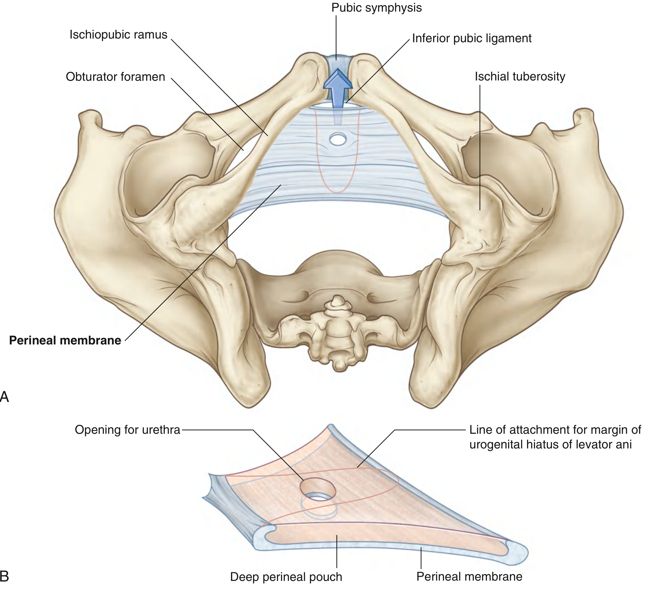
The perineal membrane (previously called the urogenital diaphragm) is a thick, triangular sheet of tough fibrous (deep) fascia that fills and spans the urogenital triangle of the perineum. It lies in a roughly horizontal plane and forms the floor of the deep perineal pouch above it and the roof of the superficial perineal pouch below it. - Gray's Anatomy for Students
Attachments
- Laterally: attached on each side to the ischiopubic rami (inferior pubic and ischial rami), spanning between the right and left pubic arch from the pubis to the ischial tuberosities.
- Posteriorly: has a free posterior border, which is anchored in the midline to the perineal body.
- Anteriorly: a small gap exists between the membrane and the inferior pubic ligament (associated with the pubic symphysis). The anterior edge of the perineal membrane is close to, but separated from, the inferior pubic ligament by this narrow interval.
Openings / Perforations
The membrane is pierced by:
- Urethra - passes through a circular hiatus as it travels from the pelvic cavity to the perineum (in both sexes).
- Vagina (in females only) - passes through a second hiatus just posterior to the urethral opening.
Deep Perineal Pouch (Deep Perineal Space)
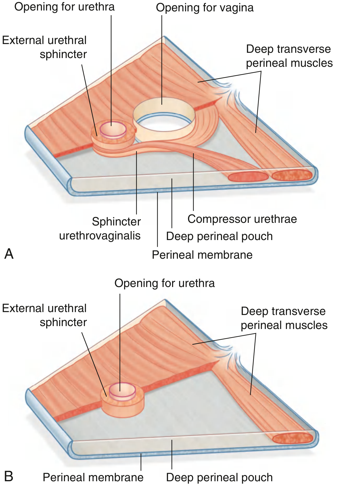
Immediately superior to the perineal membrane lies the deep perineal pouch. It is a thin region that is open superiorly - there is no distinct fascial layer separating it from pelvic structures above. It contains:
Muscles of the Deep Perineal Pouch
| Muscle | Origin | Insertion | Innervation | Function |
|---|---|---|---|---|
| External urethral sphincter | Inferior ramus of pubis (each side) + adjacent pouch walls | Surrounds membranous urethra | Perineal branches of pudendal nerve (S2-S4) | Compresses membranous urethra; relaxes during micturition |
| Deep transverse perineal | Medial aspect of ischial ramus | Perineal body | Perineal branches of pudendal nerve (S2-S4) | Stabilizes position of perineal body |
| Compressor urethrae (females only) | Ischiopubic ramus (each side) | Blends with partner anterior to urethra | Perineal branches of pudendal nerve (S2-S4) | Accessory sphincter of urethra |
| Sphincter urethrovaginalis (females only) | Perineal body | Passes forward lateral to vagina, meets partner anterior to urethra | Perineal branches of pudendal nerve (S2-S4) | Accessory sphincter of urethra; may help close vagina |
Neurovascular contents of the deep perineal pouch
- Branches of the pudendal nerve and internal pudendal vessels
- Bulbo-urethral (Cowper's) glands in males (their ducts pierce the perineal membrane to reach the urethra)
Relationships and Pouches
The perineal membrane divides the urogenital triangle into two spaces:
| Space | Location | Contents |
|---|---|---|
| Superficial perineal pouch | Below (inferior to) the perineal membrane | Roots/bulbs of external genitalia, superficial transverse perineal muscles, bulbospongiosus, ischiocavernosus, superficial perineal vessels/nerves |
| Deep perineal pouch | Above (superior to) the perineal membrane | External urethral sphincter, deep transverse perineal muscles, compressor urethrae + sphincter urethrovaginalis (females), neurovascular elements |
The Colles fascia (membranous layer of superficial fascia) attaches posteriorly to the posterior margin of the perineal membrane, creating the closed posterior wall of the superficial perineal pouch.
Functions
- Supports external genitalia - the roots/bulbs of the penis (or clitoris and vestibular bulbs) attach to the inferior surface of the membrane.
- Pelvic floor support - the parts of the membrane enclosed by the urogenital hiatus of the levator ani contribute to the pelvic floor and provide support to urogenital viscera above (bladder, uterus, vagina).
- Urethral continence - the external urethral sphincter in the deep perineal pouch compresses the membranous urethra.
- Stabilizes the perineal body - via attachment of the deep transverse perineal muscles at the posterior free border.
- Provides a passage for the urethra (and vagina in females) between the pelvis and perineum.
Perineal Body
At the midline along the free posterior border of the perineal membrane sits the perineal body - an ill-defined but functionally important fibromuscular node into which nearly every pelvic muscle and fascial structure converges:
- Deep transverse perineal muscles
- Superficial transverse perineal muscles
- External anal sphincter
- Bulbospongiosus muscles
- Sphincter urethrovaginalis (in females)
- Posterior end of the urogenital hiatus of levator ani
Damage to the perineal body (e.g. during childbirth or perineal prostatectomy) risks urinary and fecal incontinence. - Campbell-Walsh-Wein Urology
Sex Differences at a Glance
| Feature | Male | Female |
|---|---|---|
| Structures passing through | Urethra only | Urethra + vagina |
| Accessory sphincter muscles | None | Compressor urethrae + sphincter urethrovaginalis |
| Bulbo-urethral glands | Present (Cowper's glands) | Absent (greater vestibular glands are in superficial pouch) |
| Perineal membrane continuity | With prostate above | With arcus tendineus fasciae pelvis |
Describe parts blood supply nerve supply lymphatic drainage applied anatomy of stomach
Finding Sources
Finding Sources
Finding Sources
Reading File
Finding Sources
Reading File
Reading File
Reading File
Reading File
Finding Sources
Reading File
Finding Sources
Reading File
Reading File
Finding Sources
Reading File
Finding Sources
Loading Image
Loading Image
Finding Sources
Loading Image
Reading File
Now I have comprehensive information from multiple authoritative surgical and anatomy textbooks. Let me compile the full answer.
The Stomach - Complete Anatomy
1. Parts of the Stomach
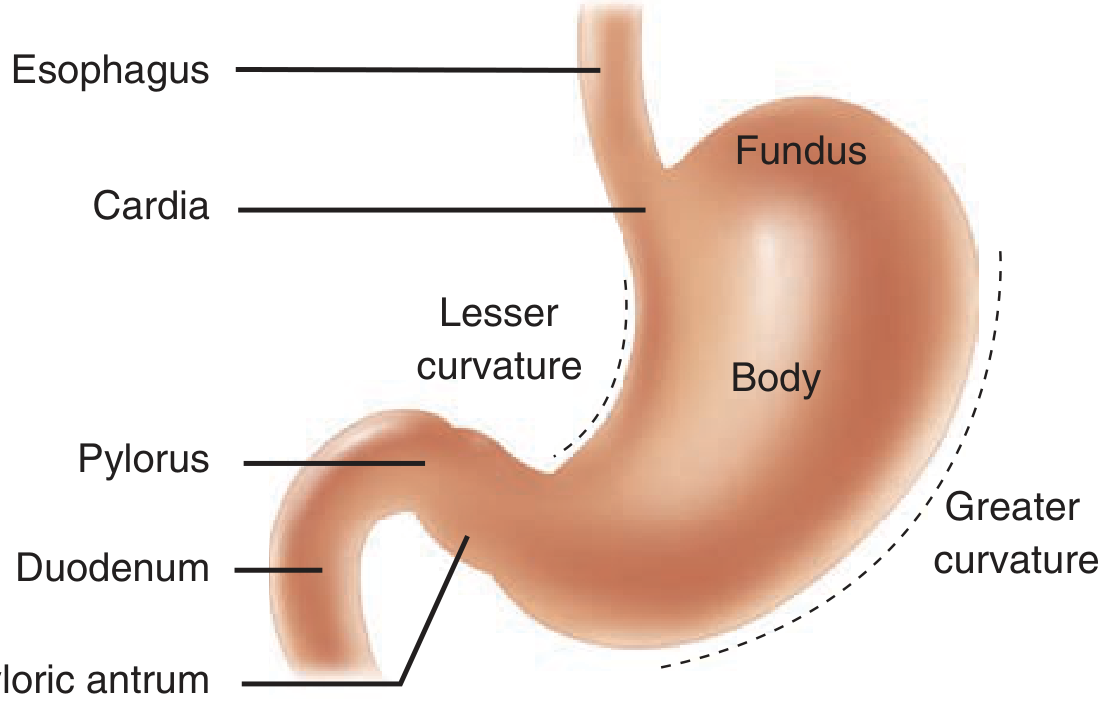
The stomach is the most dilated part of the GI tract, J-shaped, positioned in the epigastric, umbilical, and left hypochondrium regions. It has four regions: - Gray's Anatomy for Students
| Part | Description |
|---|---|
| Cardia | Surrounds the esophagogastric junction (cardial orifice) |
| Fundus | Dome-shaped region above the level of the cardial orifice; contacts the left hemidiaphragm |
| Body | The largest region, between the fundus and the pyloric part |
| Pyloric part | Divided into: (a) pyloric antrum (proximal wider part) and (b) pyloric canal (narrow distal part leading to the pylorus) |
The pylorus is the most distal point, marked by a pyloric constriction and a thickened ring of circular smooth muscle - the pyloric sphincter - which guards the pyloric orifice. It lies just to the right of midline at the transpyloric plane (L1).
Surface features:
- Lesser curvature (right/medial border) - attachment for lesser omentum; contains the angular incisure (incisura angularis), a bend that marks the junction of body and pyloric part
- Greater curvature (left/lateral border) - attachment for gastrosplenic ligament and greater omentum
- Cardiac notch - the superior angle where the esophagus enters the stomach
2. Blood Supply
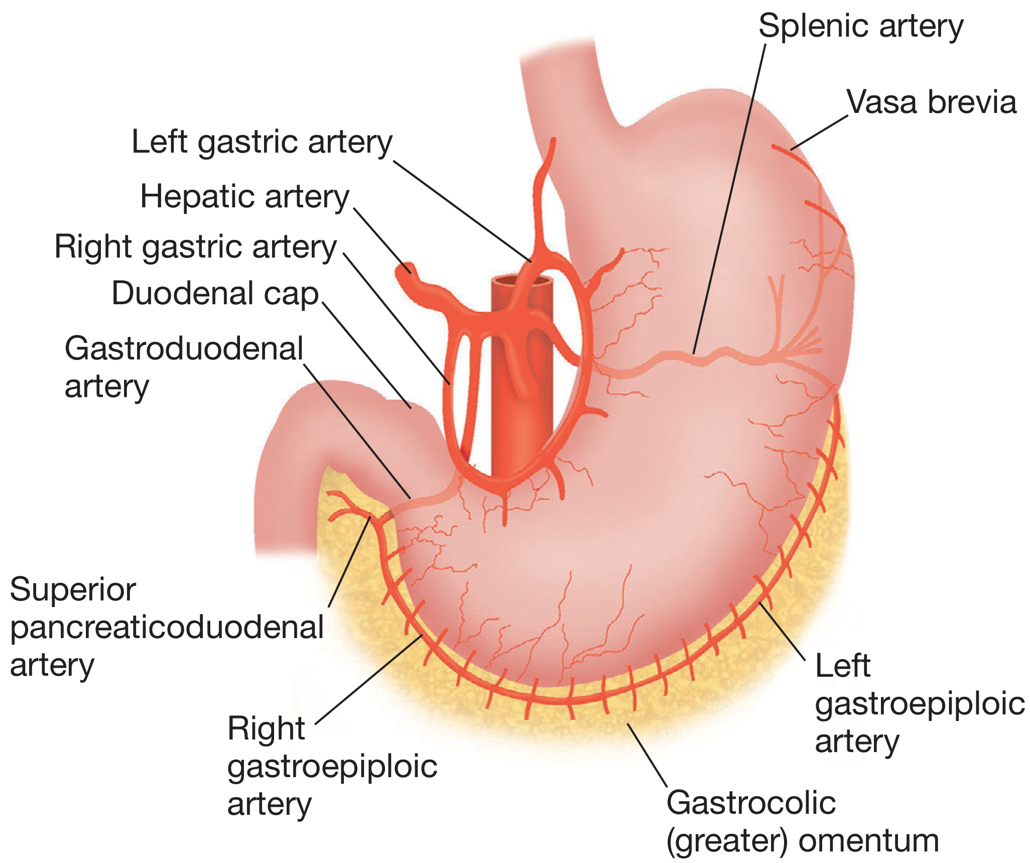
The stomach has one of the richest blood supplies of any abdominal organ, drawing from five sets of arteries, all ultimately from the celiac trunk. - Mulholland and Greenfield's Surgery
Arteries
Lesser curvature arcade:
| Artery | Origin | Territory |
|---|---|---|
| Left gastric artery | Directly from celiac trunk | Cardia and upper lesser curvature; also gives esophageal branches upward |
| Right gastric artery | Hepatic artery proper (or common hepatic artery) | Lower lesser curvature |
| These two anastomose to form the lesser curvature arcade |
Greater curvature arcade:
| Artery | Origin | Territory |
|---|---|---|
| Right gastro-omental (gastroepiploic) artery | Gastroduodenal artery (branch of common hepatic artery) | Right greater curvature |
| Left gastro-omental (gastroepiploic) artery | Splenic artery | Left greater curvature |
| These two anastomose to form the greater curvature arcade (often variably incomplete) |
Fundus:
- Short gastric arteries (vasa brevia) - 5-7 in number, arise from the terminal branches of the splenic artery and the left gastroepiploic artery; supply the fundus
Additional (variant):
- Posterior gastric artery - arises from the splenic artery (not always present); supplies the posterior gastric wall
Veins
The veins accompany the arteries and ultimately drain into the portal venous system:
- Veins along the lesser curvature (right and left gastric veins) drain into the portal vein
- Veins along the greater curvature (right and left gastroepiploic veins) drain into the splenic vein
- The left gastric (coronary) vein runs along the lesser curvature toward the esophagus, then passes left to right to join the portal vein. This vein becomes markedly dilated in portal hypertension, forming esophageal varices. - Bailey and Love's
3. Nerve Supply
The stomach has both intrinsic (enteric) and extrinsic nerve supply.
Extrinsic Innervation
Parasympathetic (Vagus Nerve - CN X)
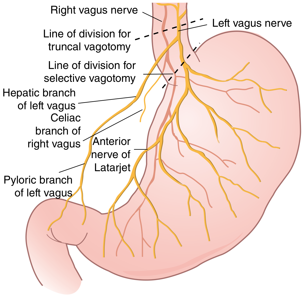
- The vagal plexus around the esophagus condenses into trunks at the esophageal hiatus
- The left (anterior) vagus - lies on the anterior surface of the stomach; gives off the hepatic branch (in gastrohepatic ligament to liver) and continues as the anterior nerve of Latarjet along the lesser curvature
- The right (posterior) vagus - lies on the posterior surface; gives off the celiac branch (to celiac plexus) and continues as the posterior nerve of Latarjet along the lesser curvature
- The nerves of Latarjet give segmental branches to the body of the stomach, then terminate near the angularis incisura as the "crow's foot," sending branches to the antro-pyloric region
- The criminal nerve of Grassi is the first branch of the right posterior vagus to the posterior fundus - it arises above the esophageal hiatus and is easily missed during vagotomy, causing recurrent ulcers if left undivided
- 90% of vagal fibers are afferent (sensory, to brain); efferent fibers originate in the dorsal nucleus of the medulla and synapse in the myenteric/submucosal plexuses
- Functions: receptive relaxation, gastric motility, gastric acid secretion, mucosal blood flow, cytoprotection - Sabiston
Sympathetic
- Originates from spinal levels T5-T10
- Travels in the greater splanchnic nerves to the celiac ganglion
- Postganglionic fibers travel along the blood vessels to innervate the stomach
- Contains afferent pain fibers (pain referred to the epigastrium / T5-T10 dermatomes) and motor fibers to the pyloric sphincter
Intrinsic (Enteric) Nervous System
| Plexus | Location | Notes |
|---|---|---|
| Myenteric plexus of Auerbach | Between circular and longitudinal muscle layers | Well-developed ganglia in antrum; relatively sparse in fundus |
| Submucosal plexus of Meissner | In submucosa | Relatively few ganglionic cells in stomach compared to rest of gut |
Neurotransmitters include: acetylcholine, serotonin, substance P, calcitonin gene-related peptide, bombesin, CCK, somatostatin - Sabiston
4. Lymphatic Drainage
Understanding gastric lymphatics is the key to radical surgery for gastric cancer. The lymphatics parallel the vasculature and drain into four zones: - Sabiston Textbook of Surgery
| Zone / Group | Drains | Primary Nodes | Ultimate Drainage |
|---|---|---|---|
| Superior gastric (left gastric) group | Upper lesser curvature, cardia, lower esophagus | Left gastric and paracardial nodes | Celiac nodes |
| Suprapyloric group | Antrum on lesser curvature, pylorus (superior) | Right gastric and right suprapancreatic nodes | Celiac nodes |
| Pancreaticolienal group | Upper greater curvature, fundus | Left gastroepiploic and splenic (hilar) nodes | Celiac nodes |
| Inferior gastric / Subpyloric group | Lower greater curvature, pylorus (inferior) | Subpyloric nodes (around gastroduodenal artery), right gastroepiploic nodes | Superior mesenteric nodes |
All four zones ultimately converge on the celiac nodes and then the thoracic duct.
Key surgical points:
- Gastric cancers may metastasize to any of the four nodal groups, regardless of tumor location
- The extensive submucosal lymphatic plexus means malignant cells can be found several centimeters beyond the gross tumor margin - this determines resection margins
- Lymphatics of the cardia communicate freely with those of the esophagus - Bailey and Love's
5. Applied Anatomy
Peptic Ulcer Disease
- Duodenal ulcers occur most often in the first part of the duodenum (duodenal cap/ampulla)
- A bleeding posterior duodenal ulcer erodes the gastroduodenal artery, which runs behind the first part of the duodenum - this is the anatomical basis for life-threatening hemorrhage
- Pain from peptic ulcers is referred to the epigastrium (T5-T10 dermatomes) via sympathetic afferents
Vagotomy (for peptic ulcer)
- Truncal vagotomy: division above the hepatic and celiac branches - removes all gastric vagal innervation but also denervates liver, biliary system, and gut; requires a drainage procedure (pyloroplasty or gastroenterostomy)
- Selective vagotomy: division below the hepatic and celiac branches - spares liver and celiac innervation but still requires drainage
- Highly selective (proximal gastric) vagotomy: divides the "crow's foot" branches to the body only, preserving antro-pyloric innervation - no drainage procedure needed
- The criminal nerve of Grassi must be divided or recurrent ulcers result
Gastric Cancer
- Spreads via: (1) direct invasion of adjacent organs (pancreas, liver, transverse colon), (2) lymphatic spread (D1/D2 node dissection), (3) haematogenous spread (liver most common), (4) transcoelomic/peritoneal spread
- Peritoneal spread may deposit on the ovaries (Krukenberg tumours) or at the umbilicus (Sister Joseph's nodule / Sister Mary Joseph nodule)
- Late detection leads to poor prognosis: overall 5-year survival is only 5-20% - Gray's Anatomy for Students
Portal Hypertension
- The left gastric (coronary) vein normally drains into the portal vein. In portal hypertension, it becomes dilated and reverses flow, creating a portosystemic anastomosis at the gastroesophageal junction - forming esophageal and gastric varices prone to catastrophic hemorrhage
Gastric Conduit (Esophageal Surgery)
- After esophagectomy, the stomach is the preferred organ for reconstruction - it is tubularized using the right gastroepiploic arcade
- The rich dual-curvature blood supply means even a tubularized stomach survives well on a single pedicle (right gastroepiploic artery)
- The left gastroepiploic arcade is often variably incomplete, which is relevant when planning the conduit
Short Gastric Vessels in Splenectomy
- The short gastric arteries (vasa brevia) run in the gastrosplenic ligament. During splenectomy they must be ligated carefully to avoid bleeding and inadvertent gastric injury
Nerve supply of tounge
Finding Sources
Finding Sources
Reading File
Reading File
Reading File
Finding Sources
Reading File
Reading File
Finding Sources
Finding Sources
Finding Sources
Reading File
Finding Sources
Loading Image
Reading File
I now have comprehensive information from multiple authoritative sources. Here is the full account.
Nerve Supply of the Tongue
The tongue has a complex nerve supply involving five cranial nerves (V, VII, IX, X, XII), reflecting its embryological origin from multiple pharyngeal arches. It is useful to divide the supply into sensory (general + special/taste) and motor.
Embryological Basis
The tongue develops from the 1st, 3rd, and 4th pharyngeal arches. Each arch brings its own nerve:
-
1st arch → mandibular nerve (V3) - general sensation, anterior 2/3
-
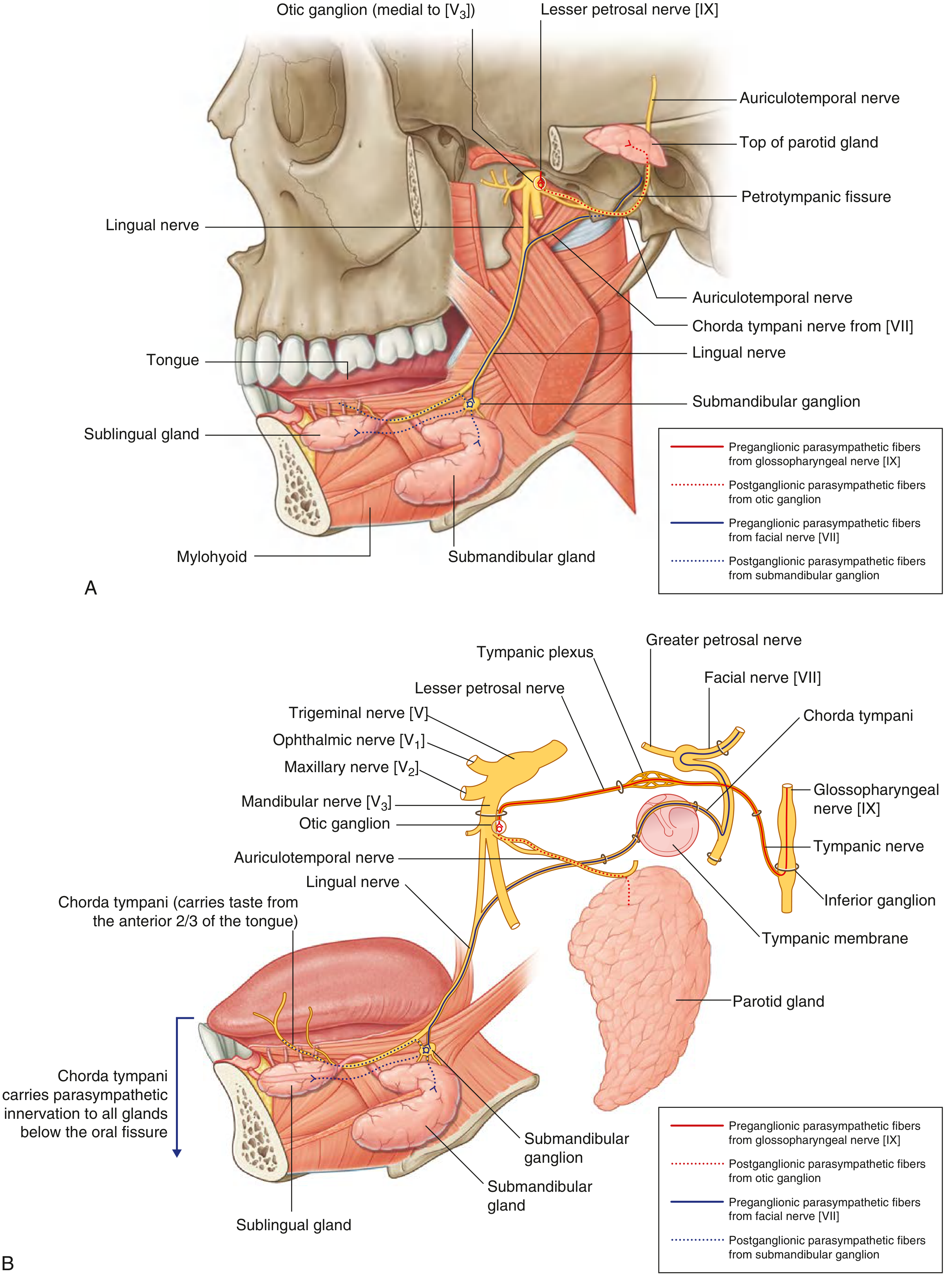
2nd arch (facial, CN VII) → chorda tympani - taste, anterior 2/3 (the 2nd arch is overgrown by the 3rd, so CN VII doesn't supply mucosa, only taste buds)
-
3rd arch → glossopharyngeal (CN IX) - sensation + taste, posterior 1/3
-
4th arch → vagus (CN X) - small area near epiglottis/valleculae
-
Occipital somite migrants bring hypoglossal (CN XII) for muscle supply
-
The Developing Human (Moore's Embryology)
I. Sensory Nerve Supply
The tongue is divided into anterior 2/3 (presulcal / oral part) and posterior 1/3 (postsulcal / pharyngeal part) by the sulcus terminalis and the row of circumvallate (vallate) papillae.
A. General Sensation (Touch, Pain, Temperature)
| Region | Nerve | Origin |
|---|---|---|
| Anterior 2/3 | Lingual nerve (branch of V3, mandibular division of trigeminal) | Infratemporal fossa |
| Posterior 1/3 | Lingual branch of glossopharyngeal nerve (CN IX) | Passes around posterior border of stylopharyngeus |
| Valleculae (recess between median and lateral glossoepiglottic folds) | Internal laryngeal nerve (branch of superior laryngeal nerve, CN X) | Pierces thyrohyoid membrane |
B. Special Sensation (Taste)
| Region | Nerve | Pathway |
|---|---|---|
| Anterior 2/3 (fungiform and foliate papillae) | Chorda tympani (branch of CN VII - facial nerve) | Taste fibers travel via lingual nerve → chorda tympani → geniculate ganglion → nucleus of tractus solitarius (NTS) |
| Circumvallate papillae (anterior to sulcus terminalis) | Glossopharyngeal (CN IX) | Despite being on anterior side of sulcus, these papillae are supplied by CN IX (pulled forward during development) |
| Posterior 1/3 (foliate and circumvallate papillae, base of tongue, vallecula, posterior oropharynx) | Glossopharyngeal (CN IX) | → inferior petrosal ganglion → NTS |
| Epiglottis and laryngeal surface | Vagus (CN X) via internal laryngeal nerve | → NTS |
- K.J. Lee's Essential Otolaryngology; Developing Human Embryology
II. Motor Nerve Supply
Hypoglossal Nerve (CN XII) - ALL tongue muscles EXCEPT palatoglossus
All intrinsic and extrinsic tongue muscles are supplied by the hypoglossal nerve (CN XII):
| Muscle | Type | Function |
|---|---|---|
| Genioglossus | Extrinsic | Protrudes tongue |
| Hyoglossus | Extrinsic | Depresses tongue |
| Styloglossus | Extrinsic | Retracts and elevates tongue |
| Superior longitudinal | Intrinsic | Shortens tongue, curls tip up |
| Inferior longitudinal | Intrinsic | Shortens tongue, curls tip down |
| Transverse | Intrinsic | Narrows and elongates tongue |
| Vertical | Intrinsic | Flattens tongue |
Course of the hypoglossal nerve:
- Exits skull via hypoglossal canal (alongside C1-2 fibers)
- Crosses over the internal and external carotid arteries
- Passes between mylohyoid and hyoglossus muscles
- Lies below the submandibular gland, submandibular duct, and lingual nerve
- Passes deep to the lingual nerve along the genioglossus toward the tip of the tongue
- Gives branches to styloglossus, hyoglossus, and genioglossus; slender rami ascend into intrinsic muscles
The lingual vein (veins of Ranine) travels with the hypoglossal nerve, making the nerve vulnerable during attempts to control lingual hemorrhage. - Scott-Brown's Otorhinolaryngology
Exception - Palatoglossus
- Palatoglossus (the muscle forming the anterior pillar of the fauces) is supplied by the vagus nerve (CN X) via the pharyngeal branch to the pharyngeal plexus - NOT the hypoglossal nerve.
- This is because palatoglossus is functionally a muscle of the soft palate, not the tongue.
III. Autonomic Supply
| Component | Nerve | Function |
|---|---|---|
| Parasympathetic to lingual glands | Chorda tympani (CN VII) → submandibular ganglion → postganglionic fibers re-enter lingual nerve | Secretomotor to submandibular and sublingual salivary glands |
| Sympathetic to lingual glands and vessels | Plexuses around lingual artery (from superior cervical ganglion via carotid plexus) | Vasoconstriction, reduced secretion |
Summary Diagram (Simplified)
TONGUE
├── Anterior 2/3
│ ├── General sensation → Lingual nerve (V3)
│ ├── Taste (fungiform/foliate) → Chorda tympani (CN VII via lingual nerve)
│ └── Taste (circumvallate) → CN IX (exception!)
│
├── Posterior 1/3
│ ├── General sensation → CN IX (glossopharyngeal)
│ └── Taste → CN IX
│
├── Valleculae / epiglottis
│ └── General + taste → Internal laryngeal nerve (CN X)
│
└── MOTOR (all muscles)
├── ALL tongue muscles → CN XII (hypoglossal)
└── Palatoglossus ONLY → CN X (vagus via pharyngeal plexus)
IV. Applied Anatomy / Clinical Correlates
1. Lingual Nerve Injury
The lingual nerve is at greatest risk during surgical removal of impacted lower third molars (wisdom teeth) using a lingual approach - it is closely applied to the periosteum of the medial mandible at the roots of the third molar. - Scott-Brown's; Gray's Anatomy
- Lesion proximal to where chorda tympani joins: Loss of general sensation (touch, pain, temperature) from anterior 2/3 of tongue + loss of taste from anterior 2/3 + reduced submandibular and sublingual salivary secretion
- Lesion distal to where chorda tympani joins (i.e. after it has already joined): Loss of general sensation from anterior 2/3 only; taste and salivary loss also occur
2. Hypoglossal Nerve Palsy (CN XII)
- Lower motor neuron lesion: tongue deviates toward the side of the lesion on protrusion (due to paralysis of ipsilateral genioglossus, which normally pushes the tongue to the opposite side)
- Chronic LMN lesion: ipsilateral wasting and fasciculation of tongue
- UMN lesion: deviation may be contralateral; no wasting
- Relevant in head and neck cancer - CN XII involvement by primary tumor or level I/II neck nodes causes tongue weakness and deviation - Mulholland and Greenfield's Surgery
3. Surgical Landmark
The lingual vein (veins of Ranine) travels alongside the hypoglossal nerve in the floor of the mouth - attempts to control lingual bleeding may inadvertently injure CN XII.
4. Taste Pathway Central Connections
All taste fibers (CN VII, IX, X) ultimately synapse in the nucleus of tractus solitarius (NTS) in the brainstem, then relay to the thalamus (VPM nucleus) and on to the cortical taste area in the parietal lobe (postcentral gyrus, area 43).
This is a shared conversation. Sign in to Orris to start your own chat.