
Can U show me the lateral view of brain stem having and structure above mid brain with ventucles in both lateral and frontal view ?
Reading File
The brainstem consists of the midbrain, pons, and medulla oblongata, with the diencephalon (thalamus/hypothalamus) sitting above it. The ventricular system includes the lateral ventricles, 3rd ventricle (between thalami), cerebral aqueduct (through midbrain), and 4th ventricle (between pons/medulla and cerebellum).
Searching sources...
Finding Sources
Finding Sources
Searching the Web
brainstem lateral view anatomy diagram midbrain pons medulla ventricles labeled
Finding Sources
Finding Sources
Loading Image
Loading Image
Reading File
Searching the Web
brain ventricles lateral view frontal view labeled diagram 3rd 4th lateral ventricle cerebral aqueduct
Loading Image
Loading Image
Loading Image
Loading Image
Loading Image
Here is a complete visual breakdown of the brainstem (lateral view) with structures above the midbrain, plus the ventricular system in both lateral and frontal views.
Brainstem - Lateral (Sagittal) View with Structures Above Midbrain
This diagram from Neuroscience: Exploring the Brain (5th Ed.) shows the midsagittal cut of the human brain, with the brainstem parts and all structures clearly labeled:
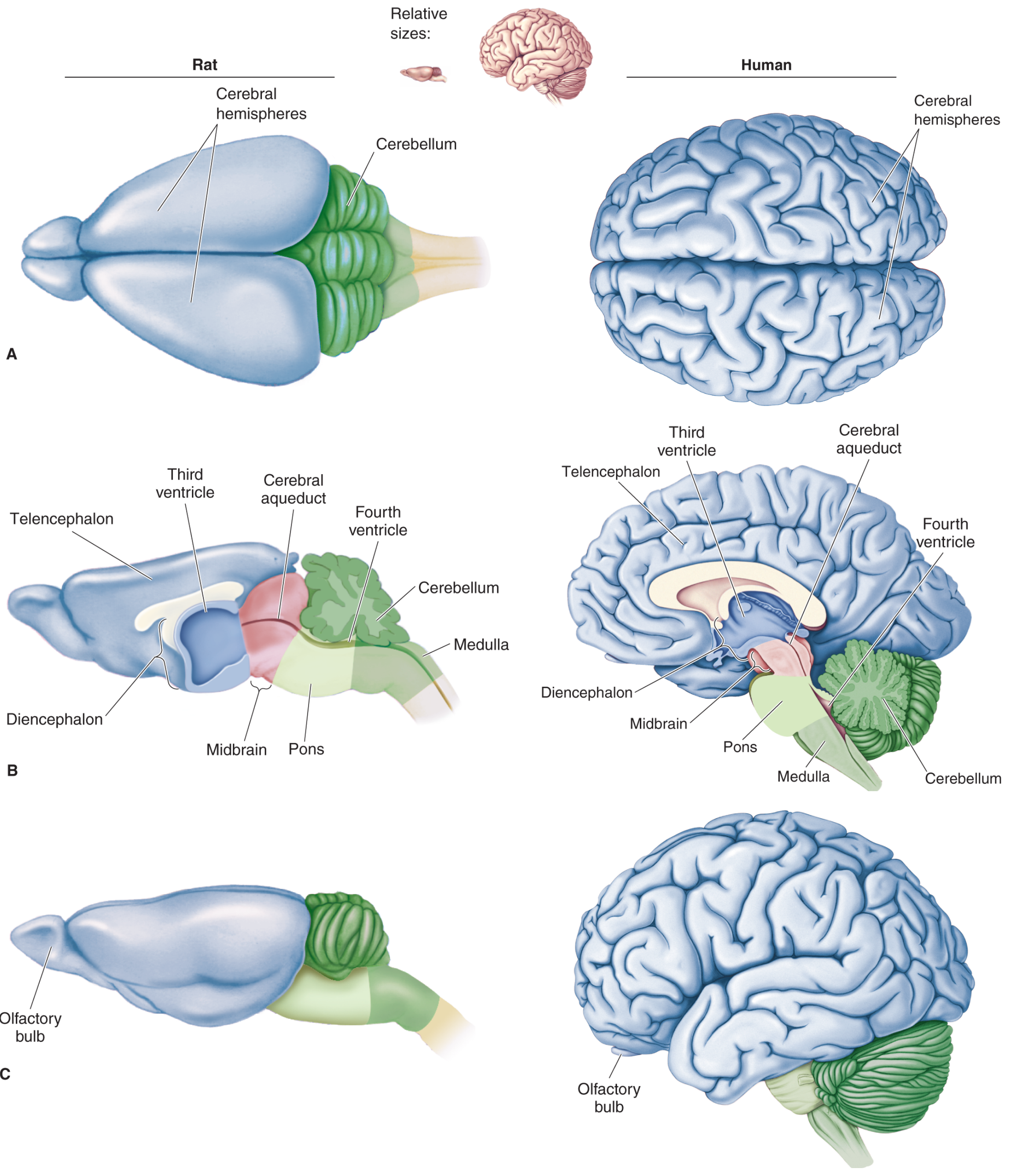
Key structures from top to bottom (medial/lateral view):
| Structure | Location | What's Inside/Associated |
|---|---|---|
| Telencephalon (Cerebral cortex) | Topmost | Lateral ventricles |
| Diencephalon (Thalamus + Hypothalamus) | Above midbrain | 3rd ventricle |
| Midbrain (Mesencephalon) | Upper brainstem | Cerebral aqueduct |
| Pons | Middle brainstem | Upper 4th ventricle |
| Medulla oblongata | Lower brainstem | Lower 4th ventricle |
| Cerebellum | Posterior | 4th ventricle (posterior wall) |
Brainstem Lateral View - Detailed (Colored)
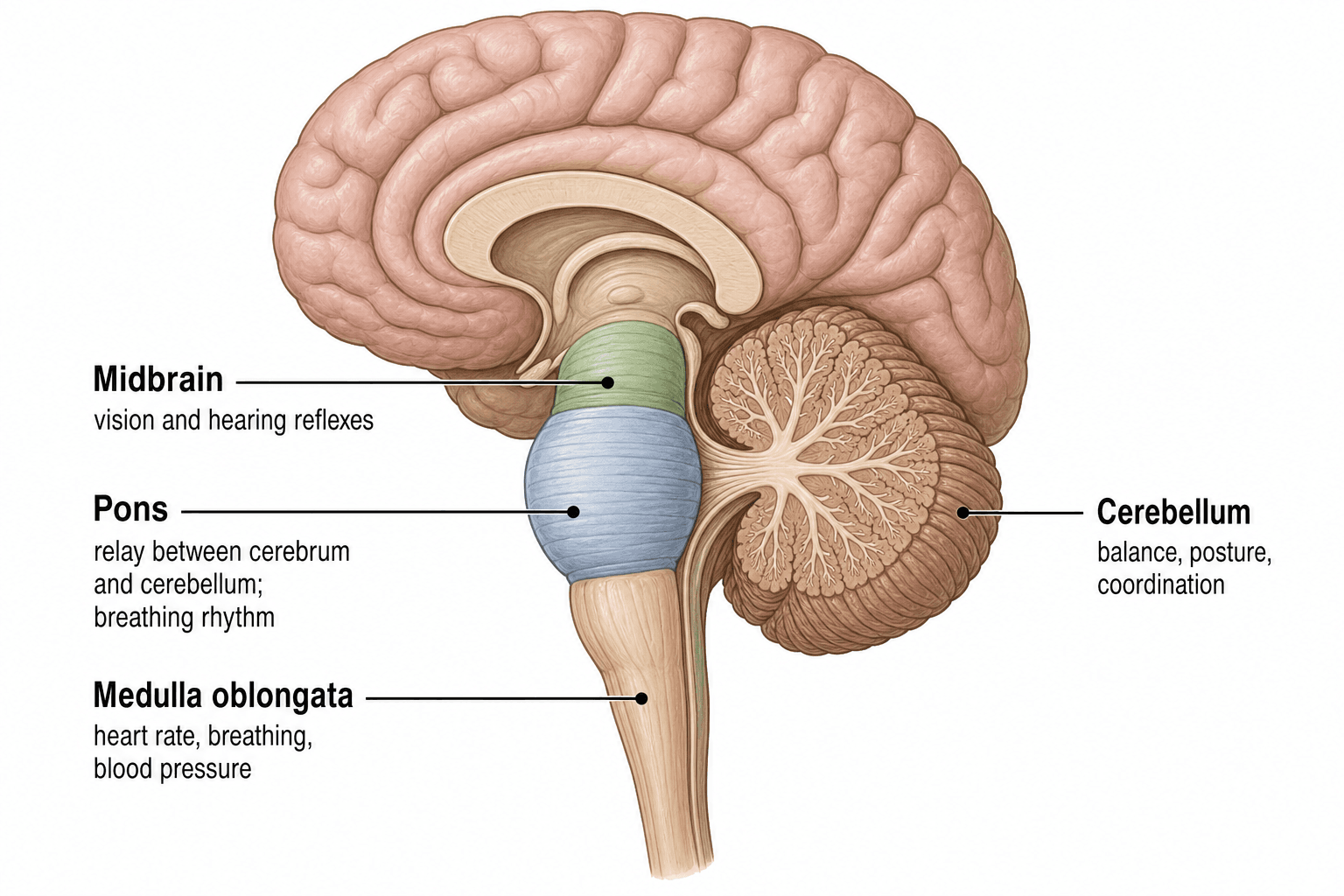
- Midbrain (green) - most superior brainstem part; sits just below the diencephalon/thalamus
- Pons (blue) - bulging anterior structure; relay between cerebrum and cerebellum
- Medulla oblongata - most inferior part; merges with spinal cord
Ventricular System - Lateral View
This diagram from TeachMeAnatomy shows all four ventricles in a lateral (sagittal) projection:
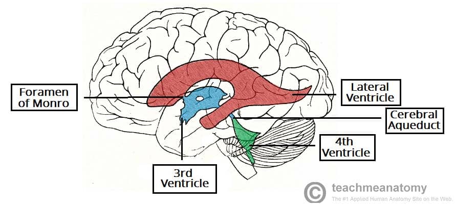
CSF Flow pathway (lateral view):
Lateral ventricles → Foramen of Monro → 3rd ventricle → Cerebral aqueduct (through midbrain) → 4th ventricle → Subarachnoid space
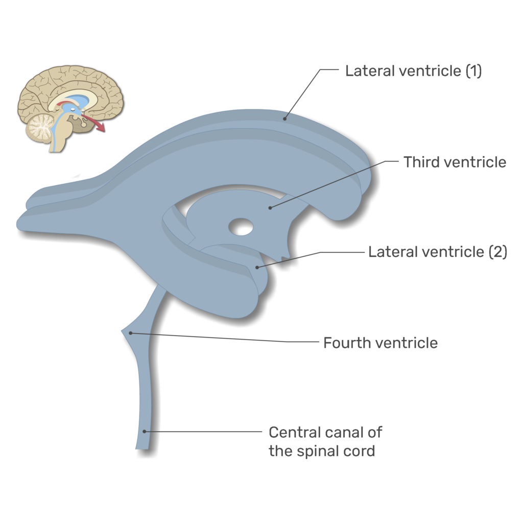
Ventricular System - 3D Frontal/Lateral Oblique View
Summary Table - Ventricular System & Corresponding Brain Structures
(From: Neuroscience: Exploring the Brain, 5th Ed., Table 7.3)
| Ventricle | Corresponding Brain Structure |
|---|---|
| Lateral ventricles (x2) | Cerebral cortex, basal telencephalon |
| 3rd ventricle | Thalamus, Hypothalamus (diencephalon) |
| Cerebral aqueduct | Tectum, Midbrain tegmentum |
| 4th ventricle | Cerebellum, Pons, Medulla |
Key clinical point: The cerebral aqueduct (of Sylvius) passing through the midbrain is the narrowest point in the entire ventricular system - obstruction here causes non-communicating (obstructive) hydrocephalus, which is why midbrain tumors or tectal lesions can rapidly cause raised intracranial pressure.
This is a shared conversation. Sign in to Orris to start your own chat.