
How to prevent tounge fall
- Place the patient in a lateral (recovery) position - this is the most basic and effective method
- Prone positioning also prevents tongue from falling back
- Avoid supine positioning in unconscious/sedated patients
- Head-tilt chin-lift - extends the neck and lifts the tongue off the posterior pharynx
- Jaw thrust - preferred when cervical spine injury is suspected
- Triple airway maneuver - combines head tilt, mouth open, and jaw thrust
- Oropharyngeal airway (Guedel airway) - holds the tongue forward mechanically
- Nasopharyngeal airway - tolerated better in semi-conscious patients
- Laryngeal mask airway (LMA) - for deeper sedation/anesthesia
- Endotracheal intubation for complete airway protection
- Prone positioning
- Nasopharyngeal airway
- Tongue-lip adhesion (surgical)
- Mandibular distraction osteogenesis
tongue fall prevention airway management glossoptosis
tongue fall airway obstruction prevention oropharyngeal airway
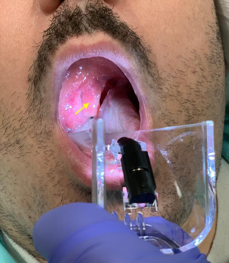
A clinical photograph captures an oropharyngeal examination of a patient using a clear plastic laryngoscope or tongue depressor. The primary finding, indicated by a yellow arrow, is a grossly edematous and erythematous uvula (uvulitis). The uvula is significantly enlarged and swollen, appearing bulbous and distorted, which contributes to significant narrowing of the oropharyngeal airway space. The surrounding mucosa of the soft palate and posterior pharynx exhibits generalized erythema, suggesting acute inflammation. This visual evidence is characteristic of conditions such as Quincke's edema (isolated uvular angioedema) or anaphylaxis-related airway involvement. The palatine tonsils and tongue are visible, with the latter being depressed to allow for a clear view of the upper airway. This image serves as a teaching tool for recognizing emergency airway obstruction signs in otolaryngology and emergency medicine.
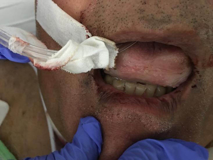
This clinical photograph displays a close-up view of a patient's oral cavity exhibiting severe macroglossia (tongue swelling). The tongue is markedly enlarged and edematous, occupying nearly the entire visible oropharyngeal space and obscuring the soft palate and uvula. This presentation is consistent with a Mallampati Class IV airway, indicating a high risk for airway obstruction. The tongue surface appears relatively smooth with focal areas of pallor and erythema. A clear medical tube, likely an endotracheal or orogastric tube, is positioned at the corner of the mouth, secured with white gauze that shows minor serosanguinous staining. The surrounding perioral region and lips also show signs of swelling. This image serves as a critical educational example of acute angioedema, frequently associated with ACE inhibitor use (such as lisinopril), highlighting the importance of emergent airway management in clinical practice.
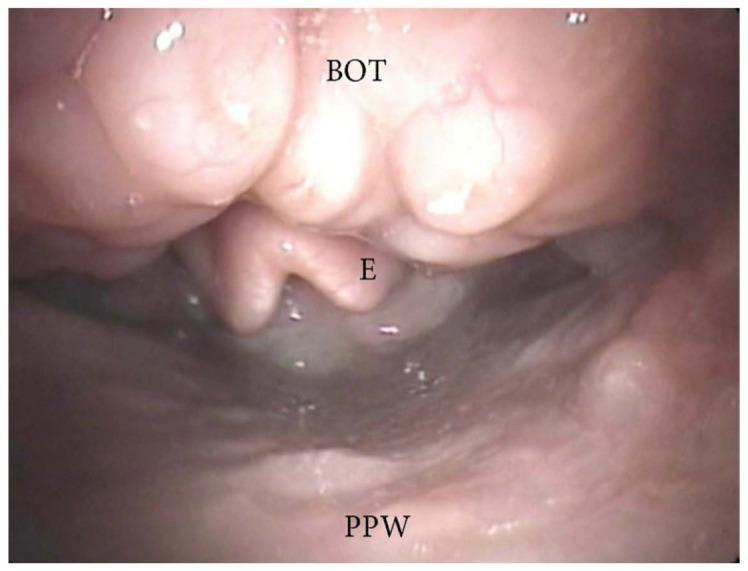
This diagnostic endoscopic image, obtained during drug-induced sleep endoscopy (DISE), illustrates the oropharyngeal airway and its anatomical landmarks. The base of the tongue (BOT) is positioned in the superior aspect of the frame, showing significant tissue hypertrophy of the lingual tonsils. This enlargement causes a marked anteroposterior narrowing of the airway space. Inferior to the BOT, the epiglottis (E) is partially visible but obscured by the overriding base of the tongue. The posterior pharyngeal wall (PPW) is seen at the bottom of the image, maintaining a smooth, pink mucosal appearance. The visual evidence confirms a pattern of airway obstruction driven by lingual tonsil hypertrophy, a common finding in patients with obstructive sleep apnea (OSA). The image demonstrates the spatial relationship between the lingual tonsillar tissue and the laryngeal inlet, highlighting the reduction in the cross-sectional area of the pharyngeal lumen.
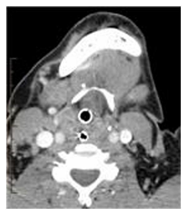
This diagnostic image is an axial computed tomography (CT) scan of the neck at the level of the oropharynx and mandible. The primary finding is a large, bulky soft tissue mass centered at the base of the tongue, which demonstrates heterogeneous density. This mass causes significant posterior displacement of tissues, resulting in near-complete obstruction of the oropharyngeal airway. An endotracheal or tracheostomy tube is visible within the narrowed airway lumen, characterized by its hyperdense circular wall and central air-filled (black) lumen, indicating active airway management. The surrounding anatomy shows the mandible anteriorly, the cervical vertebral body posteriorly, and the major vascular structures of the neck laterally. The subcutaneous fat appears appropriately hypodense, while the neck musculature maintains a typical intermediate density. No significant cervical lymphadenopathy is visualized in this specific plane. This image serves as a clinical example of upper airway obstruction secondary to base-of-tongue pathology, relevant for otolaryngology and emergency medicine curricula.
Preventing Tongue Fall (Glossoptosis)
1. Positioning Techniques
- Lateral (recovery) position - most basic and universally effective; gravity pulls the tongue forward and away from the posterior pharynx
- Prone positioning - allows the tongue to fall away from the posterior pharyngeal wall; commonly used in neonates with Pierre Robin Sequence (PRS)
- Avoid supine positioning in unconscious or sedated patients whenever possible
2. Manual Airway Maneuvers
| Maneuver | How | When |
|---|---|---|
| Head-tilt chin-lift | Tilt head back + lift chin forward | Standard first-line in non-trauma patients |
| Jaw thrust | Push mandible forward from angles | When cervical spine injury suspected |
| Triple airway maneuver | Head tilt + mouth open + jaw thrust | Deep unconsciousness, maximum effect |
3. Airway Adjuncts (Artificial Airways)
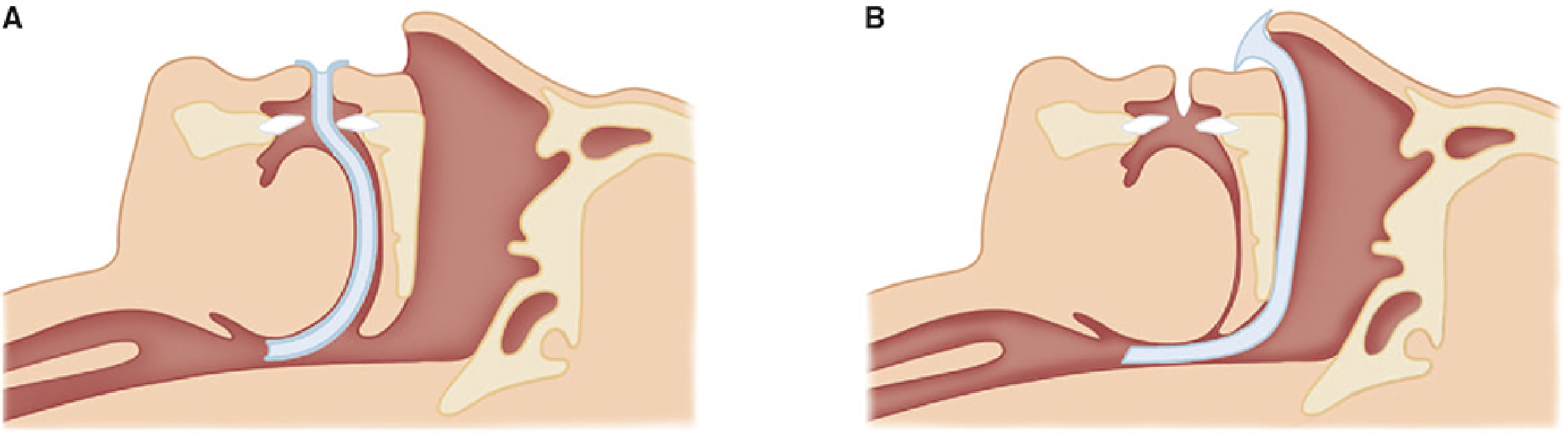
- Follows the curvature of the tongue, pulling it and the epiglottis away from the posterior pharyngeal wall
- Sizes: Small (80 mm, Guedel No. 3), Medium (90 mm, No. 4), Large (100 mm, No. 5)
- Only tolerated in unconscious patients - awake/semi-awake patients will gag or develop laryngospasm
-
Passes through the nose, extends just above the epiglottis
-
Better tolerated in semi-conscious patients
-
Length estimated as distance from nares to the ear meatus
-
Use with caution in anticoagulated patients (epistaxis risk) and basilar skull fractures
-
Morgan & Mikhail's Clinical Anesthesiology, 7e
4. Definitive Airway
- Endotracheal intubation (orotracheal preferred) - indicated for apnea, altered mental status, inability to maintain oxygenation, or impending airway compromise
- LMA (Laryngeal Mask Airway) - intermediate option in deeper sedation/anesthesia
- Cricothyroidotomy / Tracheostomy - when intubation fails or extensive facial injury precludes standard access
5. Special Situation: Neonates & Pierre Robin Sequence (PRS)
- Prone positioning - first-line; simple and effective
- Nasopharyngeal airway - augments positioning when prone positioning alone fails; requires periodic suctioning
- Glossopexy (lip-tongue adhesion) - surgical adhesion of tongue to the lower lip, used when positioning fails; dehiscence occurs in up to 56% of patients
- Mandibular distraction osteogenesis (MDO) - now advocated as primary treatment; bilateral mandibular osteotomies with distraction devices lengthen the mandible over weeks, advancing the tongue base forward and permanently relieving obstruction
- Cummings Otolaryngology Head and Neck Surgery
Summary Table
| Setting | Priority Intervention |
|---|---|
| Anesthesia recovery | Lateral position + oral/nasal airway |
| Trauma/emergency | Jaw thrust + chin lift, then intubation |
| Unconscious patient | Recovery position + oropharyngeal airway |
| Semi-conscious | Nasopharyngeal airway |
| Neonate with PRS | Prone positioning → NPA → glossopexy → MDO |
| Sleep apnea | CPAP, mandibular advancement devices |