
Give me Complete clinical, surgical and surface anatomy of the Anus and rectum
anatomy of rectum and anal canal diagram cross section
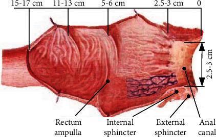
This anatomical diagram illustrates a longitudinal cross-section of the human rectum and anal canal, emphasizing spatial dimensions and clinical landmarks relevant for transrectal procedures. The illustration identifies the rectum ampulla as the proximal, dilated portion, transitioning distally toward the anal canal. Key muscular structures labeled include the internal sphincter and external sphincter. A horizontal scale at the top provides anatomical distance markers: 0 cm at the anal opening, 2.5-3 cm at the anal canal, 5-6 cm at the sphincter level, 11-13 cm at the mid-rectum, and 15-17 cm at the proximal rectal boundary. Vertical arrows also indicate the anal canal depth (2.5-3 cm). The diagram highlights the mucosal surface texture and the relative narrowness of the anal canal compared to the wider rectal ampulla, providing critical orientation for the insertion of diagnostic tools such as transrectal ultrasound (TRUS) probes used in prostate biopsies.
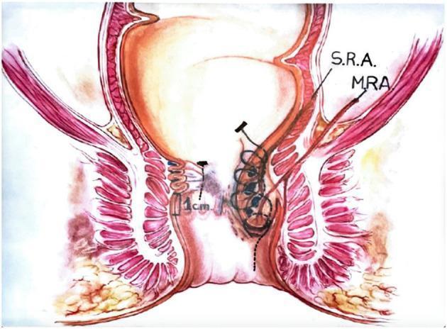
This medical illustration depicts a coronal cross-section of the rectum and anal canal, demonstrating the anatomical landmarks and surgical technique for a Transanal Hemorrhoidal Dearterialization (THD) 'Anolift' procedure. The diagram highlights the internal and external anal sphincters, the rectal mucosa, and key vascular structures including the Superior Rectal Artery (S.R.A.) and Middle Rectal Artery (M.R.A.). On the right side of the illustration, a hemorrhoidal prolapse is shown being addressed with a surgical suture. The suture is anchored superiorly with a locking button and continues as a running plication through the mucosa and submucosa, stopping proximal to the dentate line. This visualizes the mechanism of mucopexy, where the prolapsed tissue is lifted and repositioned proximally by tightening the suture to create a concertina effect on the rectal wall. A scale marker of 1 cm is provided near the anal transition zone for spatial orientation. This diagram is intended for surgical education regarding the management of Grade III and IV internal hemorrhoids.
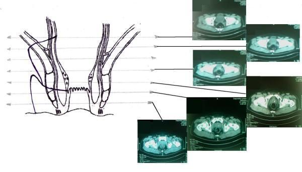
This composite educational image features a Comparison Chart and Diagnostic Image set correlating perianal anatomy with radiological findings. On the left, a coronal anatomical diagram of the rectum and anal canal illustrates two fistulous tracts originating from a shared internal orifice in the rectal wall. One tract demonstrates an extrasphincteric path, while the other shows the placement of a surgical seton. The diagram highlights key landmarks including the internal and external anal sphincters, the levator ani muscle, and the dentate line. Horizontal reference lines extend from different anatomical levels of the schematic to a series of six axial CT scans (labeled as MRI in context) on the right. These cross-sectional diagnostic images display the pelvic floor, rectal lumen, and surrounding soft tissues at sequential heights, facilitating a 3D understanding of the fistula's complex trajectory relative to the pelvic musculature. This material is designed for advanced medical education in colorectal surgery and radiology, focusing on the diagnosis and management of complex anal fistulas.
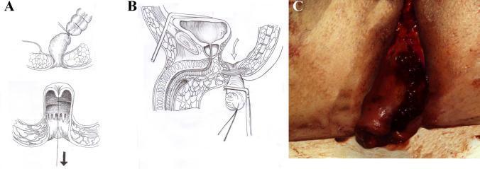
This composite educational image illustrates a surgical technique for rectal cancer resection, specifically focusing on the eversion and stapling of the rectal stump. Panel A provides a schematic diagram showing the eversion of the rectum through the anal canal, with a tumor located on the mucosal surface. Panel B presents a sagittal cross-section anatomical illustration of the male pelvis, depicting the distal rectum pulled through the perineum. A linear stapler is shown positioned to provide a safe distal clearance margin from the tumor, while preserving pelvic structures like the bladder and prostate. Panel C is an intraoperative clinical photograph demonstrating the real-world application of the technique. It shows a dark red, fleshy, and irregular tissue mass—the everted rectal stump containing the tumor—protruding from the perianal region. The image series demonstrates key steps in a modified pull-through procedure aimed at achieving an oncologically safe margin before performing a low colorectal or coloanal anastomosis. This material is designed for surgical education in colorectal oncology and gastrointestinal surgery.
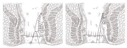
Anatomical diagram in a cross-sectional view illustrating the Transanal Hemorrhoidal Dearterialization (THD) with mucopexy procedure. The schematic is divided into two parts showing the progression of the surgical technique within the rectum and anal canal. The left side depicts a longitudinal continuous suture line being applied to the rectal mucosa and submucosa, targeting a prolapsed hemorrhoidal pile. The suture starts approximately 4-5 cm above the anal verge. The right side illustrates the effect of tying the suture, showing the clinical result where the redundant rectal tissue is plicated and lifted proximally (mucopexy), reducing the prolapse and repositioning the tissue within the distal rectum. Key anatomical features visible include the internal and external anal sphincters, the dentate line, and the rectal wall layers. This diagram serves as a surgical guide for colorectal residents and surgeons to visualize the lifting of pathologic piles during hemorrhoidal dearterialization.
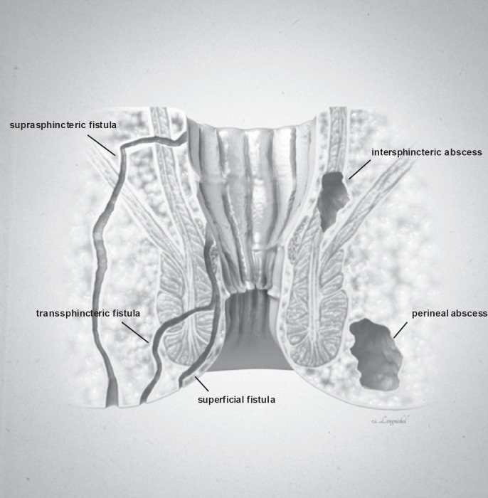
**Imaging Modality:** Medical illustration/Anatomical diagram. **Anatomical Region:** Coronal cross-section of the anorectal region, including the anal canal, rectum, internal and external anal sphincters, and surrounding perianal soft tissues. **Observed Pathology:** The illustration classifies various types of anorectal fistulas and abscesses based on their anatomical trajectory and location. **Characteristic Visual Features:** * **Fistula Tracts:** Three distinct epithelial-lined tracks are labeled. A **superficial fistula** is shown confined to the perianal skin and distal anal canal. A **transsphincteric fistula** is depicted crossing both the internal and external sphincter muscles into the ischioanal fossa. A **suprasphincteric fistula** is illustrated looping above the puborectalis muscle before descending to the skin surface. * **Abscess Formations:** Two localized collections of fluid/pus are identified. An **intersphincteric abscess** is positioned within the space between the internal and external sphincter muscles. A **perineal (perianal) abscess** is shown as a larger pocket located in the subcutaneous tissue adjacent to the anal verge. **Key Diagnostic Features:** The diagram emphasizes the relationship between inflammatory tracts and the sphincter complex, following the Parks classification system for fistula-in-ano. Suitable for clinical education on perianal sepsis and surgical planning.
pelvic floor muscles levator ani puborectalis sphincter anatomy
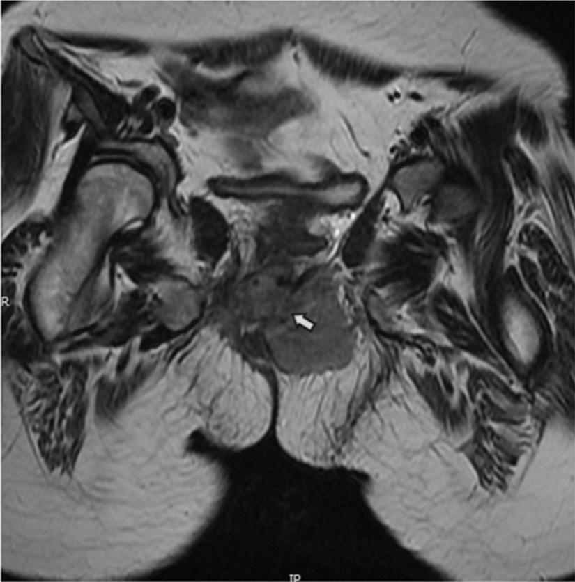
This diagnostic image is an axial T2-weighted magnetic resonance imaging (MRI) scan of the human pelvic region, specifically focusing on the pelvic floor anatomy. The image demonstrates a significant soft tissue mass representing a tumor, likely rhabdomyosarcoma, which has extensively infiltrated the pelvic musculature. Key findings include the replacement of the normal hypointense signal of the puborectalis muscle with a heterogeneous, hyperintense tumor signal. Furthermore, there is clear bilateral involvement of the levator ani muscles (indicated by the white arrow), leading to a marked loss of normal muscle morphology and anatomical symmetry. The mass is positioned centrally within the posterior pelvic floor, compressing adjacent structures. This image is clinically significant for staging pelvic soft tissue sarcomas and evaluating the invasion of the anal sphincter complex and levator ani, which are critical for determining surgical resectability and treatment planning in oncology.
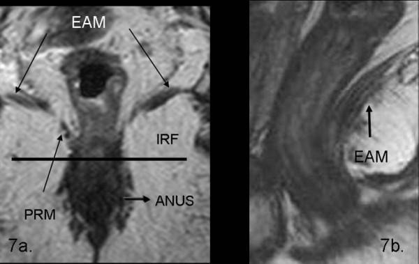
This diagnostic imaging set consists of two T2-weighted turbo spin-echo (TSE) MRI scans of the pelvic floor, demonstrating normal rectal and perianal anatomy. Image 7a is a coronal view illustrating the spatial relationship between the levator ani muscle (EAM), visible as a low-signal intensity band extending laterally and superiorly, and the puborectalis muscle (PRM) forming a muscular sling around the anal canal. The ischio-rectal fossa (IRF) is identified as a hyperintense region of fat lateral to the sphincter complex. The anal canal (ANUS) is shown in the midline. Image 7b is a sagittal view providing a longitudinal perspective of the levator ani muscle (EAM), appearing as a hypointense linear structure supporting the pelvic viscera. These images serve as an educational reference for identifying normal pelvic floor musculature, including the internal and external anal sphincters, and are critical for the staging and preoperative planning of rectal carcinomas and pelvic floor disorders.
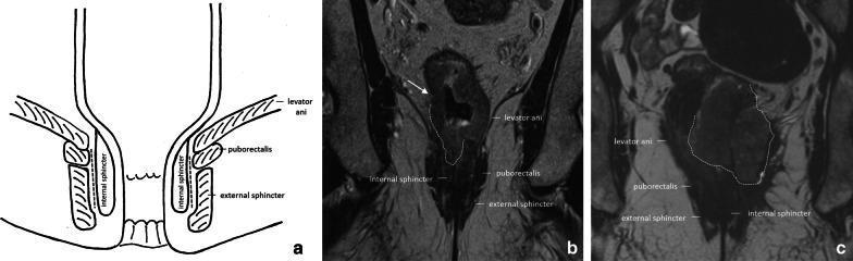
This composite educational image illustrates the pelvic floor and anal canal anatomy and its clinical relevance in rectal cancer staging. Panel (a) is an anatomical diagram depicting the coronal view of the anal canal, highlighting the levator ani, puborectalis muscle, external sphincter, and internal sphincter, with a dashed line marking the intersphincteric plane. Panel (b) is a coronal T2-weighted MRI showing a cT3a rectal tumor in a male patient. An arrow indicates irregular wall thickening with minimal (±1 mm) perirectal extension invading the right internal anal sphincter. Panel (c) is a coronal T2-weighted MRI of a female patient with a cT4b rectal tumor. The tumor (outlined by a dashed line) shows extensive infiltration, invading both internal sphincters and extending into the external sphincter, levator ani, and puborectalis muscles on the left side. Muscles and sphincters appear characteristically hypointense on T2 imaging, while the tumor presents as an intermediate-signal mass disrupting normal anatomical boundaries, essential for determining surgical management such as abdominoperineal resection.
Complete Clinical, Surgical & Surface Anatomy of the Rectum and Anus
EMBRYOLOGY
- Mulholland and Greenfield's Surgery 7e, p. 3176
PART I: THE RECTUM
1. Position & Extent
- The rectum begins at the rectosigmoid junction at the level of the sacral promontory (S3), where the three taenia coli of the sigmoid colon converge and fan out into the continuous longitudinal muscle of the rectum.
- It is 12-15 cm long, lying posterior and caudal in the pelvis, following the concave curvature of the sacrum and coccyx.
- It terminates at the anorectal ring (puborectalis sling), where it turns abruptly posteriorly and inferiorly to become the anal canal.
- Unlike the colon, the rectum lacks taeniae coli, haustra, and appendices epiploicae.
2. Curvatures (Curves & Valves of Houston)
- Lower valve (Kohlrausch's valve): 7-8 cm from the anal verge
- Middle valve: 9-11 cm from the anal verge (marks the anterior peritoneal reflection internally)
- Upper valve: 12-13 cm from the anal verge
3. Peritoneal Relations
| Segment | Peritoneal Coverage |
|---|---|
| Upper 1/3 | Anterior + both lateral surfaces (intraperitoneal) |
| Middle 1/3 | Anterior surface only |
| Lower 1/3 | Entirely extraperitoneal |
- In males, the peritoneal reflection anteriorly forms the rectovesical pouch (of Douglas) between rectum and bladder.
- In females, it forms the rectouterine pouch (of Douglas/pouch of Douglas) between rectum and uterus/posterior vaginal wall.
4. Relations of the Rectum
- Sacrum, coccyx, median sacral vessels, presacral venous plexus, sacral nerves - all invested in the presacral (Waldeyer's) fascia
- Bladder, seminal vesicles, vas deferens, prostate, posterior urethra - separated by Denonvilliers' fascia
- Uterine cervix and posterior vaginal wall
- Levator ani muscles, mesorectum, pelvic plexuses
5. The Mesorectum & Fascial Envelopes
- Terminal branches of the superior rectal artery
- Lymphatics and lymph nodes
- Autonomic nerves
- Mulholland and Greenfield's Surgery 7e, p. 3180; Sabiston Textbook of Surgery, Total Mesorectal Excision
6. The Rectal Ampulla
PART II: THE ANAL CANAL
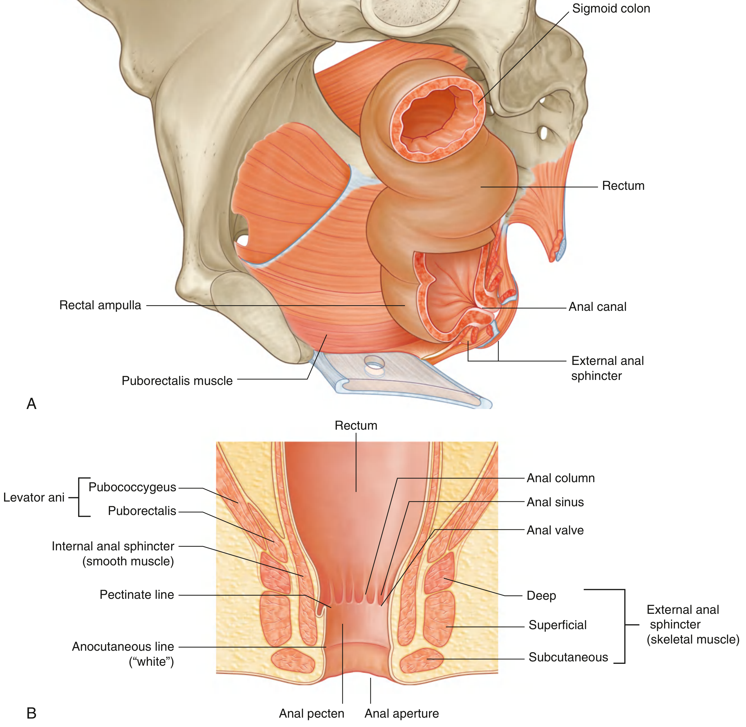
1. Definition & Length
- The anal canal is the terminal 4-5 cm of the intestinal tract, extending from the anorectal ring (where it narrows at the pelvic floor) to the anal verge.
- Surrounded along its entire length by the internal and external anal sphincters.
- The anorectal junction is pulled forward (perineal flexure) by the puborectalis, creating the anorectal angle (~90°).
2. Zones of Epithelial Lining (Superior to Inferior)
| Zone | Location | Epithelium | Clinical Relevance |
|---|---|---|---|
| Rectal mucosa | Above dentate line 1-2 cm | Columnar epithelium | Adenocarcinoma origin |
| Transition zone (ATZ) | 0-2 cm above dentate line | Mixed columnar/cuboidal/squamous | Cloacal origin; both endo/ectodermal |
| Dentate (pectinate) line | At anal columns/valves | Junction | Key surgical landmark |
| Anal pecten / Anoderm | Below dentate to white line (~1.5 cm) | Non-keratinized stratified squamous (no glands, no hair) | Richly innervated - somatic pain |
| Perianal skin | Below white line (Hilton's line) | Keratinized skin with hair and glands | True skin |
3. The Dentate (Pectinate) Line - THE Key Landmark
| Feature | Above Dentate Line | Below Dentate Line |
|---|---|---|
| Embryology | Endoderm (hindgut) | Ectoderm (proctodaeum) |
| Epithelium | Columnar/transitional | Stratified squamous |
| Arterial supply | Superior rectal a. (IMA) | Inferior rectal a. (pudendal) |
| Venous drainage | Superior rectal v. → portal | Inferior rectal v. → systemic (IVC) |
| Lymph drainage | Internal iliac / inferior mesenteric nodes | Inguinal nodes |
| Innervation | Autonomic (visceral - no pain) | Somatic (pudendal - pain sensitive) |
| Hemorrhoids | Internal hemorrhoids | External hemorrhoids |
| Abscess/fistula | Internal opening (cryptoglandular) | External components |
- Gray's Anatomy for Students; Bailey and Love's 28e, p. 1439; Mulholland & Greenfield's Surgery 7e, p. 3534
4. Anal Glands
- 6-10 anal glands open into the anal crypts at the dentate line.
- They penetrate the internal sphincter and lie in the intersphincteric plane.
- These are the source of cryptoglandular anal abscesses and fistula-in-ano (Park's classification).
PART III: MUSCULAR ANATOMY
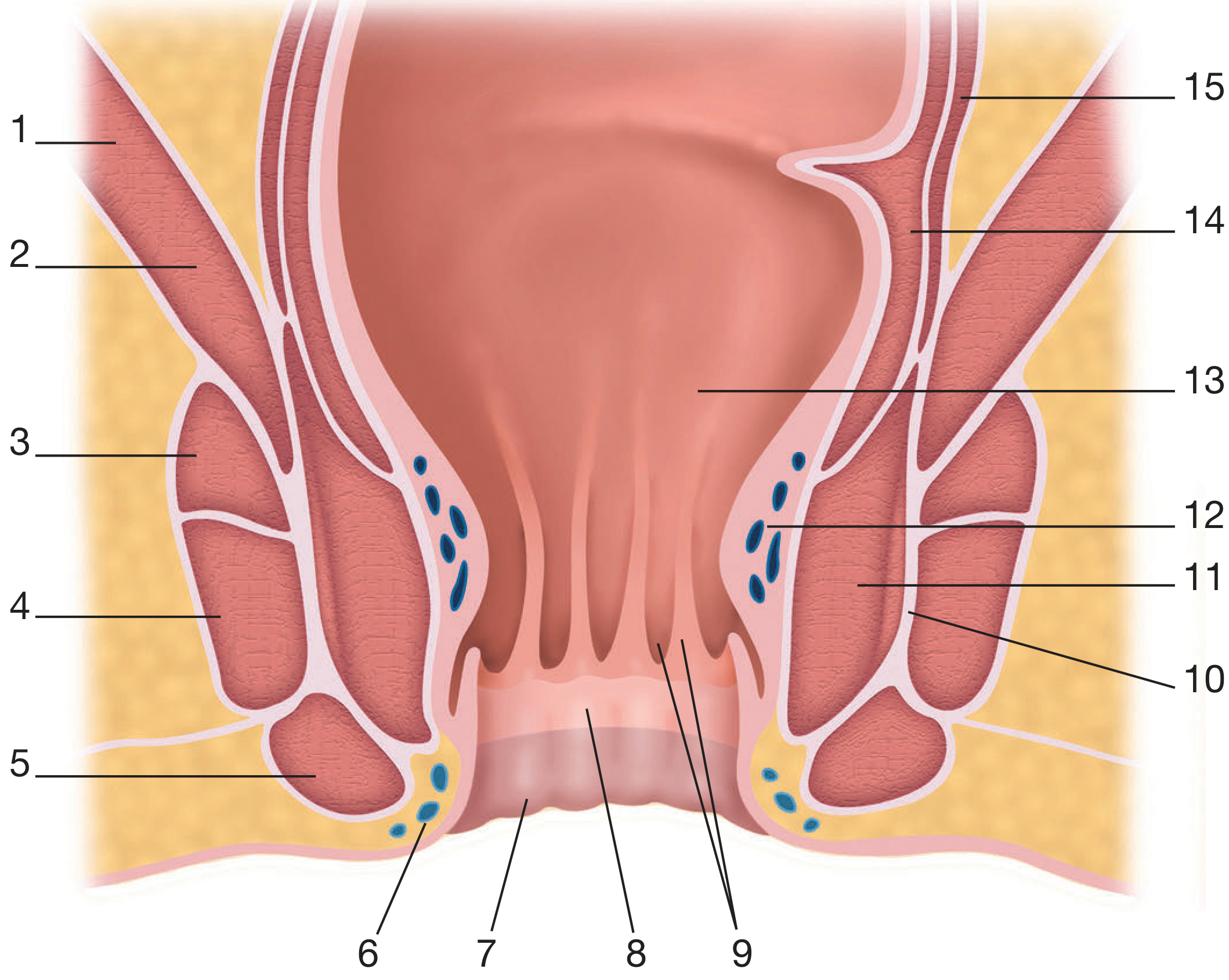
1. Internal Anal Sphincter (IAS)
- Thickened (2-5 mm) distal continuation of the circular muscle layer of the rectum
- Smooth (involuntary) muscle - autonomically innervated
- Extends from the anorectal ring to the intersphincteric groove (palpable externally), 1-2 cm above the anal verge
- Appears pearly white when exposed surgically
- Maintained in tonic contraction at rest (contributes ~80% of resting anal pressure)
- Relaxes with rectal distension (recto-anal inhibitory reflex, RAIR)
- Non-adrenergic, non-cholinergic fibres release nitric oxide causing relaxation
- Bailey and Love's 28e
2. External Anal Sphincter (EAS)
- Striated, voluntary muscle, innervated by the inferior rectal branch of the pudendal nerve (S2, S3, S4)
- Red in colour
- Although classically divided into three parts (deep, superficial, subcutaneous), functionally it is a single muscle (Goligher)
- Continuously fused with the levator ani superiorly
- Attached to the coccyx posteriorly via the anococcygeal ligament/raphe
- Anteriorly, fuses with perineal body muscles
- Contributes to the squeeze (voluntary) pressure of the anal canal
3. The Longitudinal Muscle (Conjoined Longitudinal Muscle)
- Direct continuation of the outer longitudinal smooth muscle of the rectum
- Passes caudally in the intersphincteric plane between EAS and IAS
- Splits into multiple terminal septa that fan out to:
- Surround subcutaneous EAS bundles
- Insert into skin of lower anal canal and perianal skin (corrugator cutis ani)
- Creates a fibromuscular "scaffolding" that supports the anal canal
- During defecation: widens anal lumen, flattens anal cushions, shortens the anal canal
- The septa also create pathways for infection spread (intersphincteric abscesses)
4. Levator Ani & Pelvic Floor
| Component | Origin | Action |
|---|---|---|
| Puborectalis | Posterior pubis - forms U-shaped sling around anorectal junction | Maintains anorectal angle (~90°), major continence mechanism |
| Pubococcygeus | Posterior pubis → coccyx | Pelvic floor support |
| Iliococcygeus | Arcus tendineus → coccyx | Pelvic floor support |
- Mulholland & Greenfield's Surgery 7e, p. 3534-3539
PART IV: ANORECTAL SPACES
| Space | Boundaries | Contents | Clinical Significance |
|---|---|---|---|
| Perianal space | Surrounds anal canal below dentate | EAS subcutaneous portion, inferior hemorrhoidal plexus | External hemorrhoids, perianal abscess |
| Intersphincteric space | Between IAS and EAS | Longitudinal muscle, anal glands | Intersphincteric abscess; route for cryptoglandular infection |
| Ischioanal (ischiorectal) space | Ischial tuberosities laterally; levator ani superiorly; EAS medially | Fatty areolar tissue, inferior rectal vessels/nerves | Ischiorectal abscess; horseshoe abscess |
| Supralevator space | Above levator ani, lateral to rectum | Pelvic organs | Supralevator abscess |
| Postanal space | Posterior to anal canal; deep to anococcygeal ligament | Areolar tissue | Source of horseshoe abscesses; limited by anococcygeal ligament, forcing lateral spread |
- Mulholland & Greenfield's Surgery 7e, p. 3535
PART V: BLOOD SUPPLY
Arterial Supply
| Artery | Origin | Territory |
|---|---|---|
| Superior rectal (hemorrhoidal) artery | Terminal branch of inferior mesenteric artery (IMA) | Upper rectum, bifurcates at ~S3, supplies upper anal canal |
| Middle rectal (hemorrhoidal) artery | Internal iliac artery (variable - may be absent) | Middle and lower rectum laterally |
| Inferior rectal (hemorrhoidal) artery | Internal pudendal artery (from internal iliac) | Lower anal canal, external sphincter, perianal skin |
| Median sacral artery | Posterior aorta | Small contribution to posterior rectum |
Venous Drainage - Portal-Systemic Anastomosis
| Plexus | Drains Into | System |
|---|---|---|
| Superior rectal vein | Inferior mesenteric → portal | Portal |
| Middle rectal vein | Internal iliac → IVC | Systemic |
| Inferior rectal vein | Internal pudendal → internal iliac → IVC | Systemic |
PART VI: LYMPHATIC DRAINAGE
| Region | Primary Drainage | Secondary Drainage |
|---|---|---|
| Upper and middle rectum | Inferior mesenteric (pararectal) nodes | Along superior rectal vessels |
| Lower rectum and upper anal canal | Inferior mesenteric OR internal iliac nodes | Lateral pelvic nodes |
| Anal canal below dentate line | Superficial inguinal nodes | Internal iliac; inferior mesenteric (occasionally) |
- Mulholland & Greenfield's Surgery 7e, p. 3535
PART VII: INNERVATION
Sympathetic
- Origin: T12-L3 spinal cord → synapse at preaortic plexus (inferior mesenteric ganglion)
- Nerves descend to form the hypogastric plexus (visible over top of sacrum during anterior resection)
- Function: Ejaculation (damage → retrograde ejaculation), smooth muscle contraction (IAS tone, decrease motility)
Parasympathetic (Nervi Erigentes / Pelvic Splanchnics)
- Origin: S2, S3, S4 sacral roots
- Travel medially along Denonvilliers' fascia
- Join the hypogastric plexus to form the pelvic plexus (lateral to rectum)
- Function: Erection in males (damage → impotence), increase GI motility
- Critical to preserve during TME and anterior resection
Somatic (Pudendal Nerve - S2, S3, S4)
- Innervates EAS and perineal muscles
- Carries somatic sensation from anal canal below the dentate line (pain, touch, temperature)
- Above dentate line: only visceral sensation (stretch/fullness, not pain) - hence internal hemorrhoid banding painless
Clinical Note on Pain Sensation:
-
Most sensory endings are distal to the dentate line (somatic pain)
-
However, patients may perceive pain up to 1.5 cm proximal to the dentate line
-
This dictates planning for anorectal procedures (e.g., banding of internal hemorrhoids must be above the dentate line to avoid pain)
-
Mulholland & Greenfield's Surgery 7e, p. 3538
PART VIII: SURFACE ANATOMY
Anal Verge / Anal Margin
- The most inferior visible landmark - the junction of the anoderm with true perianal skin at Hilton's white line (anocutaneous line)
- Visible externally as the puckered skin of the anal opening
Anorectal Ring
- The uppermost palpable ring on digital rectal examination
- Formed by the junction of the puborectalis + deep EAS + IAS
- At approximately 3-4 cm from the anal verge
- Critical surgical landmark: division of the anorectal ring causes complete fecal incontinence
Digital Rectal Examination (DRE) Landmarks
- At rest, the sphincter complex is palpable 1-2 cm from anal verge
- Anterior wall (male): prostate at 3-5 cm; seminal vesicles not normally palpable
- Anterior wall (female): cervix and posterior vaginal wall palpable
- Posterior wall: coccyx, sacrum, presacral space
- Lateral walls: levator ani, pelvic side walls
- Rectal content palpable up to 7-8 cm
- The normal rectum can accommodate a proctoscope / rigid sigmoidoscope
Distances from Anal Verge (Clinical Reference)
| Landmark | Distance from Anal Verge |
|---|---|
| Anocutaneous (white) line / anal verge | 0 cm |
| Dentate (pectinate) line | 1.5-2 cm |
| Anorectal ring / top of EAS | 3-4 cm |
| Anterior peritoneal reflection (male) | 7-9 cm |
| Anterior peritoneal reflection (female) | 5-7.5 cm |
| Lower valve of Houston | 7-8 cm |
| Middle valve of Houston (= peritoneal reflection) | 9-11 cm |
| Upper valve of Houston | 12-13 cm |
| Rectosigmoid junction | 15 cm |
Positions Used for Examination
- Lithotomy position: hemorrhoids described at 3, 7, 11 o'clock
- Left lateral (Sims') position: used in DRE - 12 o'clock = anterior
- Prone jack-knife position: used in most anorectal surgery
PART IX: PHYSIOLOGY & CONTINENCE MECHANISM
-
At rest: Both sphincters closed; puborectalis tightened maintaining anorectal angle ~90°. IAS provides 80% of resting tone; hemorrhoidal cushions provide fine airtight seal.
-
Rectal filling: Causes distension; IAS relaxes (RAIR) and EAS reflexly contracts. Small bolus of stool contacts the sensory-rich anoderm - sampling reflex (allows discrimination between gas, liquid, solid stool).
-
If defecation deferred: EAS and puborectalis tighten further; anorectal angle becomes more acute; stool returns to ampulla.
-
Defecation: Voluntary relaxation of EAS and puborectalis; Valsalva manoeuvre increases intraabdominal pressure; puborectalis relaxes straightening the anorectal angle; longitudinal muscle shortens the anal canal; levator ani contracts elevating the pelvic floor.
- Mulholland & Greenfield's Surgery 7e, p. 3539
PART X: CLINICAL / SURGICAL IMPORTANCE SUMMARY
| Condition | Key Anatomical Basis |
|---|---|
| Internal hemorrhoids | Prolapse of superior hemorrhoidal plexus cushions above dentate line; staged by degree of prolapse (Grades I-IV) |
| External hemorrhoids | Inferior hemorrhoidal plexus below dentate line; pain when thrombosed |
| Anal fissure | Tear in anoderm below dentate line at posterior midline; IAS spasm + reduced blood supply at 6 o'clock |
| Anorectal abscess | Cryptoglandular infection in intersphincteric plane → spreads along fascial spaces |
| Fistula-in-ano (Parks classification) | Intersphincteric, transsphincteric, suprasphincteric, extrasphincteric - based on relation to EAS |
| Rectal prolapse | Full-thickness protrusion through anal canal; involves loss of anorectal angle |
| TME (Total Mesorectal Excision) | Sharp dissection in avascular plane between fascia propria and presacral fascia; preserves pelvic autonomic nerves |
| Abdominoperineal Resection (APR) | For tumors <2 cm from anorectal ring; removes rectum and entire anal canal |
| Impotence/ejaculatory failure post-op | Injury to nervi erigentes (parasympathetic) or hypogastric nerves (sympathetic) respectively |
| Anal SCC | Arises from squamous epithelium below/at dentate line; spreads to inguinal nodes |
| Rectal adenocarcinoma | Columnar mucosa; spreads to mesorectal/internal iliac nodes; requires TME |
Key Reference Images
- Mulholland & Greenfield's Surgery: Scientific Principles and Practice, 7e - Chapter 64 & 70
- Bailey and Love's Short Practice of Surgery, 28e - Chapter 80
- Gray's Anatomy for Students - Chapter 5 (Pelvis & Perineum)
- Sabiston Textbook of Surgery - Total Mesorectal Excision chapter
- Sleisenger & Fordtran's Gastrointestinal and Liver Disease - Anal Sphincter Muscles section