
Endothelial and leukocyte adhesion molecules in inflammation in detail to wright in MD pathology exam
Endothelial and Leukocyte Adhesion Molecules in Inflammation
Introduction
- Margination and rolling
- Activation (integrin activation by chemokines)
- Firm (stable) adhesion
- Transmigration (diapedesis)
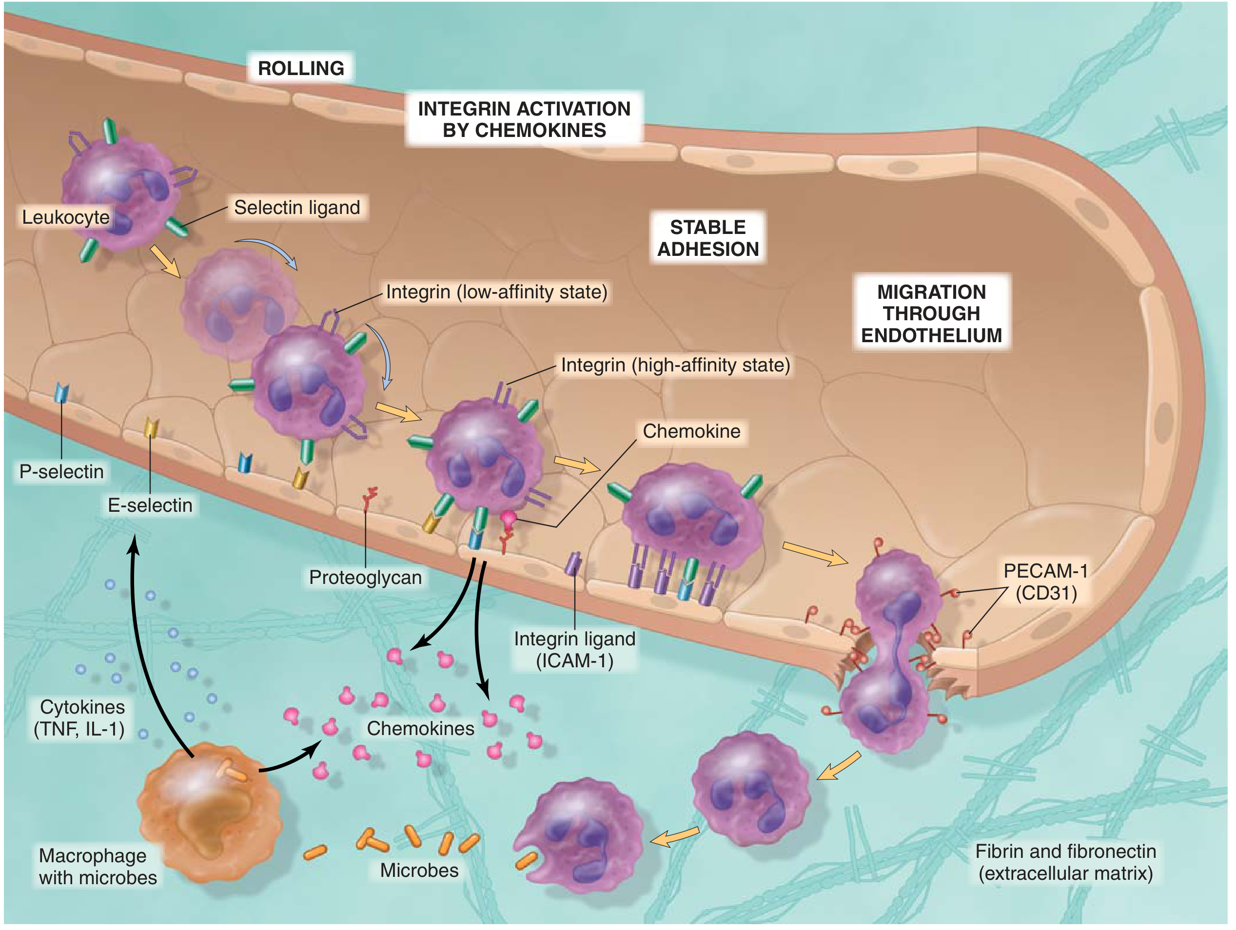
Families of Adhesion Molecules
| Family | Location | Members |
|---|---|---|
| Selectins | Leukocytes + endothelium | L-selectin, E-selectin, P-selectin |
| Integrins | Leukocytes | LFA-1, MAC-1, VLA-4 |
| Immunoglobulin (Ig) superfamily | Endothelium | ICAM-1, ICAM-2, VCAM-1, PECAM-1 |
1. SELECTINS
Structure
Members
P-Selectin (CD62P)
- Location: Endothelial cells and platelets
- Storage: Stored in cytoplasmic granules of endothelial cells called Weibel-Palade bodies (not expressed on resting endothelium)
- Induction: Rapidly mobilized to cell surface within minutes of exposure to histamine (from mast cells) or thrombin (from coagulation cascade)
- Ligands on leukocytes: Sialyl-Lewis X, particularly on P-selectin glycoprotein ligand 1 (PSGL-1) - present on neutrophils, monocytes, and some T cells
- Function: Mediates initial rolling of leukocytes on endothelium
E-Selectin (CD62E)
- Location: Activated endothelial cells only (not on resting endothelium)
- Induction: Synthesized and expressed within 1-2 hours in response to IL-1, TNF (cytokines from tissue macrophages and DCs), and bacterial LPS
- Ligands on leukocytes: Sialyl-Lewis X on PSGL-1, E-selectin ligand-1, and certain glycolipids on neutrophils, monocytes, and activated T lymphocytes
- Function: Mediates rolling and weak adhesion
L-Selectin (CD62L)
- Location: Expressed on leukocytes (not on endothelial cells) - constitutively expressed on naive T cells and neutrophils
- Ligands on endothelium: Sialomucins on endothelial cells whose expression is upregulated by IL-1, TNF; the major determinant is sialyl 6-sulfo Lewis X
- Function: Promotes adhesion of neutrophils to endothelium at sites of inflammation; also mediates homing of naive lymphocytes to lymph nodes via interaction with HEV (high endothelial venules)
Selectin Ligands Summary
- PSGL-1 (P-selectin glycoprotein ligand 1) - primary P-selectin ligand
- PSGL-1 and E-selectin ligand-1 - for E-selectin
- Sialomucins on endothelium - for L-selectin
2. INTEGRINS
Structure
Key Feature: Inside-Out Signaling (Integrin Activation)
- Low-affinity (bent) state - on resting leukocytes; do not bind their ligands effectively
- High-affinity (extended) state - after inside-out activation by chemokines
Key Members
| Integrin | Also Called | Expressed On | Ligands |
|---|---|---|---|
| LFA-1 | αLβ2, CD11aCD18 | T lymphocytes, all leukocytes | ICAM-1 (CD54), ICAM-2, ICAM-3 |
| MAC-1 | αMβ2, CD11bCD18, CR3 | Monocytes, neutrophils | ICAM-1; also iC3b (complement receptor) |
| VLA-4 | α4β1, CD49dCD29 | Lymphocytes, monocytes, eosinophils | VCAM-1 (CD106) |
| α4β7 | - | Lymphocytes homing to gut | MAdCAM-1 |
| CR4 | CD11cCD18, αXβ2 | DCs, myeloid cells | iC3b fragment |
| αEβ7 | CD103 | Intraepithelial T cells, DCs | E-cadherin |
3. IMMUNOGLOBULIN (Ig) SUPERFAMILY MOLECULES ON ENDOTHELIUM
ICAM-1 (Intercellular Adhesion Molecule-1, CD54)
- Structure: Membrane glycoprotein with immunoglobulin (Ig) domains
- Expression: Constitutively low; markedly upregulated on endothelium, lymphocytes, DCs, macrophages, fibroblasts, and epithelial cells by IL-1 and TNF
- Binds to: LFA-1 and MAC-1 on leukocytes
- Role: Mediates firm (stable) adhesion of leukocytes to endothelium; also critical for T cell interaction with APCs
ICAM-2 (CD102)
- Expression: Constitutively expressed on endothelial cells
- Binds to: LFA-1
- Role: Contributes to leukocyte adhesion and transmigration
VCAM-1 (Vascular Cell Adhesion Molecule-1, CD106)
- Expression: Induced on endothelium by IL-1, TNF
- Binds to: VLA-4 (α4β1) integrin on lymphocytes, monocytes, and eosinophils
- Clinical relevance: Important in lymphocyte and eosinophil recruitment; target of natalizumab (anti-α4 integrin antibody used in MS and Crohn's disease)
PECAM-1 (Platelet-Endothelial Cell Adhesion Molecule-1, CD31)
- Expression: Expressed on endothelial cells (particularly at intercellular junctions) and on leukocytes
- Homophilic binding: PECAM-1 on leukocytes binds PECAM-1 on endothelial junctions
- Role: Mediates transmigration (diapedesis) - leukocytes squeeze through intercellular junctions using PECAM-1 interactions. CD31 blockade inhibits leukocyte extravasation.
MAdCAM-1 (Mucosal Addressin Cell Adhesion Molecule-1)
- Expression: Expressed on endothelial cells of gut-associated lymphoid tissue
- Binds to: α4β7 integrin on gut-homing lymphocytes
- Role: Directs lymphocyte homing to intestinal Peyer's patches and lamina propria
4. THE MULTISTEP PROCESS OF LEUKOCYTE RECRUITMENT (Step-by-Step)
Step 1: Margination
- Under normal laminar flow, red cells occupy the vessel center, pushing leukocytes peripherally
- In early inflammation, as blood flow slows (stasis), leukocytes accumulate along the endothelial surface
- Called margination
Step 2: Rolling
- Molecules involved: Selectins (E-selectin and P-selectin on endothelium; L-selectin on leukocytes) binding sialyl-Lewis X carbohydrates
- P-selectin rapidly redistributes from Weibel-Palade bodies to the surface in response to histamine and thrombin (within minutes)
- E-selectin is synthesized within 1-2 hours in response to TNF and IL-1
- These weak, transient, reversible interactions cause leukocytes to slow down and roll along the endothelial surface
- Rolling allows leukocytes to survey the endothelial surface for further activation signals
Step 3: Integrin Activation by Chemokines
- Chemokines (e.g., CXCL8/IL-8 for neutrophils; CCL2 for monocytes) are produced at the inflammatory site and displayed on endothelial surface proteoglycans
- Rolling leukocytes encounter surface-bound chemokines
- Chemokine binding to GPCR-type chemokine receptors on leukocytes triggers intracellular signaling (via RAP GTPases) that converts integrins from low-affinity to high-affinity conformation
- Integrins also cluster on the leukocyte surface, increasing avidity
Step 4: Firm (Stable) Adhesion
- Molecules involved: Integrins on leukocytes (LFA-1, MAC-1, VLA-4) binding Ig-superfamily ligands on endothelium (ICAM-1, VCAM-1)
- The high-affinity integrins bind firmly to ICAM-1 and VCAM-1 (upregulated on endothelium by TNF/IL-1)
- Results in strong, stable adhesion - leukocytes stop rolling and flatten on the endothelium
Step 5: Transmigration (Diapedesis)
- Molecules involved: PECAM-1 (CD31) on both leukocytes and endothelial junctions
- Leukocytes squeeze between endothelial cells at intercellular junctions (paracellular route - most common) or rarely through the endothelial cell body (transcellular route)
- PECAM-1 homophilic binding drives migration through the junction
- After crossing the endothelium, leukocytes pierce the basement membrane using matrix metalloproteinases (collagenases)
Step 6: Chemotaxis in Tissues
- Once in the extracellular space, leukocytes migrate along a concentration gradient of chemokines and other chemoattractants toward the site of injury/infection
- Chemoattractants include: CXCL8 (IL-8), C5a, leukotriene B4 (LTB4), fMet-Leu-Phe (fMLF) bacterial peptides
5. SUMMARY TABLE: Robbins Table 2.3 (Endothelial and Leukocyte Adhesion Molecules)
| Family | Adhesion Molecule | Expressed On | Principal Ligand | Role |
|---|---|---|---|---|
| Selectin | L-selectin (CD62L) | Leukocytes | Sialyl-Lewis X on endothelium | Rolling; lymphocyte homing |
| Selectin | E-selectin (CD62E) | Activated endothelium | Sialyl-Lewis X on neutrophils, monocytes, T cells | Rolling |
| Selectin | P-selectin (CD62P) | Activated endothelium, platelets | Sialyl-Lewis X (PSGL-1) on neutrophils, monocytes, T cells | Rolling |
| Integrin | LFA-1 (αLβ2, CD11aCD18) | T cells, other leukocytes | ICAM-1 on activated endothelium | Firm adhesion |
| Integrin | MAC-1 (αMβ2, CD11bCD18) | Monocytes, other leukocytes | ICAM-1; iC3b | Firm adhesion; phagocytosis |
| Integrin | VLA-4 (α4β1) | Lymphocytes, monocytes, eosinophils | VCAM-1 on activated endothelium | Firm adhesion |
| Integrin | α4β7 | Gut-homing lymphocytes | MAdCAM-1 | Gut homing |
| Ig superfamily | ICAM-1 (CD54) | Activated endothelium | LFA-1, MAC-1 | Firm adhesion |
| Ig superfamily | VCAM-1 (CD106) | Activated endothelium | VLA-4 | Firm adhesion (lymphocytes, eos) |
| Ig superfamily | PECAM-1 (CD31) | Endothelial junctions, leukocytes | PECAM-1 (homophilic) | Transmigration |
| Ig superfamily | MAdCAM-1 | Gut endothelium | α4β7 integrin | Lymphocyte gut homing |
6. REGULATION OF ADHESION MOLECULE EXPRESSION
| Stimulus | Effect | Timeframe |
|---|---|---|
| Histamine, thrombin | P-selectin redistribution to surface (from Weibel-Palade bodies) | Minutes |
| TNF, IL-1 | E-selectin synthesis and expression | 1-2 hours |
| TNF, IL-1 | ICAM-1, VCAM-1 upregulation on endothelium | 4-6 hours |
| Chemokines (CXCL8, CCL2) | Integrin affinity/avidity switch on leukocytes | Seconds-minutes |
| LPS (bacterial) | E-selectin expression on endothelium | 1-2 hours |
7. CLINICAL RELEVANCE AND DISEASE CORRELATES
Leukocyte Adhesion Deficiency (LAD)
- Type I: Autosomal recessive deficiency of CD18 (β2 integrin subunit) → absent LFA-1 and MAC-1 → failure of leukocyte firm adhesion and transmigration → recurrent bacterial infections, impaired pus formation, delayed umbilical cord separation, leukocytosis without inflammation at sites of infection
- Type II: Deficiency of fucosyl transferase → absent sialyl-Lewis X → failure of selectin-mediated rolling (leukocytes never slow down enough to adhere)
- Type III: Deficiency in kindlin-3 (activates integrin inside-out signaling)
Therapeutic Targeting
- Anti-TNF antibodies (infliximab, etanercept): reduce upregulation of E-selectin, ICAM-1, VCAM-1 - used in rheumatoid arthritis, IBD, psoriasis
- Natalizumab (anti-α4 integrin): blocks VLA-4/VCAM-1 and α4β7/MAdCAM-1 interactions - used in multiple sclerosis and Crohn's disease
- Vedolizumab (anti-α4β7): selectively blocks gut lymphocyte trafficking - used in IBD
8. SEQUENCE OF CELLULAR INFILTRATION
- First 6-24 hours: Neutrophil predominance (respond rapidly, more numerous in blood, attach firmly to early-induced P- and E-selectin)
- After 24-48 hours: Monocyte/macrophage predominance (neutrophils undergo apoptosis; macrophages survive longer and proliferate)
- Exceptions: Pseudomonas infections - prolonged neutrophil dominance; viral infections - lymphocytes first; allergic reactions/parasitic infections - eosinophils predominant
Key Points to Remember for Exam
- Selectins mediate rolling - weak, reversible, calcium-dependent, carbohydrate-binding
- P-selectin is from Weibel-Palade bodies (rapid, minutes); E-selectin is newly synthesized (slow, 1-2 hours)
- Integrins mediate firm adhesion after activation by chemokines (inside-out signaling, conformational change)
- PECAM-1 (CD31) mediates transmigration at intercellular junctions
- ICAM-1 and VCAM-1 are the endothelial Ig-superfamily ligands upregulated by TNF/IL-1
- LAD type I = β2-integrin deficiency (absent CD18); LAD type II = absent sialyl-Lewis X
- Chemokines are displayed on endothelial surface proteoglycans - they activate integrins during rolling, not before
Effects of lead poisoning in children related to blood levels
"lead poisoning"[MeSH Terms] AND "child"[MeSH Terms] AND "blood lead"
lead poisoning children blood lead levels effects CDC 2025 guidelines
https://www.cdc.gov/lead-prevention/hcp/clinical-guidance/in…
Effects of Lead Poisoning in Children Related to Blood Lead Levels
Key Principle: No Safe Level
- Higher gastrointestinal absorption of lead (~50% in children vs. ~10% in adults)
- Pica behavior (compulsive ingestion of non-food items)
- Rapid neurodevelopment during this critical window
- Proximity to floor-level lead dust
Effects by Blood Lead Level (BLL)
BLL < 3.5 µg/dL
- Considered "normal range" for U.S. children (below the 97.5th percentile)
- However: no threshold has been established below which lead is safe
- Subtle subclinical neurodevelopmental effects may still be occurring
BLL 3.5 - 9 µg/dL (Low-Level Exposure)
- Decreased IQ (approximately 1-5 IQ points lost per µg/dL increase - effect is steepest at low levels, with the greatest IQ loss per unit occurring in the 1-10 µg/dL range)
- Reduced attention span and ability to concentrate
- Impaired academic achievement and reading scores
- Subtle behavioral changes: irritability, hyperactivity
- Decreased nerve conduction velocity (subclinical)
- Education about lead exposure sources
- Dietary counseling (iron and calcium intake - these compete with lead absorption)
- Development monitoring at well-child visits
- Follow-up BLL testing every 3 months initially, then every 6-9 months
BLL 10 - 19 µg/dL (Mild Elevation)
- More pronounced cognitive impairment - measurable IQ decline
- Reduced attention and executive function
- Impaired language development
- Slowed nerve conduction velocity
- Early inhibition of heme synthesis: elevated erythrocyte protoporphyrin (EP) and zinc protoporphyrin (ZPP) - though ZPP is unreliable below 25 µg/dL
- Subclinical impairment of renal tubular function
BLL 20 - 44 µg/dL (Moderate Elevation)
- Significant cognitive and behavioral impairment
- Anemia - microcytic hypochromic anemia from impaired heme synthesis (lead inhibits δ-aminolevulinic acid dehydratase [ALAD] and ferrochelatase)
- Basophilic stippling of red blood cells (impaired clearance of RNA degradation products)
- Elevated urinary δ-aminolevulinic acid (ALA) and coproporphyrin
- Elevated ZPP reliably detectable at this range
- Lead lines (Burtonian lines): dense metaphyseal bands on X-ray of long bones (especially knee), representing failure of bone remodeling
- Abdominal colic, constipation, vague abdominal pain
- Peripheral motor neuropathy (wrist drop, foot drop) - more prominent in adults; less common in children
- Impaired renal tubular function (Fanconi syndrome-like): aminoaciduria, glycosuria, phosphaturia
- May have irritability, anorexia, lethargy
BLL 45 - 69 µg/dL (Severe - Chelation Threshold for Children)
- All effects above, more pronounced
- Overt gastrointestinal symptoms: persistent vomiting, colicky abdominal pain
- Progressive neurological deterioration: apathy, drowsiness, ataxia, clumsiness
- Peripheral neuropathy
- Strongly positive urinary coproporphyrin test (UCP test: ether layer shows red fluorescence under Wood lamp)
- Impaired kidney function
- Succimer (dimercaptosuccinic acid, DMSA): 350 mg/m² (or 10 mg/kg) PO every 8 hours for 5 days, then every 12 hours for 14 days
- Monitor blood lead weekly (lead is mobilized from bone into blood during chelation)
- Remove child from lead source - essential
BLL 70 - 100 µg/dL (Very Severe)
- Pre-encephalopathic: persistent vomiting, severe irritability, ataxia
- Encephalopathy may develop acutely, especially in children < 2 years
- Marked anemia with basophilic stippling
- Renal tubular damage
- CaNa₂ EDTA alone (edetate calcium disodium) via continuous IV infusion: 1,500 mg/m²/day, OR
- Succimer (oral) for asymptomatic cases
BLL > 70 µg/dL in Children / Encephalopathy (Life-Threatening)
- Develops over 3-6 weeks (may evolve rapidly within a week in children < 2 years)
- Early: anorexia, irritability, reduced alertness, less playful, vague abdominal pain, intermittent vomiting
- Progressive: apathy → drowsiness → stupor alternating with hyperirritability
- Late: persistent vomiting, ataxia, seizures, coma
- CSF: elevated pressure, papilledema, mild lymphocytic pleocytosis, elevated protein, normal glucose
- Massively swollen brain with temporal lobe and cerebellar herniation
- Multiple microscopic ischemic foci in cerebrum and cerebellum
- Endothelial damage with perivascular proteinaceous deposits
- Perivascular lymphocytic/mononuclear infiltrates
- Hyperplastic changes in arteries and arterioles
- Foci of ischemic necrosis with glial reaction
- Similar vascular changes in kidneys
- Do NOT wait for blood lead level results if encephalopathy is clinically suspected
- Dimercaprol (BAL): 75 mg/m² (or 4 mg/kg) IM every 4 hours for 5 days (given first - BAL crosses the blood-brain barrier and prevents EDTA from redistributing lead into the brain)
- CaNa₂ EDTA: 1,500 mg/m²/day IV continuous infusion for 5 days; start 4 hours after first BAL dose
- Followed by oral penicillamine (40 mg/kg/day, max 1 g/day)
- Target: reduce serum lead below 40 µg/dL
- Mannitol IV for cerebral edema
- Benzodiazepines (diazepam/midazolam IV) for seizures
- Restrict IV fluids to basal requirements (to limit cerebral edema)
- Whole bowel irrigation with polyethylene glycol if radiopaque lead visible on abdominal X-ray
- Establish urinary flow before chelation
Summary Table: BLL Effects and Actions in Children
| BLL (µg/dL) | Clinical Effects | Management |
|---|---|---|
| < 3.5 | No detectable clinical signs; subclinical harm possible | Routine screening |
| 3.5 - 9 | Subtle IQ loss, attention deficit, behavioral changes | Education, dietary counseling, follow-up BLL q3 months |
| 10 - 19 | Measurable IQ decline, language delay, nerve conduction slowing, ZPP may rise | Above + environmental investigation, developmental referral, q1-3 months |
| 20 - 44 | Anemia, basophilic stippling, lead lines on X-ray, abdominal pain, renal tubular effects | Above + urgent evaluation, consider chelation consult, q2 weeks-1 month |
| 45 - 69 | Overt GI symptoms, neurological deterioration, peripheral neuropathy | Oral succimer (DMSA), remove from source |
| 70 - 100 | Pre-encephalopathic, severe anemia, renal damage | CaNa₂ EDTA alone or succimer |
| > 70 (symptomatic) / encephalopathy | Seizures, coma, cerebral edema, 5-20% mortality | Emergency BAL + CaNa₂ EDTA (dual chelation), ICU care |
Mechanisms of Lead Toxicity (Pathophysiology)
1. Heme Synthesis Inhibition
- δ-Aminolevulinic acid dehydratase (ALAD) - inhibited even at very low BLLs
- Ferrochelatase - inhibited; prevents iron incorporation into protoporphyrin IX → accumulation of zinc protoporphyrin (ZPP) (zinc substitutes for iron)
- Results in: sideroblastic-type anemia; elevated urinary ALA and coproporphyrin
2. Neurotoxicity
- Lead mimics calcium in neurons and disrupts calcium-dependent processes
- Inhibits N-methyl-D-aspartate (NMDA) receptors → impairs long-term potentiation and memory consolidation
- Disrupts protein kinase C signaling
- Inhibits synaptic neurotransmission (particularly in hippocampus and prefrontal cortex)
- Direct myelin toxicity → demyelination → peripheral neuropathy (motor > sensory)
- At high levels: disrupts blood-brain barrier, causes cerebral edema and vasculopathy
3. Renal Toxicity
- Proximal tubular damage → Fanconi syndrome: aminoaciduria, glycosuria, phosphaturia, hyperuricosuria
- With chronic high-level exposure: interstitial nephritis, progressive renal failure, gout (saturnine gout)
- Characteristic lead-acid-fast nuclear inclusions in proximal tubular cells on biopsy
4. Bone Effects
- Lead is stored in bone (> 90% of total body lead burden in adults)
- Competes with calcium in bone mineralization
- Metaphyseal lead lines on X-ray
- Bone acts as a long-term reservoir - released during pregnancy, illness, fever, immobilization, or hyperthyroidism
5. Cardiovascular
- Inhibits Na⁺/K⁺-ATPase → hypertension
- Associated with increased risk of cardiovascular disease in adults exposed as children
Diagnosis: Key Laboratory Findings
| Test | Significance |
|---|---|
| Venous blood lead level | Gold standard; confirm any capillary screen ≥ 3.5 µg/dL with venous sample |
| Zinc protoporphyrin (ZPP) | Elevated in moderate-severe exposure; also elevated in iron deficiency |
| Urinary ALA and coproporphyrin | Elevated; UCP test (ether + Wood lamp) strongly positive when BLL > 80 µg/dL |
| CBC | Microcytic hypochromic anemia; basophilic stippling (also in thalassemia, arsenic poisoning) |
| X-ray long bones | "Lead lines" at metaphyses (dense bands) - represents failure of bone remodeling |
| Abdominal X-ray | Radiopaque material in GI tract after acute ingestion |
| CaNa₂ EDTA mobilization test | Lead excretion > 500 µg in 24 hours confirms plumbism |
Sources of Lead Exposure in Children
- Lead paint in homes built before 1940 (universal) and before 1960 (common) - primary source via pica and paint chips/dust
- Lead-contaminated soil and household dust
- Imported toys, jewelry, ceramics with lead-containing glazes
- Lead pipes (contaminated drinking water - Flint, Michigan crisis)
- E-waste recycling areas
- Traditional/folk remedies and cosmetics (e.g., surma/kohl, azarcon, greta)
- Parental occupational exposure (secondary contamination of home)
Prevention
- Primary prevention (before any exposure): eliminate lead hazards from housing; ban leaded gasoline, paint, and solder
- Secondary prevention: universal screening at ages 12 and 24 months (Medicaid-enrolled children); targeted screening by risk assessment
- Nutritional: adequate calcium and iron intake significantly reduce GI lead absorption
- Remove child from the lead source - fundamental to all management
Pathophysiology of respiratory distress syndrome in same format
"respiratory distress syndrome" AND "newborn" AND "surfactant"
Pathophysiology of Respiratory Distress Syndrome
Overview: Two Distinct Syndromes Under One Name
| Feature | Neonatal RDS (Hyaline Membrane Disease) | ARDS (Adult/Acute RDS) |
|---|---|---|
| Age | Preterm neonates | Any age; usually adults |
| Core defect | Surfactant deficiency | Alveolar-capillary barrier damage |
| Edema type | Atelectasis + plasma leak | Increased-permeability (non-cardiogenic) edema |
| Histology | Hyaline membranes, atelectasis | Diffuse alveolar damage (DAD) |
| Inciting cause | Prematurity (immature type II pneumocytes) | Sepsis, trauma, aspiration, etc. |
| Treatment | Exogenous surfactant + CPAP/ventilation | Lung-protective ventilation; treat cause |
PART 1: NEONATAL RDS (Hyaline Membrane Disease)
1. Epidemiology and Risk Factors
- ~60% of infants born at < 28 weeks' gestation
- ~30% of infants born at 28-34 weeks' gestation
- < 5% of infants born after 34 weeks' gestation
- Prematurity (principal factor)
- Male gender
- Maternal diabetes (fetal hyperinsulinism suppresses surfactant synthesis)
- Cesarean section before onset of labor (labor normally stimulates surfactant release)
- Perinatal asphyxia
- Intrauterine stress / fetal growth restriction (increases fetal corticosteroid output)
- Antenatal maternal glucocorticoids (betamethasone/dexamethasone)
- Thyroid hormones
2. Surfactant: Normal Function and Composition
- ~80% phospholipids - principally dipalmitoylphosphatidylcholine (DPPC / lecithin), the major surface-tension-reducing component
- ~10% neutral lipids (cholesterol)
- ~10% surfactant-associated proteins: SP-A, SP-B, SP-C, SP-D
- SP-B and SP-C: hydrophobic; essential for surface tension reduction and rapid surface adsorption
- SP-A and SP-D: hydrophilic; important for innate immune defense in the lung
- Congenital loss-of-function mutations in SP-B gene → lethal neonatal respiratory failure
- L/S ratio < 1.5 → high risk of RDS
- L/S ratio ≥ 2.0 → lung maturity (< 2% risk of RDS)
- Phosphatidylglycerol (PG) in amniotic fluid further confirms maturity
3. Pathophysiology of Neonatal RDS (Step-by-Step Cascade)
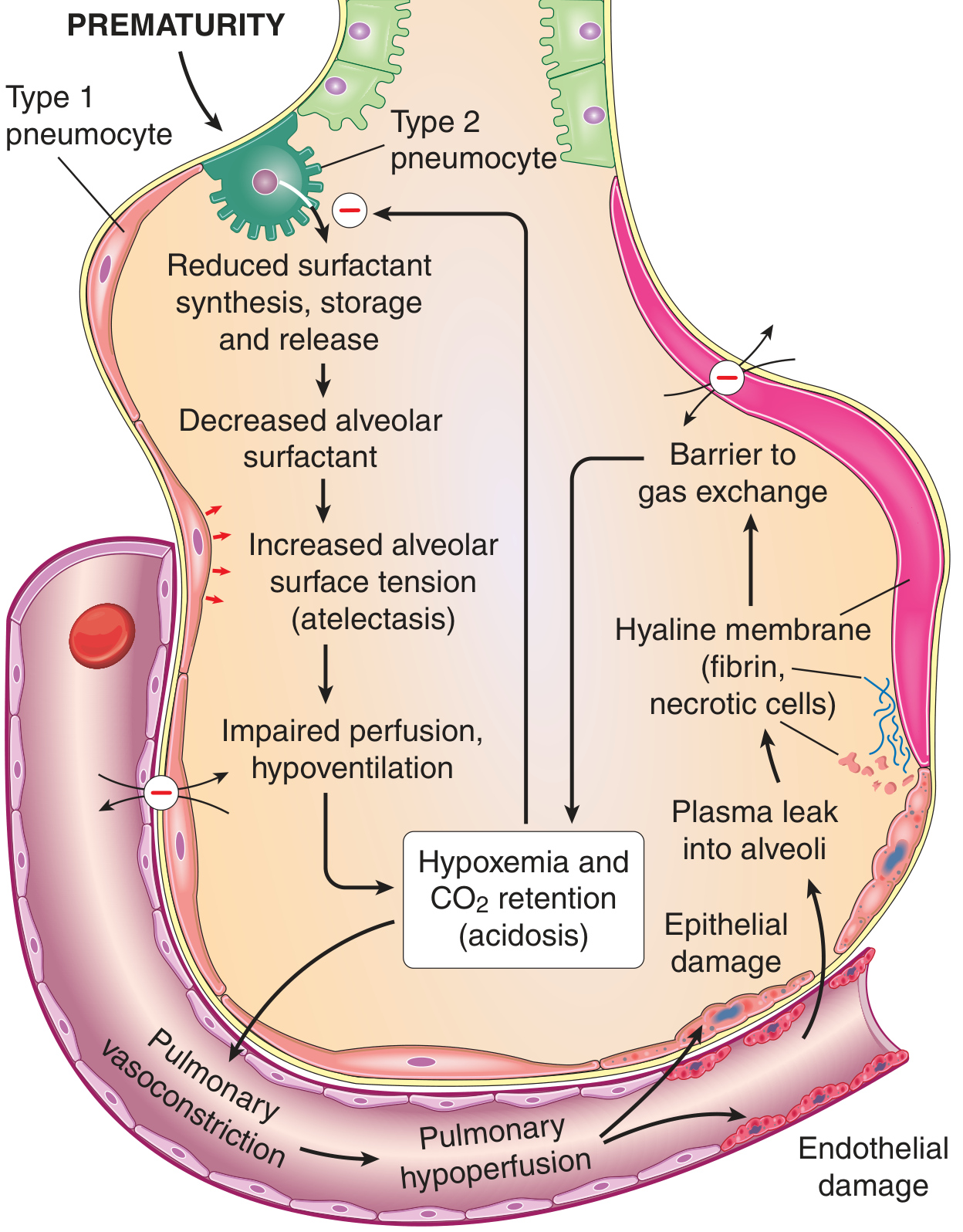
Step 1: Surfactant Deficiency
- Immature type II pneumocytes in preterm lungs have reduced capacity for surfactant synthesis, lamellar body storage, and secretion
- Result: insufficient surfactant coating of alveolar surfaces
Step 2: Increased Alveolar Surface Tension → Atelectasis
- Without surfactant, alveolar surface tension increases dramatically
- On expiration, small alveoli collapse (atelectasis) because pressure required to re-open them exceeds the infant's inspiratory effort
- Each breath requires huge effort to re-expand collapsed alveoli
- The infant tires rapidly → generalized progressive atelectasis
Step 3: Hypoventilation and V/Q Mismatch
- Collapsed alveoli are perfused but not ventilated → intrapulmonary shunt (right-to-left)
- This is the primary cause of hypoxemia in RDS
- Hypoxemia → respiratory and metabolic acidosis
Step 4: Pulmonary Vasoconstriction
- Hypoxemia and acidosis trigger pulmonary arterial vasoconstriction
- This causes pulmonary hypoperfusion
- Persisting fetal circulation pattern: right-to-left shunting through patent ductus arteriosus and foramen ovale worsens systemic hypoxemia
Step 5: Endothelial and Epithelial Damage
- Hypoxemia and acidosis cause ischemic damage to:
- Pulmonary capillary endothelium → increased vascular permeability
- Alveolar epithelium (type I and II pneumocytes) → cell necrosis
- Plasma proteins (including fibrinogen) leak into alveolar spaces
Step 6: Hyaline Membrane Formation
- Leaked plasma proteins + necrotic cellular debris (particularly necrotic type II pneumocytes) deposit along the walls of respiratory bronchioles, alveolar ducts, and alveoli
- Fibrinogen polymerizes into fibrin
- This forms the characteristic eosinophilic hyaline membranes (amorphous, glassy appearance on H&E)
- Hyaline membranes act as a further barrier to gas exchange, worsening hypoxemia
Step 7: Vicious Cycle
- Hyaline membranes + atelectasis → worsening hypoxemia → more vascular/epithelial damage → more membrane formation → further impaired gas exchange
- This self-perpetuating cycle leads to death if untreated
4. Morphology (Gross and Microscopic)
- Lungs are normal or small in size but heavy and relatively airless
- Mottled, purple-red appearance (congested and collapsed)
- Sink in water (unlike normal aerated lungs)
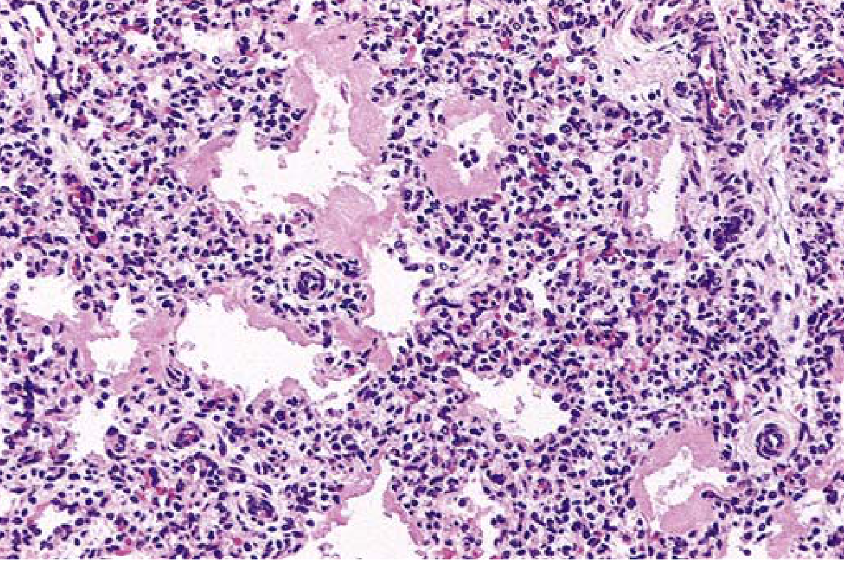
- Alternating atelectasis (collapsed alveoli) and dilated alveolar ducts
- Eosinophilic hyaline membranes lining respiratory bronchioles and alveolar ducts (fibrin + necrotic cells)
- Membranes contain necrotic type II pneumocytes mixed with extravasated plasma proteins (mainly fibrinogen/fibrin)
- Paucity of neutrophilic inflammation (unlike pneumonia)
- In infants dying within hours: only necrotic debris in terminal bronchioles; no fully formed membranes yet
- In surviving infants after several days: proliferation of type II pneumocytes and interstitial fibrosis (reparative phase)
- Not seen in stillborns (requires live breathing + plasma leak)
5. Hormonal Regulation of Surfactant Production
| Factor | Effect on Surfactant |
|---|---|
| Glucocorticoids (cortisol, betamethasone) | Stimulate synthesis of surfactant lipids and SP proteins - accelerate lung maturation |
| Thyroid hormones | Potent stimulators of surfactant production |
| Insulin (elevated in diabetic mothers) | Suppresses surfactant synthesis → increased RDS risk in infants of diabetic mothers |
| Labor | Stimulates surfactant secretion → cesarean before labor increases risk |
| Intrauterine stress / growth restriction | Increases fetal cortisol → accelerates surfactant synthesis → lowers RDS risk |
6. Fetal Lung Maturity Assessment
- L/S ratio in amniotic fluid (lecithin/sphingomyelin): L/S ≥ 2.0 indicates maturity
- Phosphatidylglycerol (PG): presence further confirms maturity
- Lamellar body count (LBC): lamellar bodies (surfactant storage organelles) shed into amniotic fluid
7. Clinical Features
- Onset: within hours of birth (premature infant)
- Rapid, labored breathing (tachypnea >60/min)
- Intercostal, subcostal, and sternal retractions
- Expiratory grunting (attempts to generate PEEP to prevent alveolar collapse)
- Nasal flaring, cyanosis
- Increasing oxygen requirements
- Chest X-ray: diffuse ground-glass haziness, air bronchograms, "white-out" lungs
8. Treatment and Complications
Treatment
- Antenatal corticosteroids (betamethasone IM to mother before 34 weeks): most effective intervention; reduces RDS incidence, severity, and mortality
- Exogenous surfactant replacement therapy at birth (especially for < 28 weeks): dramatically reduces mortality; prophylactic in extremely preterm infants
- CPAP (continuous positive airway pressure): keeps alveoli open, reduces atelectasis
- Mechanical ventilation with lung-protective strategies (low tidal volume, adequate PEEP)
Complications of Oxygen Therapy / Ventilation
- Phase I (hyperoxic): high O₂ suppresses VEGF → retinal endothelial apoptosis and vessel obliteration
- Phase II (return to room air): rebound VEGF surge → retinal neovascularization (abnormal vessel proliferation) → vitreous hemorrhage, retinal detachment, blindness
- Chronic lung disease of prematurity
- Pathology: decreased alveolar septation (large, simplified alveoli), dysmorphic capillary configuration
- Contributing factors: hyperoxia, barotrauma, prematurity, inflammatory cytokines, vascular maldevelopment
- Clinical: oxygen dependence beyond 28 days or 36 weeks postmenstrual age
- Patent ductus arteriosus (PDA)
- Intraventricular hemorrhage (IVH)
- Necrotizing enterocolitis (NEC)
PART 2: ACUTE RESPIRATORY DISTRESS SYNDROME (ARDS)
1. Definition (Berlin Definition 2012)
- Acute onset: within 1 week of a known clinical insult or new/worsening respiratory symptoms
- Bilateral radiographic opacities consistent with pulmonary edema (not explained by effusions, collapse, or nodules)
- Not fully explained by cardiac failure or fluid overload (left atrial hypertension excluded by echocardiography or PCWP ≤ 18 mmHg)
- Hypoxemia (with PEEP ≥ 5 cm H₂O):
| Severity | PaO₂/FiO₂ (P/F Ratio) |
|---|---|
| Mild | 200 - 300 mmHg |
| Moderate | 100 - 200 mmHg |
| Severe | ≤ 100 mmHg |
2. Causes / Precipitating Events
| Direct (Pulmonary) Injury | Indirect (Extrapulmonary) Injury |
|---|---|
| Aspiration of gastric contents | Sepsis (most common overall cause) |
| Pneumonia (bacterial, viral, fungal) | Severe trauma with shock |
| Near-drowning | Burns |
| Toxic gas/smoke inhalation | Blood transfusions (TRALI) |
| Lung contusion | Pancreatitis |
| Reperfusion injury after transplant | Drug overdose (heroin, aspirin) |
3. Pathophysiology of ARDS
Core Mechanism: Increased Permeability Pulmonary Edema
Phases of ARDS / Diffuse Alveolar Damage (DAD)
Phase 1: Exudative Phase (Days 1-7)
- Macrophages release pro-inflammatory cytokines: TNF-α, IL-1β, IL-6, IL-8 (CXCL8)
- IL-8 is a potent neutrophil chemoattractant
- Neutrophils are sequestered in pulmonary capillaries in massive numbers
- Neutrophils transmigrate into alveoli and release:
- Reactive oxygen species (ROS)
- Proteases (elastase, matrix metalloproteinases)
- Platelet-activating factor (PAF)
- Leukotrienes
- Thromboxanes (cause pulmonary vasoconstriction)
- These mediators destroy the capillary endothelium and alveolar epithelium (type I cells)
- Increased vascular permeability → protein-rich, fibrin-containing fluid floods alveoli and interstitium
- Loss of type I pneumocytes (cover 95% of alveolar surface) → denuded basement membrane
- Damage to type II pneumocytes → loss of surfactant production → increased surface tension → atelectasis
- Inactivation of residual surfactant by leaked plasma proteins (especially fibrin and albumin)
- Hyaline membranes form: fibrin + necrotic cells line alveolar walls (morphological hallmark = Diffuse Alveolar Damage / DAD)
- Alveolar flooding → V/Q mismatch → intrapulmonary shunt → refractory hypoxemia
- Decreased FRC (functional residual capacity) as alveoli collapse
- Decreased lung compliance (stiff lungs - "baby lung" concept: only ~20-30% of lung is aerated)
- Increased work of breathing
- Heterogeneous injury on CT: edema and collapse predominate in dependent lung regions (gravity-dependent compression atelectasis from the weight of edematous lung above); non-dependent regions may remain relatively aerated
- Pulmonary hypertension: from hypoxic vasoconstriction + thromboxanes + microvascular thrombosis
- CO₂ retention in severe cases (decreased minute ventilation)
Phase 2: Proliferative Phase (Days 7-21)
- If the patient survives, resolution begins
- Type II pneumocytes proliferate to cover denuded basement membrane
- Type II cells differentiate into type I cells (re-epithelialization)
- Macrophages phagocytose alveolar debris and fibrin
- Edema fluid is reabsorbed (driven by sodium-potassium ATPase in type II cells)
- Lung compliance begins to improve; oxygen requirements decrease
- Fibroblasts begin to infiltrate alveolar walls
Phase 3: Fibrotic Phase (After Day 21, some patients)
- In severe or prolonged ARDS, excessive collagen deposition occurs
- Interstitial and intra-alveolar fibrosis develops (organizing pneumonia pattern)
- Progressive fibrosis → permanently stiff, honeycomb-like lung
- Poor prognosis; may require prolonged ventilation
- CT: cystic changes, traction bronchiectasis
4. Morphology of ARDS (Diffuse Alveolar Damage)
- Lungs are heavy, firm, red-purple, and "beef-liver" consistency
- Microscopically: hyaline membranes lining alveolar ducts and alveolar walls
- Interstitial and alveolar edema
- Congestion, microthrombi in capillaries
- Type I pneumocyte necrosis
- Neutrophilic infiltrate (unlike neonatal RDS which has minimal inflammation)
- Type II pneumocyte hyperplasia (cuboid cells lining alveoli)
- Interstitial fibroblast proliferation
- Progressive collagen deposition
5. Physiological Consequences Summary
| Abnormality | Mechanism |
|---|---|
| Refractory hypoxemia | Intrapulmonary right-to-left shunt (flooded, unventilated alveoli) |
| Decreased lung compliance | Loss of surfactant + alveolar flooding + edema |
| Increased work of breathing | Stiff lungs + increased airflow resistance |
| Pulmonary hypertension | Hypoxic vasoconstriction + thromboxanes + microvascular thrombosis |
| Decreased FRC | Alveolar collapse (atelectasis) and flooding |
| CO₂ retention (severe) | Decreased effective alveolar ventilation |
6. Treatment Principles (Pathophysiology-Based)
| Intervention | Rationale |
|---|---|
| Lung-protective ventilation (low tidal volume 6 mL/kg IBW; limit plateau pressure ≤ 30 cmH₂O) | Prevents ventilator-induced lung injury (volutrauma/barotrauma) to remaining aerated units; reduces mortality (ARDSNet trial) |
| PEEP (positive end-expiratory pressure) | Keeps alveoli open at end expiration; recruits collapsed alveoli; improves FRC |
| Permissive hypercapnia | Accepts CO₂ rise to minimize inflation pressures |
| Prone positioning | Recruits dependent lung; improves V/Q matching; reduces mortality in severe ARDS (P/F < 150) |
| Fluid restriction / conservative fluid strategy | Reduces hydrostatic driving pressure for edema formation |
| Treat underlying cause | Removes the inciting trigger (antibiotics for sepsis, etc.) |
| Dexamethasone | Reduces inflammatory cytokine storm; evidence of reduced duration of ventilation |
7. Comparison Summary: Neonatal RDS vs. ARDS
| Feature | Neonatal RDS | ARDS |
|---|---|---|
| Primary defect | Surfactant deficiency (immature type II cells) | Alveolar-capillary barrier destruction |
| Edema type | Plasma leak into atelectatic lung | High-permeability (protein-rich) non-cardiogenic edema |
| Surfactant | Deficient from the start | Initially normal; secondarily inactivated by plasma proteins |
| Neutrophil role | Minimal | Central (major mediator of barrier damage) |
| Hyaline membranes | Yes - fibrin + necrotic type II cells | Yes (DAD) - fibrin + cellular debris + neutrophils |
| Phases | Single event → recovery with treatment | Three phases: exudative → proliferative → fibrotic |
| Mortality | < 5% with modern treatment | ~35-40% overall (varies by severity and cause) |
| Definitive treatment | Exogenous surfactant + ventilation | No specific treatment; lung-protective ventilation |
Key Points to Remember for Exam
- Neonatal RDS fundamental defect = surfactant deficiency in immature type II pneumocytes
- Surfactant is DPPC + SP proteins (SP-B deficiency = lethal); L/S ratio ≥ 2 = lung maturity
- Pathophysiology cascade: Surfactant deficiency → increased surface tension → atelectasis → V/Q mismatch → hypoxemia → acidosis → pulmonary vasoconstriction → endothelial/epithelial damage → plasma leak → hyaline membrane formation → vicious cycle
- Hyaline membranes = fibrin + necrotic type II cells (eosinophilic on H&E); absent in stillborns
- Insulin suppresses surfactant (infants of diabetic mothers at risk); corticosteroids stimulate surfactant
- ARDS = increased permeability edema (non-cardiogenic); loss of oncotic protection of the barrier
- Neutrophil-mediated endothelial and epithelial destruction is the core of ARDS pathophysiology
- Three phases of DAD: exudative (days 1-7), proliferative (days 7-21), fibrotic (> 21 days)
- Berlin classification: P/F ratio < 300/200/100 for mild/moderate/severe ARDS with PEEP ≥ 5
- Lung-protective ventilation (6 mL/kg tidal volume) is the only intervention proven to reduce ARDS mortality
Disease caused by trinucleotide repeat mutations
Diseases Caused by Trinucleotide (Triplet) Repeat Mutations
1. Introduction and Concept of Dynamic Mutations
- The repeat copy number is unstable - it changes between generations (and sometimes within an individual)
- The repeat tends to expand (increase in length) during gametogenesis
- Expansion above a threshold number causes disease
- Repeat number correlates with disease severity and age of onset
- Repeats can be located in coding regions, 5' UTR, 3' UTR, or intronic regions - and the location determines the mechanism of pathogenesis
2. Anticipation
- Appears at an earlier age of onset in successive generations
- Shows more severe clinical features with each generation
3. Classification of Pathogenic Mechanisms
| Class | Location of Repeat | Mechanism | Example Diseases |
|---|---|---|---|
| Class 1 | Noncoding (5' UTR, intron) | Loss of protein expression (gene silencing) | Fragile X syndrome, Friedreich ataxia |
| Class 2 | Noncoding (3' UTR, intron) | Toxic RNA gain-of-function (RNA sequestration) | Myotonic dystrophy (DM1, DM2) |
| Class 3 | Coding region (CAG = glutamine codon) | Toxic protein gain-of-function (polyglutamine expansion) | Huntington disease, SCAs, SBMA |
4. Individual Diseases in Detail
A. FRAGILE X SYNDROME (Class 1 - Loss of Protein)
| Category | CGG Repeats | Clinical Status |
|---|---|---|
| Normal | < 45 | Stable; no risk of expansion |
| Intermediate (gray zone) | 45 - 54 | Can expand to premutation range in offspring |
| Premutation | 55 - 200 | Carriers - risk of expansion to full mutation in offspring |
| Full mutation | > 200 | Fragile X Syndrome |
- Females: ~20% risk of premature ovarian failure (POI)
- Males: Fragile X Tremor/Ataxia Syndrome (FXTAS) - late-onset (>50 years) cerebellar ataxia, intention tremor, parkinsonism, cognitive decline; caused by toxic RNA gain-of-function from overexpressed FMR1 mRNA (not loss of FMRP)
- CGG repeat > 200 copies → excessive methylation of cytosines in the FMR1 promoter
- Epigenetic silencing: the FMR1 mRNA containing the expanded CGG repeat hybridizes with the complementary CGG-repeat sequence of the FMR1 gene itself, forming an RNA:DNA duplex → triggers transcriptional silencing
- Loss of FMRP (Fragile X Mental Retardation Protein) - an RNA-binding protein that associates with polyribosomes and suppresses translation of target mRNAs
- FMRP regulates proteins involved in cytoskeletal structure, synaptic transmission, and neuronal maturation
- Without FMRP: increased density of abnormally long, immature dendritic spines → impaired synaptic plasticity → intellectual disability
- Most common cause of inherited intellectual disability (in males)
- Moderate to severe intellectual disability, learning deficits, behavioral problems (autism spectrum features)
- Macroorchidism (postpubertal; most consistent physical sign in males)
- Long face, large ears, prominent jaw (macrognathia), high-arched palate
- Connective tissue dysplasia: hyperextensible joints, mitral valve prolapse
- Fragile X appearance on karyotype: when cells are cultured in folate-deficient medium, chromosome Xq27.3 appears as a fragile, constricted site (how the disease got its name)
- Males more severely affected than females (females have a second normal X chromosome)
B. MYOTONIC DYSTROPHY (Class 2 - Toxic RNA)
| Feature | DM1 (Steinert Disease) | DM2 |
|---|---|---|
| Gene | DMPK (myotonic dystrophy protein kinase) | CNBP (ZNF9) |
| Chromosome | 19q13.3 | 3q21 |
| Repeat | (CTG)n in 3' UTR | (CCTG)n in intron 1 |
| Normal | < 37 | < 26 |
| Mild DM1 | 50 - 150 | - |
| Classical DM1 | > 100 | - |
| Congenital DM1 | > 1,000 | - |
| Expansion in | Oogenesis (maternal) | - |
| Inheritance | Autosomal dominant | Autosomal dominant |
- Expanded (CUG)n repeats in the DMPK mRNA form hairpin secondary structures
- These aberrant mRNA molecules sequester RNA-binding proteins (particularly MBNL1 - Muscleblind-like proteins, and upregulate CELF1/CUG-BP proteins)
- MBNL1 proteins are regulators of alternative splicing for multiple pre-mRNAs
- Sequestration of MBNL1 → widespread splicing abnormalities in > 12 different pre-mRNAs, including:
- Cardiac troponin T → cardiac abnormalities
- Insulin receptor → insulin resistance and diabetes
- Chloride channel → myotonia
- This is why myotonic dystrophy is called a "spliceopathy" - the multiorgan involvement reflects the broad array of RNA-binding proteins affected
- Myotonia: sustained, involuntary muscle contraction (grip myotonia - cannot release handshake); pathognomonic
- Progressive distal muscle weakness and wasting (distinctive pattern: face, neck flexors, distal limbs affected early)
- Ptosis, facial weakness, "hatchet face" (temporalis wasting)
- Cardiac conduction defects: arrhythmias, heart block (leading cause of death)
- Cataracts (posterior subcapsular - almost universal; Christmas tree cataracts on slit lamp)
- Endocrine: testicular atrophy/infertility in males; insulin resistance/diabetes; adrenal insufficiency
- Cognitive/behavioral involvement (CNS affected)
- Frontal baldness in males
- Congenital DM1 (> 1000 repeats, maternally transmitted): severe neonatal hypotonia, respiratory failure, club foot, intellectual disability - most severe form; caused by extreme maternal expansion during oogenesis
C. HUNTINGTON DISEASE (Class 3 - Toxic Protein / Polyglutamine)
| Repeats | Status |
|---|---|
| < 27 | Normal |
| 27 - 35 | Intermediate (no disease; but at risk for expansion in next generation) |
| 36 - 39 | Reduced penetrance (may or may not develop disease) |
| ≥ 40 | Full penetrance - Huntington disease certain |
| > 60 | Juvenile HD (onset < 20 years) |
- CAG repeat expansion → polyglutamine (polyGln/polyQ) tract in the huntingtin protein
- The polyQ expansion confers novel toxic properties on huntingtin ("gain of toxic function")
- Mutant huntingtin misfolds → forms insoluble aggregates that accumulate as nuclear inclusions in neurons (containing huntingtin + ubiquitin + other polypeptides)
- The aggregates may be protective rather than directly pathogenic (a current debate)
- Multiple cellular processes are disrupted:
- Impaired transcription (mutant huntingtin binds transcription factors)
- Mitochondrial dysfunction: abnormal association with DRP1 (GTPase dynamin-related protein 1) → impaired mitochondrial fission → oxidative stress
- Excitotoxicity: polyQ complex renders striatal neurons sensitive to glutamate
- Disrupted vesicular transport and synaptic transmission
- Selective vulnerability: Striatal neurons (especially medium spiny neurons of caudate and putamen) are most severely affected - the reason is not fully explained, as huntingtin deposits are found more abundantly in cortical neurons
- Repeat length determines age of onset (longer repeat → younger onset); the longer allele is the determinant of onset age
- Gross: Bilateral atrophy of the caudate nucleus and putamen → characteristic "box-car ventricles" (enlarged lateral ventricles from caudate atrophy); diffuse cortical atrophy
- Microscopic: Loss of medium spiny neurons in striatum, replacement gliosis; neuronal intranuclear inclusions (immunoreactive for huntingtin and ubiquitin)
- Anterior striatum affected more than posterior
- Onset: typically 35-45 years (adult-onset, insidious)
- Choreiform movements (involuntary, random, dance-like); later rigidity
- Dementia (subcortical pattern: slowed thinking, memory loss)
- Psychiatric disturbances: depression, personality changes, psychosis, anxiety (often precede motor symptoms by years)
- Progressive neurological decline → dysphagia, death typically 15-20 years after onset
- No cure; no disease-modifying treatment available currently
D. FRIEDREICH ATAXIA (Class 1 - Loss of Protein)
| Status | GAA Repeats |
|---|---|
| Normal | 5 - 33 |
| Premutation | 34 - 65 |
| Disease | > 66 (usually > 100) |
- GAA repeat expansion → altered chromatin structure in intron 1 (heterochromatin formation)
- Transcriptional silencing → reduced frataxin (FXN) protein expression
- Frataxin is a mitochondrial inner membrane protein involved in:
- Assembly of iron-sulfur (Fe-S) cluster enzymes of mitochondrial complexes I, II, and III
- Regulation of mitochondrial iron levels
- Loss of frataxin → decreased mitochondrial oxidative phosphorylation + free iron accumulation in mitochondria
- Free iron generates reactive oxygen species via Fenton reaction → oxidative damage to neurons, myocardium, and pancreatic β-cells
- Most cases: GAA expansion on both alleles (homozygous); occasionally one allele has point mutation + other allele has expansion
- Onset: first decade of life (earliest onset of all trinucleotide repeat diseases)
- Progressive gait ataxia (cerebellar + sensory)
- Hand clumsiness, dysarthria
- Absent deep tendon reflexes (peripheral neuropathy) + extensor plantar response (Babinski sign) - unique combination of upper and lower motor neuron signs
- Loss of joint position and vibratory sense (dorsal column degeneration)
- Pes cavus (high-arched foot) and kyphoscoliosis
- Hypertrophic cardiomyopathy (in ~90%) → arrhythmias, heart failure → principal cause of death
- Diabetes mellitus (in up to 25%)
- Wheelchair-bound within ~5 years of onset; life expectancy ~40-50 years
E. SPINOCEREBELLAR ATAXIAS (SCAs) (Class 3 - Polyglutamine)
| SCA Type | Gene | Chromosome | Repeat | Normal | Disease | Key Features |
|---|---|---|---|---|---|---|
| SCA1 | ATXN1 | 6p22 | CAG | < 39 | > 40 | Cerebellar ataxia, pyramidal signs |
| SCA2 | ATXN2 | 12q24 | CAG | < 32 | > 33 | Ataxia, slow saccades, peripheral neuropathy |
| SCA3 (MJD) | ATXN3 | 14q32 | CAG | < 45 | > 60 | Most common SCA worldwide; ataxia, dystonia, parkinsonism |
| SCA6 | CACNA1A | 19p13 | CAG | < 20 | > 20 | Pure cerebellar ataxia; milder course |
| SCA7 | ATXN7 | 3p14 | CAG | < 36 | > 37 | Cerebellar ataxia + retinal degeneration |
| SCA17 | TBP | 6q27 | CAG | < 43 | > 49 | Huntington-like features |
F. SPINAL AND BULBAR MUSCULAR ATROPHY (SBMA) / KENNEDY DISEASE (Class 3)
- Adult-onset (typically 40-60 years)
- Progressive weakness of bulbar muscles (swallowing, speech), facial, and limb muscles
- Fasciculations (especially perioral)
- Gynecomastia, testicular atrophy, reduced fertility (androgen insensitivity from partial loss of AR function)
- No upper motor neuron signs (unlike ALS)
- Elevated serum CK, sensory neuropathy
- Slowly progressive; near-normal lifespan
5. Master Summary Table
| Disease | Gene | Chromosome | Repeat | Location | Normal | Premutation | Disease Threshold | Inheritance | Mechanism Class | Key Feature |
|---|---|---|---|---|---|---|---|---|---|---|
| Fragile X Syndrome | FMR1 | Xq27.3 | CGG | 5' UTR | < 45 | 55-200 | > 200 | X-linked | Class 1 (loss of FMRP) | ID, macroorchidism; oogenesis expansion |
| FXTAS | FMR1 | Xq27.3 | CGG | 5' UTR | < 45 | 55-200 | 55-200 (premutation) | X-linked | Class 2 (toxic RNA) | Late-onset ataxia/tremor in carrier males |
| Myotonic Dystrophy 1 | DMPK | 19q13 | CTG | 3' UTR | < 37 | - | > 50 (mild); > 100 (classic); > 1000 (congenital) | AD | Class 2 (toxic RNA) | Myotonia, cataracts, cardiac; spliceopathy |
| Huntington Disease | HTT | 4p16 | CAG | Exon 1 (coding) | < 27 | 27-35 | ≥ 40 | AD | Class 3 (polyQ protein) | Chorea, dementia, psychiatric; striatal atrophy |
| Friedreich Ataxia | FXN | 9q13 | GAA | Intron 1 | 5-33 | 34-65 | > 66 | AR | Class 1 (loss of frataxin) | Ataxia, cardiomyopathy, diabetes; childhood onset |
| SCA1,2,3,6,7,17 | ATXN1 etc. | Various | CAG | Coding | Variable | Variable | Variable | AD | Class 3 (polyQ protein) | Cerebellar ataxia; type-specific features |
| Kennedy Disease (SBMA) | AR | Xq11 | CAG | Exon 1 (coding) | < 36 | - | ≥ 38 | X-linked recessive | Class 3 (polyQ protein) | Bulbar weakness, gynecomastia |
6. Key Concepts for the Exam
Location Determines Mechanism:
- Coding CAG (polyQ) → toxic protein → Huntington, SCAs, SBMA
- 5' UTR CGG → gene silencing → Fragile X
- 3' UTR CTG → toxic RNA / spliceopathy → Myotonic dystrophy
- Intronic GAA → chromatin silencing → Friedreich ataxia (autosomal recessive)
Sex-Specific Expansion (Which Parent Transmits Expansion):
- Fragile X: Expansion only during oogenesis (maternal). Premutation fathers → pass premutation to daughters (stable). Premutation mothers → risk of expansion to full mutation
- Myotonic Dystrophy: Congenital form always from maternal transmission (vast maternal expansion)
- Huntington Disease: Expansion preferentially during spermatogenesis (paternal). Juvenile HD is usually paternally inherited
Anticipation (Earlier onset / more severe in each generation):
The "Premutation" Concept:
- Below the disease threshold but above normal
- May have its own distinct phenotype (FXTAS in Fragile X premutation carriers; POI in female carriers)
- At risk to expand to full mutation in next generation
Why Neurons Are Selectively Vulnerable in polyQ Diseases:
- Neurons are post-mitotic (non-dividing) → cannot dilute aggregates
- Neurons are long-lived and have high metabolic demands
- The conformation of expanded polyQ may interact with specific neuronal proteins
Macrophage interactions in chronic inflammation
Macrophage Interactions in Chronic Inflammation
1. Introduction: Macrophages as the Dominant Cell of Chronic Inflammation
- Infiltration by mononuclear cells - macrophages, lymphocytes, plasma cells
- Tissue destruction induced by persistent offending agents or inflammatory cells
- Attempts at repair - angiogenesis and fibrosis (scarring)
- Destroying foreign invaders and damaged tissues (phagocytosis)
- Secreting cytokines, growth factors, and other mediators
- Activating other cells, notably T lymphocytes (and being activated by them in return)
2. Origin and Distribution of Macrophages (Mononuclear Phagocyte System)
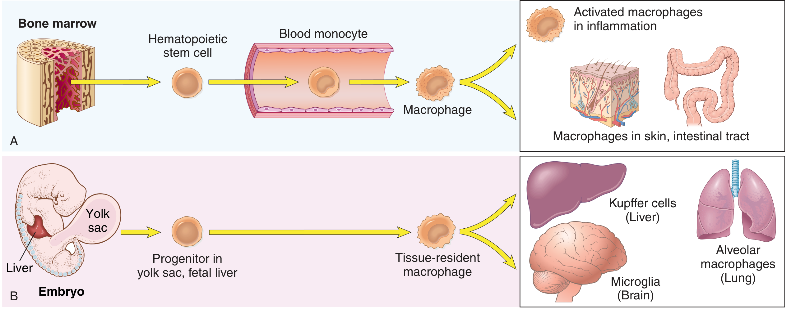
Two Populations of Tissue Macrophages:
- Hematopoietic stem cells → committed progenitors → blood monocytes (10-15 µm, bean-shaped nuclei, granular cytoplasm; half-life ~1 day in blood) → extravasate into tissues → differentiate into macrophages
- At sites of inflammation, monocytes begin emigrating early; within 48 hours they may be the predominant cell type
- Extravasation governed by same adhesion molecules and chemotactic factors as neutrophils
- Arise from progenitors in the yolk sac and fetal liver early in embryogenesis
- Migrate to their home organs and persist throughout life as stable, self-renewing populations
- Lifespan: months to years (vs. 1 day for monocytes)
| Organ | Resident Macrophage Name |
|---|---|
| Liver | Kupffer cells |
| Brain/CNS | Microglia |
| Lung | Alveolar macrophages |
| Spleen/lymph nodes | Sinus histiocytes |
| Skin | Langerhans cells (dendritic cells/macrophage lineage) |
| Bone | Osteoclasts |
| Connective tissues | Histiocytes |
3. Causes of Chronic Inflammation (Context for Macrophage Involvement)
-
Persistent infections by organisms that resist eradication - mycobacteria (M. tuberculosis, M. leprae), certain viruses, fungi, parasites → T-cell mediated delayed-type hypersensitivity → granulomatous reaction
-
Hypersensitivity / immune-mediated diseases:
- Autoimmune diseases: autoantigens trigger self-perpetuating T- and B-cell reactions (rheumatoid arthritis, multiple sclerosis, Hashimoto thyroiditis)
- Allergic diseases: excessive responses against environmental antigens (bronchial asthma)
- IBD: dysregulated immune responses against gut microbes
-
Prolonged exposure to toxic agents:
- Exogenous: silica particles (silicosis), asbestos fibers
- Endogenous: cholesterol crystals (atherosclerosis), urate crystals (gout)
-
Emerging associations: neurodegenerative diseases (Alzheimer), metabolic syndrome, type 2 diabetes - where "sterile" chronic inflammation with macrophage activation plays a pathogenic role
4. Two Pathways of Macrophage Activation: M1 and M2
Classical Activation (M1 Macrophages)
- Microbial products engaging Toll-like receptors (TLRs) - e.g., LPS (lipopolysaccharide), bacterial cell wall components
- IFN-γ (interferon-gamma) produced by activated Th1 CD4+ T cells and NK cells
- Foreign substances: silica crystals, particulate matter
- Nitric oxide (NO) via inducible NO synthase (iNOS) → microbicidal
- Reactive oxygen species (ROS) via NADPH oxidase → oxidative burst to kill microbes
- Lysosomal enzymes (protease, elastase, collagenase) → enhanced microbicidal capacity
- Pro-inflammatory cytokines: IL-1β, IL-6, IL-12, TNF-α, IL-23
- Chemokines → recruit more leukocytes to the site
- Prostaglandins and leukotrienes → amplify inflammation
- Present antigens via MHC class II → activate more T cells
Alternative Activation (M2 Macrophages)
- IL-4 and IL-13 (from Th2 CD4+ T cells, mast cells, eosinophils, epithelial cells)
- IL-10 (anti-inflammatory cytokine)
- TGF-β (transforming growth factor beta)
- Glucocorticoids, immune complexes
- Growth factors: TGF-β, PDGF (platelet-derived growth factor), FGF → stimulate fibroblasts, collagen synthesis, scar formation
- VEGF (vascular endothelial growth factor) → promote angiogenesis
- IL-10, TGF-β → suppress inflammation (anti-inflammatory)
- Mannose receptor, scavenger receptors → phagocytose debris and apoptotic cells (efferocytosis)
- Arginase (instead of iNOS) → produces ornithine/polyamines → tissue repair
M1 vs. M2 Comparison Table
| Feature | M1 (Classical) | M2 (Alternative) |
|---|---|---|
| Activating signals | LPS + IFN-γ (TLRs) | IL-4, IL-13, IL-10, TGF-β |
| Source of activating signals | Th1 T cells, microbes | Th2 T cells, mast cells |
| Main products | NO, ROS, IL-1, IL-6, IL-12, TNF | IL-10, TGF-β, VEGF, growth factors |
| Key enzyme | iNOS (inducible NO synthase) | Arginase |
| Function | Microbicidal, pro-inflammatory | Anti-inflammatory, tissue repair |
| Role in disease | Killing bacteria/fungi; tissue damage in autoimmunity | Fibrosis; wound healing; tumor progression |
| Markers | MHC II↑, CD80, CD86, iNOS | CD163, CD206 (mannose receptor), arginase-1 |
Important caveat: The M1/M2 classification is a conceptual framework. In reality, macrophages exhibit a spectrum of phenotypes, and many intermediate states exist that cannot be cleanly categorized as M1 or M2. Some authorities prefer the terms "M1-like" and "M2-like."
5. Functions of Activated Macrophages in Chronic Inflammation
A. Phagocytosis and Killing
- Engulf microbes, cell debris, apoptotic cells, foreign particles
- Intracellular killing via reactive oxygen species, reactive nitrogen species (NO), and lysosomal enzymes
- Opsonin-dependent phagocytosis enhanced by antibodies (via Fc receptors) and complement (via CR1, CR3 receptors)
- Note: Organisms like M. tuberculosis evade killing by preventing phagosome-lysosome fusion
B. Secretion of Inflammatory Mediators (Products of Activated Macrophages)
| Category | Mediators | Effect |
|---|---|---|
| Cytokines | IL-1β, TNF-α, IL-6 | Fever, acute phase response, leukocyte activation |
| IL-12, IL-23 | Activate Th1 and Th17 T cells | |
| IL-10, TGF-β (M2) | Anti-inflammatory, fibrosis | |
| Chemokines | IL-8 (CXCL8), MCP-1 (CCL2), MIP-1α | Recruit neutrophils, monocytes, lymphocytes |
| Arachidonic acid metabolites | PGE₂, TXA₂, LTB₄ | Vasodilation, pain, fever, chemotaxis |
| Reactive species | NO, ROS (superoxide, H₂O₂) | Microbicidal; tissue damage |
| Lysosomal enzymes | Protease, elastase, collagenase, MMP | Matrix degradation, tissue destruction |
| Growth factors | TGF-β, PDGF, FGF, VEGF | Fibroblast activation, angiogenesis, fibrosis |
| Complement components | C3, C4, C5, factor B | Opsonization, chemotaxis, membrane attack |
| Coagulation factors | Factor V, VII, X, thromboplastin | Fibrin deposition, walling off of infection |
C. Antigen Presentation
- Process and display antigens via MHC class II molecules to CD4+ T helper cells
- Express costimulatory molecules (B7/CD80, CD86) necessary for full T-cell activation
- Secrete IL-12 → drives differentiation of naïve CD4+ T cells toward Th1 phenotype
D. Tissue Repair and Fibrosis
- M2 macrophages secrete growth factors (TGF-β, PDGF) → activate fibroblasts → collagen synthesis
- Promote angiogenesis via VEGF
- Efferocytosis (phagocytosis of apoptotic cells) → releases anti-inflammatory signals → resolution
- Excessive M2 activation → pathological fibrosis (liver cirrhosis, pulmonary fibrosis, keloid)
6. Bidirectional Macrophage-Lymphocyte Interactions (Central to Chronic Inflammation)
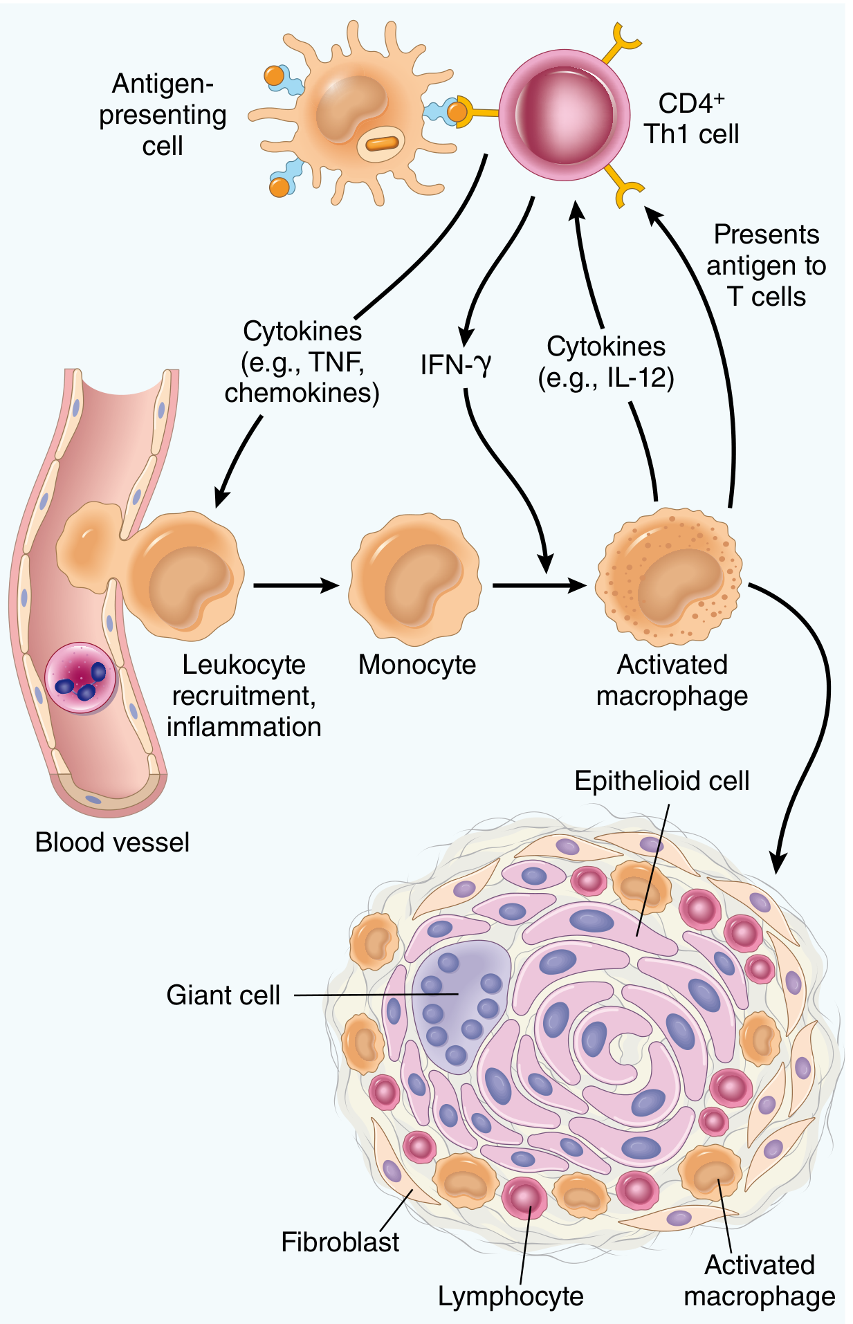
Macrophage → T Lymphocyte Signals:
- Antigen presentation via MHC class II + TCR engagement → T cell activation
- Costimulation via CD80/CD86 (on macrophage) binding CD28 (on T cell)
- IL-12 secretion → drives naïve CD4+ T cell differentiation → Th1 cells (IFN-γ producers)
- IL-23 → promotes and maintains Th17 cells
- IL-1, TNF → non-specific T cell activation and survival
T Lymphocyte → Macrophage Signals:
- IFN-γ → most potent macrophage activator → classical (M1) activation → enhanced killing, more cytokine production (self-amplifying loop)
- CD40L (CD154) → binds CD40 on macrophage → further macrophage activation
- IL-4, IL-13 → alternative (M2) macrophage activation → tissue repair, fibrosis
- IL-5 → recruits and activates eosinophils
- IL-17 → induces chemokine secretion (CXCL1, CXCL8) → neutrophil and monocyte recruitment
- Important in defense against extracellular bacteria/fungi and in autoimmune diseases (RA, IBD, psoriasis)
- IL-10, TGF-β → suppress macrophage activation → dampen chronic inflammation (defect in Tregs perpetuates chronic inflammation)
The Self-Perpetuating Cycle:
Persistent antigen → Macrophage presents antigen → T cell activation → T cells secrete IFN-γ → Macrophage classical activation → More cytokines → More T cell activation and monocyte recruitment → More activated macrophages → Tissue damage → Release of more antigens → Cycle perpetuates
7. Macrophage Interactions with Other Cells in Chronic Inflammation
With B Lymphocytes and Plasma Cells
- Macrophages present antigen to B cells (via MHC II)
- B cells differentiate into plasma cells → secrete antibodies
- Antibodies opsonize targets → antibody-dependent cellular cytotoxicity (ADCC) and complement-dependent phagocytosis via Fc receptors and complement receptors on macrophages
- At sites of prolonged chronic inflammation: tertiary lymphoid structures form (lymphoid follicle-like aggregates with germinal centers) → seen in RA synovium, Hashimoto thyroiditis, tumor microenvironments
With Fibroblasts
- M2-activated macrophages secrete TGF-β, PDGF, FGF → fibroblast proliferation and activation
- Fibroblasts synthesize and deposit collagen → fibrosis and scarring
- Excessive and unresolved M2 activation → pathological fibrosis (hepatic cirrhosis, pulmonary fibrosis, renal fibrosis)
With Endothelial Cells (Angiogenesis)
- Macrophage-derived VEGF, FGF → endothelial proliferation and new vessel formation (angiogenesis)
- New blood vessels supply the growing inflammatory tissue with nutrients and allow continued leukocyte ingress
With Neutrophils
- In chronic bacterial infection (e.g., osteomyelitis) and irritant exposures (cigarette smoke), macrophage-derived IL-8, IL-17, TNF continue to recruit neutrophils even in the chronic phase
- Neutrophil proteases + macrophage enzymes → combined tissue destruction
With Eosinophils (in Th2-type inflammation)
- Th2 cytokines (IL-4, IL-13) activate M2 macrophages AND promote eosinophil survival via IL-5 from Th2 cells
- Eotaxin (CCL11) - produced by macrophages and epithelial cells → recruits eosinophils
- Seen in: parasitic infections, allergic diseases, eosinophilic granulomatosis
With Mast Cells
- Mast cells degranulate → histamine, prostaglandins, cytokines → stimulate macrophage activation
- Macrophages secrete SCF (stem cell factor) → promote mast cell survival and proliferation
8. Granulomatous Inflammation - The Extreme Form of Macrophage-Mediated Chronic Inflammation
- Occurs when the inciting agent resists digestion by macrophages
- Persistent antigen stimulation → sustained Th1 CD4+ T cell response → IFN-γ → chronic macrophage activation
- Accumulated macrophages transform into epithelioid cells (enlarged, pink, epithelial-like cells with indistinct cell borders; low phagocytic but high secretory capacity) under the influence of IFN-γ
- Epithelioid cells fuse → form multinucleated giant cells (Langerhans-type: nuclei in horseshoe/peripheral arrangement; or foreign body type: nuclei randomly distributed)
Types of Granulomas:
| Type | Necrosis | Cause | Example |
|---|---|---|---|
| Caseating | Yes (central caseous/cheesy necrosis) | Mycobacterium tuberculosis, fungi | TB, histoplasmosis |
| Non-caseating | No | Sarcoidosis, Crohn's disease, foreign bodies, beryllium | Sarcoidosis |
Components of a Granuloma (center to periphery):
- Central zone: caseous necrosis (in TB) or no necrosis (in sarcoidosis)
- Epithelioid macrophages - aggregated, activated macrophages
- Multinucleated giant cells - Langerhans type (macrophage fusion)
- Rim of lymphocytes (mainly CD4+ Th1 cells secreting IFN-γ)
- Surrounding fibroblasts and collagen (M2-derived growth factors)
Role of Cytokines in Granuloma Formation:
- TNF-α (from macrophages): essential for granuloma formation and maintenance - anti-TNF therapy (infliximab) in RA/IBD can reactivate latent TB by disrupting granulomas
- IFN-γ (from Th1 cells): drives macrophage → epithelioid cell transformation
- IL-12 (from macrophages): sustains Th1 differentiation and IFN-γ production
9. Systemic Consequences of Macrophage Activation in Chronic Inflammation
| Systemic Effect | Mediator | Mechanism |
|---|---|---|
| Fever | IL-1β, TNF, IL-6 (endogenous pyrogens) | → COX → PGE₂ → hypothalamic temperature set point raised |
| Leukocytosis | TNF, IL-1, G-CSF, M-CSF | → accelerated release from bone marrow; CSF stimulates monocyte/granulocyte production |
| Acute phase response | IL-6 (+ IL-1, TNF) | Liver → ↑CRP, fibrinogen, SAA, complement; ↓albumin, transferrin |
| Anemia of chronic disease | IL-1, TNF, IL-6 → ↑hepcidin | Iron sequestration; impaired erythropoiesis; shortened RBC lifespan |
| Cachexia | TNF-α (cachectin), IL-1 | Suppress appetite, mobilize fat and protein stores |
| Amyloidosis | IL-6 → ↑SAA | SAA deposited as AA amyloid in chronic inflammatory diseases |
10. Role of Macrophages in Specific Chronic Inflammatory Diseases
| Disease | Macrophage Role |
|---|---|
| Tuberculosis | M1-activated macrophages attempt to kill M. tuberculosis; IFN-γ drives granuloma formation; TNF maintains granuloma integrity |
| Rheumatoid Arthritis | Synovial macrophages secrete TNF, IL-1, IL-6 → joint destruction; M2 macrophages drive pannus fibrosis |
| Atherosclerosis | Macrophages ingest oxidized LDL → foam cells → form the fatty streak; secrete MMPs → plaque instability |
| Silicosis/Asbestosis | Macrophages ingest particles → frustrated phagocytosis → persistent activation → cytokine release → fibrosis |
| Crohn's Disease | Non-caseating granulomas (transmural); macrophage-derived TNF is the key therapeutic target (anti-TNF therapy) |
| Tumor Microenvironment | Tumor-associated macrophages (TAMs) - mainly M2-like; secrete VEGF (angiogenesis), TGF-β (immunosuppression), MMPs (invasion) → promote tumor growth |
| Alzheimer Disease | Microglia (CNS macrophages) activated chronically → neuroinflammation; IL-1, TNF contribute to neuronal death |
11. Summary Diagram of Macrophage Interactions
PERSISTENT ANTIGEN
↓
Blood monocyte extravasates → TISSUE MACROPHAGE
↓
Phagocytosis + antigen processing
↓
┌────────────────┴────────────────┐
↓ ↓
ANTIGEN PRESENTATION CYTOKINE SECRETION
(MHC II + costimulators) (IL-12, TNF, IL-1)
↓ ↓
CD4+ T CELL ←──────IFN-γ released──────┐
│ │
┌─────────┼─────────┐ │
↓ ↓ ↓ │
Th1 Th2 Th17 │
(IFN-γ) (IL-4,13) (IL-17) │
↓ ↓ ↓ │
M1 macro M2 macro Neutrophil │
(killing) (repair) recruitment │
↓ ↓ │
Tissue Fibrosis ─────────────────────────────┘
damage Scar
│
↓ (if antigen persists + IFN-γ sustained)
GRANULOMA FORMATION
(Epithelioid cells + Giant cells + Lymphocyte rim + Fibrosis)
Key Points to Remember for Exam
- Macrophages are the dominant cells in chronic inflammation - they initiate, amplify, perpetuate, and attempt to resolve it
- Two lineages: inflammatory macrophages (from BM monocytes, short-lived) and tissue-resident macrophages (from yolk sac/fetal liver, long-lived - Kupffer, microglia, alveolar)
- M1 (classical, IFN-γ driven): microbicidal - NO, ROS, pro-inflammatory cytokines; tissue damage
- M2 (alternative, IL-4/IL-13 driven): anti-inflammatory; tissue repair; fibrosis; VEGF
- Macrophage-lymphocyte interaction is bidirectional: macrophage presents antigen + IL-12 → Th1 → IFN-γ → activates macrophage → more IL-12 → self-perpetuating cycle
- TNF is essential for granuloma maintenance - anti-TNF therapy can reactivate latent TB
- Granuloma = activated macrophages (epithelioid) + giant cells + lymphocytes + ± caseation - represents the extreme end of Th1/macrophage-mediated chronic inflammation
- Systemic effects of macrophage cytokines: fever (IL-1, TNF), leukocytosis (G-CSF), acute phase proteins (IL-6), anemia (hepcidin), cachexia (TNF/cachectin)
- Foam cells in atherosclerosis = macrophages engorged with oxidized LDL → secrete MMPs → plaque vulnerability
- Tertiary lymphoid structures form in long-standing RA synovium, Hashimoto thyroiditis - lymphoid follicles at sites of chronic inflammation
Virchow triad
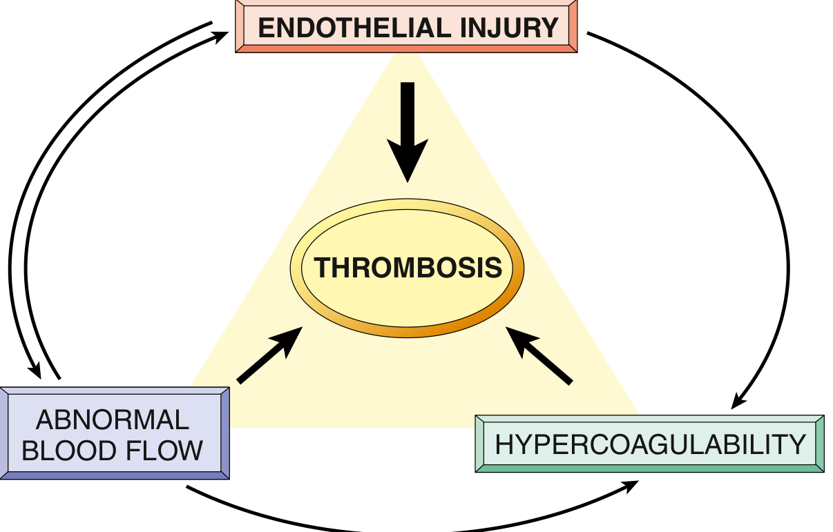
Virchow's Triad
Introduction
- Endothelial injury
- Abnormal blood flow (stasis or turbulence)
- Hypercoagulability of blood
Component 1: Endothelial Injury
Significance
Mechanisms of Endothelial Injury
- Exposure of subendothelial collagen and von Willebrand factor (vWF) → platelet adhesion and activation
- Exposure of tissue factor (thromboplastin) → initiation of the extrinsic coagulation cascade
- Examples: myocardial infarction (endocardial damage), ulcerated atherosclerotic plaques, vasculitis, trauma, cardiac surgery
- Cytokines and inflammatory mediators (TNF-α, IL-1)
- Infectious agents (viral, bacterial endotoxins)
- Abnormal blood flow (shear stress)
- Metabolic abnormalities: hypercholesterolemia, homocysteinemia (homocysteine directly damages endothelium)
- Toxins: cigarette smoke components
- Hypertension
- Radiation injury
Prothrombotic Changes from Endothelial Activation
| Normal Endothelial Function (Antithrombotic) | Activated Endothelial Change (Prothrombotic) |
|---|---|
| Thrombomodulin expression | Downregulated → less thrombin inactivation |
| Endothelial protein C receptor | Downregulated → less Protein C activation |
| Tissue factor pathway inhibitor (TFPI) | Downregulated → less inhibition of extrinsic pathway |
| Prostacyclin (PGI₂) secretion | Decreased → less platelet inhibition |
| t-PA secretion (fibrinolysis) | Decreased |
| Tissue factor (TF/thromboplastin) | Upregulated → activates extrinsic coagulation cascade |
| Plasminogen activator inhibitor (PAI-1) | Increased secretion → impairs fibrinolysis |
| vWF (stored in Weibel-Palade bodies) | Released → promotes platelet adhesion |
Summary: Activated/injured endothelium loses its normal anticoagulant, antiaggregatory, and fibrinolytic properties, and gains procoagulant properties that tip the balance toward thrombosis.
Clinical Examples (Endothelial Injury-Dominant Thrombosis)
- Coronary artery thrombosis overlying a ruptured atherosclerotic plaque → acute MI
- Mural thrombus on endocardium after MI (infarcted myocardium causes endocardial injury)
- Thrombotic vegetations (infective endocarditis, Libman-Sacks endocarditis in SLE)
- Vasculitis (arteritis)
- Trauma, burns, surgical incisions
Component 2: Abnormal Blood Flow (Stasis or Turbulence)
Normal vs. Abnormal Flow
- Blood cells (including platelets) flow centrally
- Fresh clotting factors delivered continuously; activated factors diluted and cleared
- Antithrombotic substances from endothelium maintain non-thrombotic state
| Flow Abnormality | Location | Predominant Mechanism |
|---|---|---|
| Stasis (sluggish flow) | Veins, cardiac chambers | Allows platelet-endothelium contact; depletes fresh antithrombotic factors; promotes local hypoxia and endothelial activation |
| Turbulence (chaotic flow) | Arteries, cardiac valves | Causes endothelial injury/activation; creates countercurrents and local pockets of stasis |
How Abnormal Flow Promotes Thrombosis
- Disrupts laminar flow → platelets contact endothelium instead of flowing centrally
- Prevents dilution of activated clotting factors by fresh flowing blood
- Retards inflow of clotting factor inhibitors
- Promotes endothelial activation → hypoxia activates endothelium → prothrombotic gene expression
- Turbulence creates downstream stasis pockets (eddies) in addition to direct endothelial damage
Clinical Examples (Abnormal Blood Flow-Dominant Thrombosis)
- Deep vein thrombosis (DVT): prolonged bed rest, immobilization (e.g., long-haul flights), plaster casts, paralysis → slows venous return
- Cardiac stasis: atrial fibrillation (loss of atrial contraction → atrial appendage stasis → thrombus), dilated cardiomyopathy, post-MI left ventricular aneurysm
- Varicose veins: dilated, stagnant venous flow
- Polycythemia vera: increased blood viscosity → reduced flow
- Sickle cell disease: rigid, sticky RBCs → impede flow
- Atherosclerosis: irregular intimal surface → turbulent flow around plaques
- Aortic/arterial aneurysms: turbulence at the aneurysm neck + local stasis within the sac → thrombus on inner wall (mural thrombi)
- Abnormal cardiac valves: stenosed/calcified valves, prosthetic valves → turbulent flow
Component 3: Hypercoagulability
Definition
Classification
A. Primary (Inherited / Genetic) Hypercoagulability
| Defect | Frequency | Mechanism | Thrombosis Risk |
|---|---|---|---|
| Factor V Leiden mutation (Arg506Gln) | 2-15% of European ancestry | Point mutation makes factor Va resistant to cleavage by activated Protein C → loss of anticoagulant feedback | Heterozygotes: 3-4× ↑ risk of DVT; Homozygotes: 25-50× ↑ risk |
| Prothrombin G20210A mutation | 1-2% of general population | Substitution in 3'-UTR of prothrombin gene → increased prothrombin expression → more thrombin generated | ~3× ↑ risk of venous thrombosis |
| Increased factor VIII, IX, or XI | Variable | Excess procoagulant | Increased DVT risk |
| Hyperfibrinogenemia | Variable | Increased clotting substrate | Increased thrombosis risk |
| Defect | Mechanism | Clinical Presentation |
|---|---|---|
| Antithrombin III deficiency | Loss of major inhibitor of thrombin and factors Xa, IXa | Recurrent DVT and pulmonary embolism in adolescence/young adulthood |
| Protein C deficiency | Cannot inactivate factors Va and VIIIa | Recurrent venous thrombosis; neonatal purpura fulminans in homozygotes |
| Protein S deficiency | Protein S is a cofactor for Protein C | Same as Protein C deficiency |
- Fibrinolysis defects (e.g., t-PA deficiency, excess PAI-1)
- Homozygous homocystinuria (cystathionine β-synthetase deficiency → very high homocysteine → both arterial and venous thrombosis)
Key clinical note: In young patients (<50 years) with DVT especially without a precipitating cause, inherited causes of hypercoagulability must be excluded. Factor V Leiden is present in up to 60% of those with recurrent DVT.
B. Secondary (Acquired) Hypercoagulability
| Risk Level | Acquired Conditions |
|---|---|
| High risk | Prolonged bed rest/immobilization; MI; atrial fibrillation; tissue injury (surgery, fracture, burn); cancer (especially mucin-secreting adenocarcinomas); prosthetic cardiac valves; DIC; heparin-induced thrombocytopenia (HIT); antiphospholipid antibody syndrome |
| Elevated risk | Cardiomyopathy; nephrotic syndrome; hyperestrogenic states (pregnancy, oral contraceptive use); inflammatory bowel disease; smoking; obesity; sickle cell anemia; increasing age |
Two Clinically Important Acquired Hypercoagulable States:
- Occurs in up to 5% of patients on unfractionated heparin
- Mechanism: autoantibodies form against complexes of heparin + platelet factor-4 (PF4)
- These antibody-PF4-heparin complexes bind Fc receptors on platelets → platelet activation, aggregation, and clearance
- Paradox: Thrombocytopenia (low platelets) + prothrombotic state (paradoxical thrombosis despite anticoagulation)
- Low-molecular-weight heparin causes HIT less frequently but can still trigger it if antibodies already exist
- Autoantibodies against phospholipid-binding proteins (e.g., β₂-glycoprotein I, prothrombin)
- Previously called "lupus anticoagulant" (because antibodies prolong in vitro coagulation tests but cause thrombosis in vivo)
- Can occur in SLE (secondary APS) or without another autoimmune disease (primary APS)
- Clinical manifestations: recurrent arterial and venous thromboses, recurrent pregnancy loss (placental thrombosis), thrombocytopenia
- Mechanism: antibodies activate platelets and endothelium, and interfere with natural anticoagulant mechanisms
- Cancer: Release of procoagulant tumor products (e.g., mucin from adenocarcinoma that directly activates factor X); tissue factor expression by tumor cells → Trousseau syndrome (migratory thrombophlebitis)
- Oral contraceptives/pregnancy: Increased hepatic synthesis of clotting factors II, V, VII, VIII, X; reduced antithrombin and Protein S
- Nephrotic syndrome: Loss of antithrombin III in urine + increased hepatic synthesis of fibrinogen
- Smoking/obesity: Unknown precise mechanisms; promote hypercoagulability
- Advancing age: Increased platelet aggregation + reduced PGI₂ from endothelium
Morphology of Thrombi
General Features
- Thrombi are attached to the vessel wall at the point of initiation (attachment point = "head" of thrombus)
- The thrombus extends downstream (tail/body)
- Lines of Zahn: Alternating pale (platelets + fibrin) and dark (erythrocyte-rich) layers visible in arterial thrombi; represent successive waves of platelet aggregation and red cell entrapment; only present in thrombi formed in flowing blood (helps distinguish antemortem thrombus from postmortem clot)
Arterial ("White") Thrombi
- Rich in platelets (platelet-dominant, pale/gray)
- Firm, adherent to the vessel wall
- Most commonly associated with endothelial injury overlying atherosclerotic plaques
- Locations: coronary arteries, cerebral arteries, aorta
- Typically occlusive
Venous ("Red") Thrombi - Phlebothrombosis
- Rich in erythrocytes ("red" or "stasis" thrombi) enmeshed in fibrin
- More fibrin, fewer platelets (formed under low shear stress)
- Almost always occlusive
- Propagate toward the heart (against the direction of blood flow in veins)
- Form a long cast within the lumen → prone to embolize
- Locations: deep veins of lower limbs (90%), upper extremities, dural sinuses, portal vein, hepatic veins
Distinguishing Thrombus from Postmortem Clot
| Feature | Antemortem Thrombus | Postmortem Clot |
|---|---|---|
| Attachment to wall | Focally attached | Not attached |
| Consistency | Firm | Gelatinous |
| Lines of Zahn | Present | Absent |
| Color | Gray-red with gray fibrin strands | Dark red (dependent) + yellow "chicken fat" (upper) |
| Red cell settling | Even distribution | Settled (dependent red layer) |
Special Locations of Thrombi
- Mural thrombi: On cardiac chamber walls (after MI, dilated cardiomyopathy, atrial fibrillation) or inside aortic aneurysms
- Vegetations: Thrombi on cardiac valves
- Infective endocarditis: Large, irregular, destructive vegetations (bacteria/fungi)
- Non-bacterial thrombotic endocarditis (NBTE): Small, sterile vegetations in hypercoagulable states (cancer, uremia)
- Libman-Sacks endocarditis: Sterile verrucous vegetations in SLE (on both surfaces of mitral valve leaflets)
Fates of Thrombi
1. Propagation
- Thrombus enlarges by accretion of more platelets and fibrin
- Increases risk of vascular occlusion and embolization
2. Embolization
- Part or all of the thrombus dislodges → transported in bloodstream → lodges at a distant site
- Most common: DVT → pulmonary embolism (70-80% of PE originates from leg DVT)
- Also: left heart mural thrombus → systemic arterial embolism (stroke, limb ischemia, renal infarction)
3. Dissolution (Fibrinolysis)
- Newly formed thrombi → plasminogen activators (t-PA, u-PA) → plasmin → fibrin degradation → thrombus dissolves
- Clinical implication: thrombolytic therapy (t-PA, streptokinase) is effective only if given within hours of formation; older thrombi have extensive fibrin cross-linking → resistant to plasmin (not responsive to thrombolysis)
4. Organization and Recanalization
- Older thrombi are invaded by:
- Endothelial cells (from adjacent endothelium)
- Smooth muscle cells
- Fibroblasts
- New capillary channels form within the thrombus → recanalization → partial restoration of blood flow
- Eventually, the thrombus is converted into a vascularized mass of connective tissue → incorporated into the vessel wall
- Sometimes, instead of organizing, the center of the thrombus undergoes enzymatic digestion (lysosomal enzymes from entrapped leukocytes) → central softening
Interaction Between the Three Components of Virchow's Triad
ENDOTHELIAL INJURY
↓
Releases tissue factor + vWF
+ Loses PGI₂, thrombomodulin (anticoagulant)
↓ ↓
Platelet activation Coagulation cascade
↓ ↓
Platelet aggregation ──────── Fibrin formation
↓
THROMBUS
ABNORMAL BLOOD FLOW
↓
Stasis → Platelets contact endothelium
→ Activated factors accumulate (not diluted)
→ Local hypoxia → endothelial activation
→ ↑ Procoagulant, ↓ Anticoagulant expression
→ HYPERCOAGULABILITY (secondary)
Turbulence → Endothelial injury/activation
→ Downstream stasis pockets
HYPERCOAGULABILITY
↓
Excess thrombin generation (Factor V Leiden, prothrombin mutation)
or Loss of anticoagulant control (AT III, Protein C/S deficiency)
or Platelet hypersensitivity (APS, HIT)
↓
Overwhelms normal antithrombotic mechanisms of endothelium
↓
THROMBOSIS even without gross endothelial injury
Summary Table: Virchow's Triad at a Glance
| Component | Key Mechanism | Common Causes | Thrombus Type | Location |
|---|---|---|---|---|
| Endothelial Injury | Loss of antithrombotic surface; exposure of subendothelial TF and vWF | MI, atherosclerotic plaque rupture, vasculitis, trauma, hypertension, homocysteinemia | Platelet-rich (white) | Arteries, heart |
| Abnormal Blood Flow | Stasis: platelets contact wall, activated factors accumulate; Turbulence: endothelial injury | Bed rest, AF, cardiac failure, aneurysms, varicosities, polycythemia | Red/fibrin-rich (venous) | Veins; cardiac chambers; aneurysms |
| Hypercoagulability | Excess procoagulants or deficient anticoagulants | Factor V Leiden, prothrombin mutation, AT III/Protein C/S deficiency, OCP, cancer, APS, HIT | Both arterial and venous | Veins primarily; also arteries (in APS) |
Key Points for the Exam
- Virchow's triad = Endothelial injury + Stasis/turbulence + Hypercoagulability - first described in 1856
- Endothelial injury is most important for arterial/cardiac thrombosis (platelet-rich, white thrombi)
- Stasis is most important for venous thrombosis (red, fibrin-rich thrombi); turbulence causes endothelial injury in arteries
- Factor V Leiden (most common hereditary thrombophilia) = resistant to Protein C → up to 60% of recurrent DVT cases
- Lines of Zahn = alternating pale (platelets/fibrin) and dark (red cell) layers in arterial thrombi = proof of antemortem formation in flowing blood
- Postmortem clot = gelatinous, unattached, "chicken fat" appearance - no Lines of Zahn
- Four fates of thrombus: Propagation → Embolization → Dissolution (fibrinolysis) → Organization and Recanalization
- Thrombolysis works only in early thrombus (hours) - old thrombi have cross-linked fibrin resistant to plasmin
- HIT paradox: Heparin therapy → thrombocytopenia + thrombosis (paradoxical); due to anti-PF4-heparin antibodies
- Antiphospholipid syndrome: Arterial AND venous thromboses + recurrent pregnancy loss; "lupus anticoagulant" prolongs PTT in vitro but causes thrombosis in vivo
- Trousseau syndrome (migratory thrombophlebitis) = paraneoplastic hypercoagulability in visceral cancers; mucin from adenocarcinoma activates factor X
- All three components interact - abnormal flow causes endothelial dysfunction (= secondary hypercoagulability); hypercoagulability overwhelming normal endothelium can cause thrombosis without structural injury
Pathophysiology of septic shock
Pathophysiology of Septic Shock
1. Definition and Epidemiology
- Persisting hypotension requiring vasopressors to maintain MAP ≥ 65 mmHg
- Serum lactate > 2 mmol/L despite adequate fluid resuscitation
-
750,000 cases/year in the USA; incidence rising
- 2% of all hospital admissions; 50% require ICU
- Mortality: 20-40% despite modern treatment
- Most common trigger: Gram-positive bacteria (now #1), then gram-negative bacteria, then fungi
- Emerging: viral infections (SARS-CoV-2)
2. Causes and Triggering Organisms
| Category | Examples |
|---|---|
| Gram-positive bacteria (most common) | Staphylococcus aureus (MRSA), Streptococcus pneumoniae, Enterococcus - trigger via exotoxins (superantigens), lipoteichoic acid, peptidoglycan |
| Gram-negative bacteria | E. coli, Klebsiella, Pseudomonas, Neisseria - trigger via LPS (lipopolysaccharide / endotoxin) |
| Fungi | Candida, Aspergillus - trigger via fungal cell wall β-glucans, mannans |
| Viral | SARS-CoV-2, influenza - trigger via nucleic acids, viral proteins |
- LPS (lipid A of gram-negative cell wall) - most potent trigger
- Lipoteichoic acid (gram-positive)
- Peptidoglycans
- Fungal β-glucans, mannans
- Bacterial flagellin
- Bacterial and viral nucleic acids (CpG DNA, dsRNA)
3. Pathogenesis: Overview and Cascade
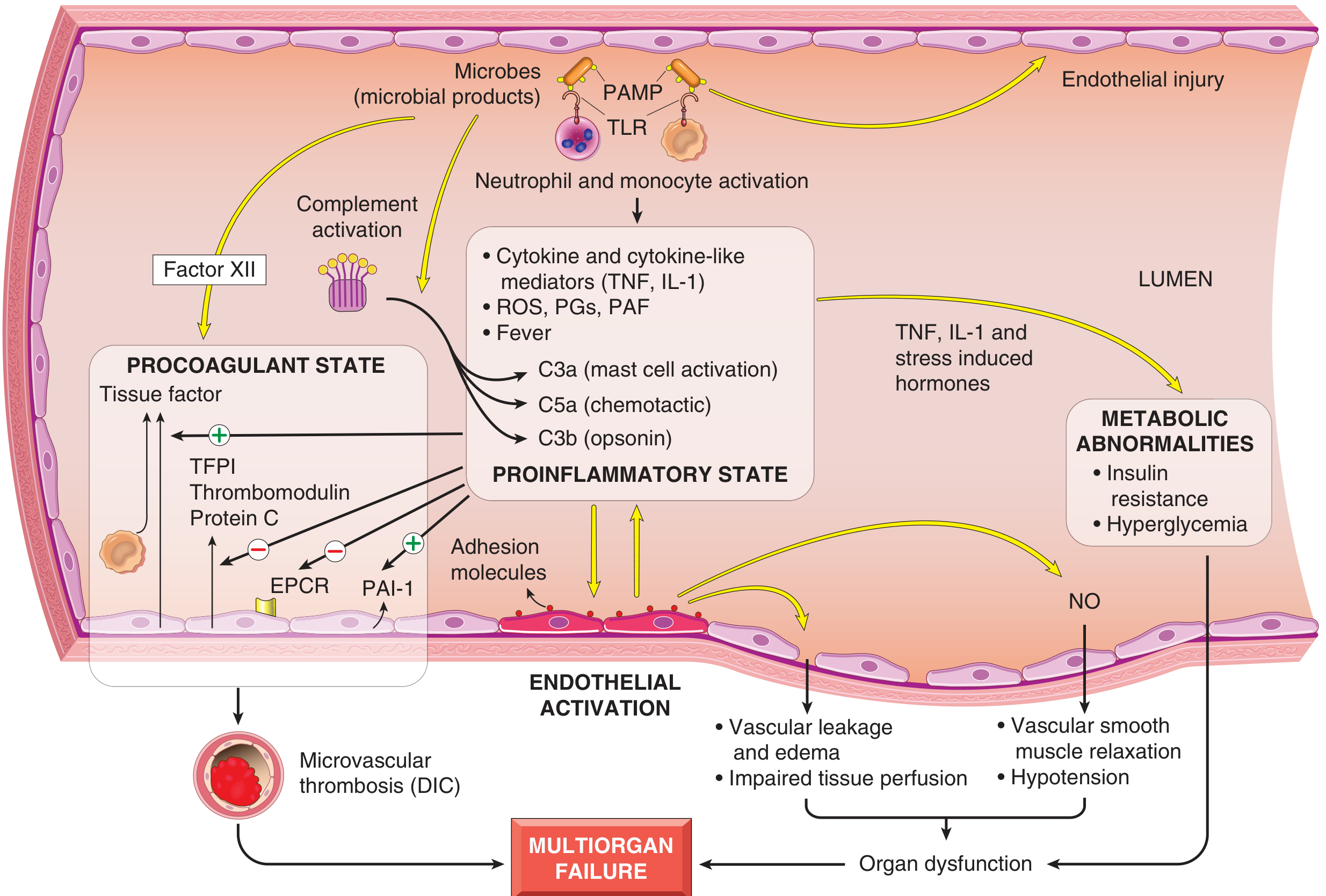
4. Step-by-Step Pathophysiological Cascade
STEP 1: Pattern Recognition - PAMPs Engage Innate Immune Receptors
-
Toll-like receptors (TLRs) - most critical
- TLR4 (with co-receptor MD-2 and LBP): recognizes LPS/endotoxin of gram-negative bacteria
- TLR2: recognizes peptidoglycan (gram-positive) and lipoteichoic acid
- TLR5: recognizes flagellin
- TLR3, TLR7, TLR9: recognize viral/bacterial nucleic acids
- LPS-binding protein (LBP) in plasma binds LPS → presents to CD14 on monocytes → CD14 presents to TLR4/MD-2 complex → signal transduction
-
G protein-coupled receptors - detect bacterial formyl peptides (fMLF)
-
C-type lectin receptors (Dectins) - recognize fungal cell wall β-glucans and mannans
- TLR ligation → recruitment of MyD88 adaptor protein → activation of NF-κB (nuclear factor-kappa B) transcription factor → nuclear translocation → transcription of pro-inflammatory genes
- Also activates MAPK (mitogen-activated protein kinase) pathways → AP-1 → further cytokine gene expression
STEP 2: Cytokine Storm - The Proinflammatory State
| Mediator | Source | Key Effects in Sepsis |
|---|---|---|
| TNF-α (tumor necrosis factor) | Macrophages (main source) | Fever; endothelial activation; ↑ tissue factor; ↓ thrombomodulin; promotes DIC; hypotension; negative cardiac inotropy; insulin resistance |
| IL-1β | Macrophages, endothelium | Synergizes with TNF; fever; endothelial activation; coagulation activation |
| IL-6 | Macrophages, T cells | Acute phase protein synthesis (CRP, fibrinogen, procalcitonin); lymphocyte activation |
| IL-12 | Macrophages, DCs | Activates NK cells and T cells → IFN-γ production |
| IFN-γ | T cells, NK cells | Amplifies macrophage activation |
| IL-18 | Macrophages | Synergizes with IL-12 to drive IFN-γ production |
| HMGB1 | Macrophages, necrotic cells | Late mediator of sepsis; promotes inflammation; damages endothelium |
| IL-8 (CXCL8) | Macrophages, endothelium | Neutrophil chemotaxis → massive neutrophil recruitment |
- Prostaglandins (PGE₂, PGI₂): vasodilation, fever, increased permeability
- Thromboxane A₂ (TXA₂): vasoconstriction, platelet aggregation (contributes to DIC)
- Platelet-activating factor (PAF): platelet aggregation, vasodilation, increased permeability, bronchoconstriction
- Leukotrienes: bronchoconstriction, increased permeability
- Superoxide (O₂•⁻), H₂O₂, OH• - produced by activated neutrophils and macrophages
- Directly damage endothelial cells, parenchymal cells → tissue injury
- iNOS induced in vascular smooth muscle by TNF, IL-1, IFN-γ
- Massive NO production → profound vasodilation → hypotension (a hallmark of septic shock)
- NO also has direct cytotoxic effects on mitochondria (inhibits cytochrome oxidase)
STEP 3: Complement Activation
| Complement Fragment | Effect in Sepsis |
|---|---|
| C3a | Mast cell activation → histamine → vasodilation, ↑ permeability |
| C5a | Potent chemotaxis for neutrophils; neutrophil and macrophage activation; mast cell activation |
| C3b | Opsonization → facilitates phagocytosis |
| C5b-9 (MAC) | Direct microbial lysis; endothelial injury |
STEP 4: Endothelial Activation and Injury
- Upregulation of adhesion molecules (E-selectin, ICAM-1, VCAM-1) → massive leukocyte adhesion and transmigration → leukocyte-mediated tissue damage
- ↑ Production of tissue factor (TF/thromboplastin) → triggers extrinsic coagulation cascade
- ↑ PAI-1 (plasminogen activator inhibitor-1) → impairs fibrinolysis
- Loss of endothelial thrombomodulin, TFPI, endothelial protein C receptor (EPCR) → loss of anticoagulant mechanisms
- Increased vascular permeability → massive third spacing of fluid → edema, hypovolemia
- Loss of vasomotor tone (via NO) → vasodilation → hypotension
- Endothelial apoptosis (via TNF, ROS) → further barrier disruption
Result: A "leaky" vascular system that cannot maintain blood pressure or keep fluid intravascular.
STEP 5: Procoagulant State and DIC
- Tissue factor expression by endothelium and monocytes → extrinsic pathway activation → thrombin generation → fibrin clot formation throughout microcirculation
- Direct coagulation activation via factor XII (Hageman factor) by microbial products → intrinsic pathway
- Loss of natural anticoagulants:
- Thrombomodulin (normally inactivates thrombin) → decreased
- Protein C pathway (normally inactivates Va, VIIIa) → impaired (EPCR lost, thrombomodulin lost)
- Antithrombin III → consumed
- TFPI → reduced
- Impaired fibrinolysis: TNF/IL-1 upregulate PAI-1 → t-PA inactivated → fibrin clots not cleared
- Platelet activation: TXA₂, PAF → platelet aggregation → microvascular thrombi
- Microvascular thrombosis in multiple organs → ischemic injury → multiorgan failure
- Consumptive coagulopathy → depletion of clotting factors and platelets → paradoxical bleeding tendency (hemorrhagic diathesis)
- Fibrin degradation products (FDPs/D-dimers) accumulate → further impair hemostasis
STEP 6: Metabolic Abnormalities
- Insulin resistance and hyperglycemia (despite adequate insulin; impairs neutrophil function, worsens outcomes)
- Lactic acidosis: tissue hypoperfusion → anaerobic metabolism → lactate accumulation → ↓ pH → blunts vasomotor response (arterioles cannot constrict → further pooling)
- Mitochondrial dysfunction: NO inhibits cytochrome oxidase (complex IV) → impaired oxidative phosphorylation → cytopathic hypoxia (cells cannot use O₂ even if delivered) → cellular death
- Hypoalbuminemia (liver diverts protein synthesis to acute-phase reactants; third spacing of albumin)
- Adrenal insufficiency (relative or absolute) → impairs vasopressor response
STEP 7: Counterregulatory Immunosuppression (Late Phase)
- Shift from Th1 to Th2 cytokine profile (IL-10, TGF-β predominate; IL-12, IFN-γ decrease)
- Production of anti-inflammatory mediators: soluble TNF receptors, IL-1 receptor antagonist (IL-1Ra), IL-10
- Lymphocyte apoptosis (massive loss of CD4+ T cells, B cells, dendritic cells → impaired adaptive immunity)
- Immunosuppressive effects of apoptotic cells → phagocytes shift to M2/anti-inflammatory phenotype
- Cellular anergy: immune cells unresponsive to further stimulation
5. Stages of Shock (Common to All Types)
Stage 1: Non-Progressive (Compensated) Phase
- Baroreceptor reflexes detect ↓ BP → sympathetic activation
- Release of catecholamines (epinephrine, norepinephrine) → tachycardia, vasoconstriction
- ADH (vasopressin) release → water retention
- Renin-angiotensin-aldosterone (RAA) axis activation → Na⁺ and water retention → ↑ blood volume
- Generalized sympathetic stimulation → redistribution of blood flow
- Tachycardia, peripheral vasoconstriction → "shocky" skin (cool, pale, clammy)
- Exception in septic shock: initial peripheral vasodilation (NO-driven) → patient may present with warm, flushed skin ("warm shock" - key distinguishing feature of early septic shock)
- Blood flow maintained to heart and brain (coronary and cerebral vessels relatively insensitive to sympathetic vasoconstriction)
- If underlying cause corrected at this stage → full recovery
Stage 2: Progressive Phase
- Persistent O₂ deficit → aerobic → anaerobic glycolysis → lactic acidosis
- Lactic acidosis → blunted arteriolar vasomotor response → arteriolar dilation despite sympathetic signals
- Blood pools in the microcirculation (venous pooling + arteriolar failure) → ↓ cardiac output
- Endothelial ischemic injury → DIC begins
- Vital organs begin to fail (early organ dysfunction)
Stage 3: Irreversible Phase
- Lysosomal enzyme leakage from dying cells → accelerate cell injury ("autolysis")
- Myocardial contractility worsens (NO, TNF, acidosis all depress cardiac function)
- Gut ischemia → mucosal barrier failure → intestinal flora translocate into circulation → bacteremia superimposed on existing shock → further cytokine release
- Acute kidney injury (ischemic ATN)
- Despite treatment, death inevitable
6. Multiorgan Dysfunction Syndrome (MODS) / Multiorgan Failure (MOF)
- Tissue hypoperfusion → ischemic injury
- Microvascular thrombosis (DIC) → ischemic necrosis
- Direct cytokine/ROS-mediated injury
- Mitochondrial dysfunction (cytopathic hypoxia)
- Neutrophil-mediated collateral damage
Organ-Specific Consequences:
| Organ | Pathological Change | Clinical Manifestation |
|---|---|---|
| Kidney | Acute tubular necrosis (ATN) - ischemic + toxic | Oliguria → anuria; rising creatinine; acute kidney injury (AKI) |
| Lung | Diffuse alveolar damage (DAD) = "shock lung" = ARDS | Bilateral infiltrates, ↓ PaO₂/FiO₂, respiratory failure |
| Liver | Ischemic hepatitis ("shock liver"); Kupffer cell activation | ↑ transaminases, jaundice, coagulopathy |
| Brain | Ischemic encephalopathy; microthrombi | Confusion, delirium, coma → "septic encephalopathy" |
| Adrenals | Cortical lipid depletion; occasionally adrenal hemorrhage (Waterhouse-Friderichsen in meningococcemia) | Relative/absolute adrenal insufficiency; vasopressor-refractory hypotension |
| GI tract | Ischemic enterocolitis; mucosal ulceration; barrier failure | Ileus, translocation of bacteria → bacteremia |
| Heart | Septic cardiomyopathy (TNF + IL-1 + NO → myocardial depression) | ↓ ejection fraction; "warm shock" with low SVR |
| Coagulation | DIC | Bleeding + microvascular thrombosis |
| Skin | Peripheral vasoconstriction in late shock; purpuric lesions in DIC | Cool, mottled skin; petechiae, ecchymoses |
7. Morphology in Shock (Autopsy Findings)
| Organ | Morphological Change |
|---|---|
| Kidney | Acute tubular necrosis - tubular epithelial cell necrosis (straight portion, loop of Henle); cast formation; tubular regeneration in survivors |
| Lung | Diffuse alveolar damage (hyaline membranes, edema, type II pneumocyte proliferation) - "shock lung" |
| Liver | Central hemorrhagic necrosis (centrilobular); fatty change |
| Brain | "Ischemic encephalopathy" - ischemic neurons in hippocampus and Purkinje cells |
| Adrenals | Cortical lipid depletion (reflects stress response - rapid use of stored lipids for steroid synthesis); Waterhouse-Friderichsen syndrome in severe gram-negative sepsis (bilateral adrenal hemorrhage) |
| GI tract | Superficial mucosal hemorrhages, stress ulcers (Curling ulcers in shock/burns) |
| Kidney glomeruli | Fibrin thrombi in glomerular capillaries (most readily visualized thrombi in DIC) |
Note: Except for neuronal and cardiomyocyte loss (irreversibly lost), affected tissues may recover completely if the patient survives.
8. Current Definitions (Sepsis-3, 2016)
| Term | Definition |
|---|---|
| Sepsis | Life-threatening organ dysfunction caused by dysregulated host response to infection; identified by SOFA score increase ≥ 2 |
| Septic shock | Sepsis + persistent hypotension requiring vasopressors to maintain MAP ≥ 65 mmHg + serum lactate > 2 mmol/L despite adequate fluid resuscitation |
| qSOFA (quick SOFA) | Bedside screen: altered mentation + tachypnea (RR >22) + hypotension (SBP <100); ≥2 criteria warrants further workup |
9. Summary: Key Pathophysiological Steps
MICROBIAL INFECTION (gram-positive > gram-negative > fungi)
↓
PAMPs recognized by TLRs (TLR4 for LPS) on macrophages/neutrophils/DCs
↓
NF-κB activation → CYTOKINE STORM
(TNF-α, IL-1β, IL-6, IL-12, IFN-γ, IL-8, HMGB1)
↙ ↓ ↘
ENDOTHELIAL COMPLEMENT COAGULATION
ACTIVATION ACTIVATION ACTIVATION
↓ (C3a, C5a, C3b) ↓
• ↑ permeability ↑ Tissue factor
• ↑ adhesion molecules ↓ Thrombomodulin
• ↓ anticoagulants ↓ Protein C
• ↑ PAI-1 ↓
↓ DIC (microvascular thrombosis
↑ Leukocyte recruitment + consumptive coagulopathy)
↓ ↓
Leukocyte-mediated tissue damage ↓
↓ ↓
↓NO (via iNOS) ↓
VASODILATION + HYPOTENSION ↓
(warm shock → low SVR) ↓
↓ ↓
THIRD SPACING (edema) ↓
(↑ vascular permeability) ↓
↓ ↓
↓ ─────────────────────────→↓
TISSUE HYPOPERFUSION
↓
ANAEROBIC METABOLISM
→ LACTIC ACIDOSIS
↓
MITOCHONDRIAL DYSFUNCTION
(NO inhibits cytochrome oxidase)
↓
MULTIORGAN FAILURE
(Kidneys-ATN, Lungs-ARDS, Brain-encephalopathy,
Liver-shock hepatitis, Heart-cardiomyopathy, DIC)
↓
DEATH
10. Key Points for the Exam
- Most common trigger: Gram-positive bacteria (#1 today), gram-negative bacteria (#2), fungi
- Core initiating event: PAMP + TLR4 (LPS/TLR4 is the classical pathway) → NF-κB → cytokine storm
- Three limbs of Virchow's triad are all activated in sepsis: endothelial injury + stasis + hypercoagulability → DIC
- TNF-α is the master cytokine in septic shock: fever, vasodilation, ↑ TF, ↓ thrombomodulin, cardiac depression, cachexia
- Nitric oxide (NO) from iNOS = central mechanism of vasodilation and hypotension in septic shock
- "Warm shock" (warm, flushed skin) in early sepsis = peripheral vasodilation (NO) - distinguishes from other types of shock (cold, clammy)
- Septic shock unique hemodynamics: ↓ SVR (low resistance) + ↑ cardiac output initially (hyperdynamic) → later ↓ CO as myocardium fails
- DIC = microvascular thrombosis + consumptive coagulopathy - fibrin thrombi most visible in glomerular capillaries
- Lactic acidosis = both a marker (tissue hypoperfusion) and a pathological mechanism (blunts vasomotor response)
- Three stages of shock: Non-progressive (compensated) → Progressive → Irreversible
- Adrenal cortical lipid depletion = morphological hallmark of stress response at autopsy
- Mitochondrial dysfunction (cytopathic hypoxia): cells die even when O₂ is delivered - NO inhibits cytochrome oxidase complex IV
- Immunosuppression phase of sepsis (lymphocyte apoptosis, IL-10, Th2 shift) = major cause of late mortality via secondary infections
Oncometabolites in cancer cells
"oncometabolites" AND "cancer" AND "IDH"
oncometabolites 2-hydroxyglutarate succinate fumarate cancer mechanisms epigenetics 2024 2025
https://pmc.ncbi.nlm.nih.gov/articles/PMC12424225
PMID: 40931345
Oncometabolites in Cancer Cells
1. Definition
- 2-Hydroxyglutarate (2-HG) - produced by mutant IDH1/IDH2
- Succinate - accumulates in SDH (succinate dehydrogenase) loss-of-function mutations
- Fumarate - accumulates in FH (fumarate hydratase) loss-of-function mutations
Conceptual core: All three canonical oncometabolites are structurally similar to α-ketoglutarate (α-KG) and act as competitive inhibitors of α-KG-dependent dioxygenases (α-KGDDs) - a broad enzyme family that uses α-KG as a co-substrate for oxidative reactions critical to epigenetics, HIF regulation, and DNA repair.
2. The TCA Cycle and Normal Enzyme Functions
Glucose → Pyruvate → Acetyl-CoA
↓
Oxaloacetate + Acetyl-CoA → Citrate
↓
cis-Aconitate ← [itaconate branched here by ACOD1]
↓
Isocitrate
↓ ← IDH1/IDH2 (normal: Isocitrate → α-KG + CO₂)
α-KG (α-ketoglutarate)
↓
Succinyl-CoA
↓
Succinate ← [SDH converts Succinate → Fumarate]
↓
Fumarate ← [FH converts Fumarate → Malate]
↓
Malate
↓
Oxaloacetate
- IDH1/IDH2 (gain-of-function neomorphic mutation): instead of Isocitrate → α-KG, mutant IDH converts α-KG → 2-HG (a "neomorphic" reaction)
- SDH (loss-of-function): succinate cannot be converted to fumarate → succinate accumulates
- FH (loss-of-function): fumarate cannot be converted to malate → fumarate accumulates
3. Oncometabolite 1: 2-Hydroxyglutarate (2-HG)
Mutation: IDH1 and IDH2
| Feature | IDH1 | IDH2 |
|---|---|---|
| Location | Cytosol and peroxisome | Mitochondria |
| Normal function | Isocitrate → α-KG + CO₂ (part of TCA cycle) | Same, in mitochondria |
| Hotspot mutation | R132H (Arg132His) - most common | R172K, R140Q |
| Mutation type | Gain-of-function neomorphic | Same |
| Product | 2-HG (D-enantiomer, also called R-2-HG) | Same |
| Key tumors | Diffuse gliomas (grades 2, 3, secondary GBM), AML, intrahepatic cholangiocarcinoma, chondrosarcoma | AML (R140Q more common in AML) |
- This is termed "neomorphic" because the mutant enzyme acquires a new enzymatic activity not possessed by the wild-type enzyme
- 2-HG accumulates to millimolar concentrations (>100-fold above normal) in tumors with IDH mutations
- Two enantiomers: D-2-HG (R-2-HG) from IDH1/IDH2 mutations (oncogenic) and L-2-HG (S-2-HG) from L2HGDH mutations (also oncogenic in certain brain/renal tumors)
Associated Cancers
| Cancer | IDH Mutation | Key Notes |
|---|---|---|
| Diffuse gliomas (Grade 2-3) | IDH1 R132H (~90%) | WHO 2021 requires IDH testing for all diffuse gliomas; IDH-mutant gliomas have better prognosis than IDH-wildtype |
| Secondary GBM | IDH1 mutation | Arises from lower grade IDH-mutant glioma |
| AML | IDH1 or IDH2 | ~20% of AML; targetable with enasidenib (IDH2) and ivosidenib (IDH1) |
| Intrahepatic cholangiocarcinoma | IDH1 (~20%) | FDA-approved: ivosidenib (IDH1i) for IDH1-mutant CCA |
| Chondrosarcoma | IDH1/IDH2 | Central/enchondroma subtypes |
| Angioimmunoblastic T-cell lymphoma (AITL) | IDH2 R172 |
4. Oncometabolite 2: Succinate
Mutation: SDH (Succinate Dehydrogenase) - Loss-of-Function
| SDH subunit gene | Cancers associated |
|---|---|
| SDHB | Paraganglioma, pheochromocytoma, GIST, renal cell carcinoma |
| SDHC | Paraganglioma (head/neck) |
| SDHD | Paraganglioma (head/neck) - paternal inheritance (imprinting) |
| SDHA | Paraganglioma, GIST, pituitary adenoma |
| SDHAF2 (assembly factor) | Paraganglioma |
5. Oncometabolite 3: Fumarate
Mutation: FH (Fumarate Hydratase) - Loss-of-Function
- Autosomal dominant germline FH mutation
- Cutaneous and uterine leiomyomas + aggressive type 2 papillary renal cell carcinoma
- FH-deficient RCC is highly aggressive, even when small
6. Core Molecular Mechanism: Inhibition of α-KG-Dependent Dioxygenases (α-KGDDs)
Normal α-KGDD reaction:
Substrate + α-KG + O₂ → Hydroxylated product + Succinate + CO₂
Inhibited by 2-HG / Succinate / Fumarate (compete with α-KG binding site)
| α-KGDD Enzyme | Normal Function | Effect of Inhibition |
|---|---|---|
| TET1/TET2/TET3 (DNA demethylases) | Convert 5-methylcytosine → 5-hydroxymethylcytosine → demethylation | DNA hypermethylation → CpG island methylator phenotype (CIMP) → silencing of tumor suppressor genes |
| JHDM/KDMs (Jumonji C domain histone lysine demethylases) | Remove methyl groups from histones (H3K4me3, H3K27me3, H3K36me2, H3K9me3) | Histone hypermethylation → altered chromatin → transcriptional reprogramming → differentiation block |
| PHD1/2/3 (Prolyl hydroxylases) | Hydroxylate HIF-1α proline residues → VHL recognition → proteasomal degradation | Failure to degrade HIF-1α → pseudohypoxia → ↑VEGF, ↑GLUT1, ↑glycolytic genes → angiogenesis + Warburg effect |
| FTO (RNA demethylase) | Removes N6-methyladenosine (m6A) from mRNA → mRNA stability regulation | ↑m6A methylation → altered mRNA stability and translation |
| ALKBH family (DNA repair) | Oxidative demethylation of alkylated DNA bases | Impaired DNA repair → genome instability |
| TET + KDM co-inhibition | Maintain differentiation programs | Epigenetic "lock" → block in differentiation → cells trapped in progenitor/stem-like state |
7. Three Major Oncogenic Mechanisms
Mechanism 1: Epigenetic Reprogramming (DNA + Histone Hypermethylation)
- TET enzymes normally catalyze the first step of active DNA demethylation: 5-methylcytosine (5mC) → 5-hydroxymethylcytosine (5hmC)
- Inhibition → CpGs remain methylated → CpG Island Methylator Phenotype (CIMP)
- In IDH-mutant gliomas: glioma CIMP (G-CIMP) - hypermethylation of hundreds of CpG islands → silencing of tumor suppressors, differentiation genes
- In IDH-mutant AML: hypermethylation blocks myeloid differentiation
- Accumulation of H3K9me2/3 → heterochromatin formation → gene silencing
- Accumulation of H3K27me3 → PRC2-like repression of differentiation genes
- Combined DNA + histone hypermethylation creates a comprehensive epigenetic lock that traps cells in an undifferentiated, proliferative state
Mechanism 2: Pseudohypoxia and HIF-1α Stabilization
- PHD inhibition → HIF-1α proline residues not hydroxylated → VHL (E3 ubiquitin ligase) cannot bind → HIF-1α not ubiquitinated → HIF-1α escapes proteasomal degradation
- HIF-1α dimerizes with HIF-1β → transcription of hypoxia-response genes even under normoxia: "pseudohypoxia"
- HIF-1α target genes upregulated:
- VEGF → angiogenesis
- GLUT1/GLUT3 → ↑glucose uptake (Warburg)
- LDHA → lactate dehydrogenase A → ↑glycolysis
- PDK1 → pyruvate dehydrogenase kinase → diverts pyruvate away from mitochondria
- EPO → erythropoietin (in renal tumors)
Mechanism 3: Redox Imbalance and Post-Translational Modifications
- Reverse electron transport (RET): succinate accumulation drives electrons backward through Complex I → massive ROS production → genomic instability, mitochondrial damage
- Retrograde signaling: succinate leaks from mitochondria → acts as a signaling molecule → binds GPR91 (SUCNR1) receptor on cell surface → activates STAT3 and ERK → ↑VEGF expression → pro-angiogenic
- Protein succinylation: succinate reacts with protein lysines via succinyl-CoA → alters protein function
- Succination: fumarate reacts non-enzymatically with free cysteine residues on proteins → forms S-(2-succinyl)-cysteine (2-SC) → irreversible post-translational modification
- Targets include: KEAP1 succination → KEAP1 inactivation → NRF2 released → NRF2 target gene activation → antioxidant response + NRF2-driven oncogenesis
- ALKBH5 succination → impaired RNA demethylation
- GSH succination → impaired glutathione function → oxidative stress
- Also: fumarate causes DNA fragmentation and chromosomal instability
- 2-HG depletes NADPH (consumed in the neomorphic IDH reaction) → impaired glutathione synthesis (NADPH-dependent) → ↑oxidative stress
- O-2-HGylation: 2-HG can modify protein serine/threonine residues → alters cytoskeletal regulation and DNA repair
8. Differentiation Block - A Key Oncogenic Output
Normal hematopoietic progenitor
↓ (TET2 + KDMs drive methylation changes enabling differentiation)
Mature blood cell
IDH-mutant cell:
TET2 inhibited + KDMs inhibited
↓
DNA/histone hypermethylation → differentiation gene promoters silenced
↓
Progenitor cell CANNOT differentiate → remains immortalized/self-renewing
↓
Accumulation of undifferentiated blasts → AML
9. Effects on the Tumor Microenvironment (TME)
| Cell Type in TME | Effect of Oncometabolites |
|---|---|
| T cells | 2-HG and succinate inhibit TET enzymes → hypermethylation of T cell effector gene loci (IFN-γ, perforin, granzyme B) → T cell exhaustion and immunosuppression |
| Dendritic cells | 2-HG impairs DC maturation and antigen presentation → reduced priming of anti-tumor T cells |
| Macrophages | Succinate stabilizes HIF-1α in macrophages → polarizes toward M1 (pro-inflammatory) and ↑IL-1β production; but in tumor context can also promote M2-like immunosuppression |
| NK cells | 2-HG impairs NK cell cytotoxicity |
| Endothelial cells | Succinate activates GPR91 on endothelium → ↑VEGF → angiogenesis |
| Tumor stroma | Fumarate succination of extracellular proteins → altered ECM composition |
Itaconate: The "Immunometabolite" Oncometabolite
- Anti-inflammatory: inhibits SDH → limits NLRP3 inflammasome activation; activates NRF2 (via KEAP1 modification) → antioxidant response; inhibits IκBζ → ↓IL-6, IL-12
- Pro-tumorigenic in TME: suppresses macrophage anti-tumor activity; promotes immune evasion in certain contexts
- Represents the concept that metabolic reprogramming of immune cells shapes tumor immunity
10. Comparison Table: Three Canonical Oncometabolites
| Feature | 2-Hydroxyglutarate (2-HG) | Succinate | Fumarate |
|---|---|---|---|
| Enzyme mutated | IDH1 (R132) / IDH2 (R172, R140) | SDH (A, B, C, D subunits) | FH |
| Mutation type | Gain-of-function (neomorphic) | Loss-of-function | Loss-of-function |
| Normal product lost | α-KG (but α-KG still made by other pathways) | Fumarate (TCA disrupted) | Malate (TCA disrupted) |
| Primary cancers | Gliomas, AML, CCA, chondrosarcoma | Paraganglioma, pheochromocytoma, GIST, RCC | HLRCC (leiomyoma + papillary RCC type 2) |
| Inheritance | Usually somatic; germline rare | Autosomal dominant germline (hereditary paraganglioma syndrome) | Autosomal dominant germline (HLRCC/Reed syndrome) |
| α-KGDD inhibition | Yes (competitive) | Yes (competitive) | Yes (competitive) |
| Dominant epigenetic effect | DNA + histone hypermethylation (CIMP) | DNA + histone hypermethylation | DNA + histone hypermethylation |
| Pseudohypoxia | Yes (PHD inhibition) | Yes (PHD inhibition) - prominent | Yes (PHD inhibition) |
| Unique mechanism | NADPH depletion; O-2-HGylation | GPR91 signaling; RET-mediated ROS; protein succinylation | KEAP1 succination → NRF2 activation; 2SC formation |
| Targeted therapy available | Yes - Ivosidenib (IDH1i), Enasidenib (IDH2i), Vorasidenib (IDH1/2i for glioma) | No approved agent yet | No approved agent yet |
| Diagnostic biomarker | 2-HG by MRS (brain); serum 2-HG in AML; IHC for IDH R132H | IHC for SDHB loss; SDH mutation sequencing | IHC for 2SC (succinated proteins) |
11. Warburg Effect and Metabolic Reprogramming Context
- Warburg effect: Cancer cells preferentially use aerobic glycolysis (glucose → lactate, even in O₂) rather than oxidative phosphorylation. Must upregulate glycolysis ~200-fold to compensate for lower ATP yield.
- Oncometabolites = "transforming activities" in the classification of metabolic reprogramming (Sabiston, Chapter 13): directly tied to cell transformation, in contrast to "enabling activities" (e.g., KRAS-driven nutrient acquisition) and "neutral activities"
- IDH-mutant tumors show:
- Increased reliance on pyruvate carboxylase (anaplerosis via oxaloacetate)
- Lipid depletion (NADPH consumed by mutant IDH reaction → less reductive lipid synthesis)
- Shift to glutaminolysis (glutamine → α-KG) to compensate for TCA cycle disruption
12. α-KG as Tumor Suppressor - The Counter-Metabolite
- Supporting TET and KDM activity → DNA and histone demethylation → maintaining differentiation programs
- Supporting PHD activity → degrading HIF-1α → preventing pseudohypoxia
- α-KG supplementation is a therapeutic strategy being investigated to reverse oncometabolite-driven epigenetic changes
- Low α-KG / High 2-HG, succinate, fumarate → oncogenic
- High α-KG / Low oncometabolites → tumor suppressive
13. Therapeutic Targeting
| Drug | Target | Cancer | Status |
|---|---|---|---|
| Ivosidenib (AG-120) | IDH1 inhibitor | AML (IDH1-mutant), CCA | FDA approved |
| Enasidenib (AG-221) | IDH2 inhibitor | AML (IDH2-mutant) | FDA approved |
| Vorasidenib (AG-881) | Pan-IDH1/2 inhibitor (CNS penetrant) | IDH-mutant glioma | FDA approved 2024 - first CNS-penetrant IDH inhibitor |
| α-KG supplementation (cell-permeable forms) | Restore α-KGDD activity | Multiple | Preclinical/early clinical |
| NRF2 inhibitors | Block NRF2 (upregulated in FH-deficient tumors via KEAP1 succination) | HLRCC-RCC | Preclinical |
| HIF-1α/VEGF pathway inhibitors | Target pseudohypoxia consequence | Paraganglioma, RCC | Belzutifan (HIF-2α inhibitor, FDA-approved for VHL disease/RCC) |
14. Key Points for the Exam
- Definition: Oncometabolites = small molecules accumulated abnormally (due to enzyme mutations) that directly drive oncogenesis - not just metabolic byproducts
- Three canonical oncometabolites: 2-HG (IDH1/2 gain-of-function), Succinate (SDH loss), Fumarate (FH loss) - all TCA cycle intermediates
- Central mechanism: Structural mimicry of α-KG → competitive inhibition of α-KG-dependent dioxygenases (α-KGDDs) → multiple downstream effects
- Key α-KGDDs inhibited: TET1/2/3 (DNA demethylation), KDMs (histone demethylation), PHDs (HIF-1α prolyl hydroxylases)
- Dominant epigenetic result: DNA + histone hypermethylation → gene silencing → differentiation block (CIMP phenotype)
- Pseudohypoxia: PHD inhibition → HIF-1α stabilization → VEGF, GLUT1 upregulation → angiogenesis + aerobic glycolysis
- IDH mutation is neomorphic (gain of new function) - generates 2-HG from α-KG; this distinguishes it from SDH/FH which are simple loss-of-function
- Fumarate's unique mechanism: non-enzymatic protein succination (2SC formation) → KEAP1 inactivation → NRF2-driven oncogenesis
- Succinate's unique mechanism: GPR91 receptor signaling → VEGF, STAT3/ERK; reverse electron transport → ROS
- IDH-mutant gliomas: better prognosis, G-CIMP phenotype, R132H most common; diagnosed by IDH R132H IHC ± sequencing (WHO 2021)
- SDH tumors: hereditary paraganglioma syndrome (autosomal dominant); SDHB most common in malignant paraganglioma
- FH tumors: HLRCC = cutaneous leiomyoma + uterine fibroids + aggressive papillary RCC type 2
- Targeted therapy: Ivosidenib (IDH1i), Enasidenib (IDH2i), Vorasidenib (pan-IDH1/2, CNS-penetrant for glioma - approved 2024)
- TME effects: oncometabolites promote immune evasion (T cell exhaustion, DC dysfunction, NK cell impairment)
- α-KG is a tumor suppressor metabolite - inverse of oncometabolites
Describe the metastatic cascade
The Metastatic Cascade
1. Definition and Overview
2. Routes of Metastatic Spread
| Route | Description | Most common for |
|---|---|---|
| Lymphatic | Travel via lymphatics to regional then distant nodes; Virchow's node (left supraclavicular) in GI/lung cancer | Carcinomas (epithelial) |
| Hematogenous | Enter blood vessels (veins > arteries, due to thinner walls) → disseminate systemically | Sarcomas; also carcinomas |
| Seeding of body cavities | Direct extension into peritoneal, pleural, pericardial, or subarachnoid cavities | Ovarian, GI, mesothelioma |
| Perineural spread | Track along nerve sheaths | Head/neck, prostate cancers |
3. The Metastatic Cascade - Step by Step
- Phase 1: Invasion - local and into vasculature
- Phase 2: Vascular dissemination, homing, and colonization at distant sites
PHASE 1: INVASION OF THE EXTRACELLULAR MATRIX
- The epithelial basement membrane (type IV collagen, laminin, fibronectin, perlecan)
- The interstitial connective tissue (type I/III collagens, fibronectin, proteoglycans)
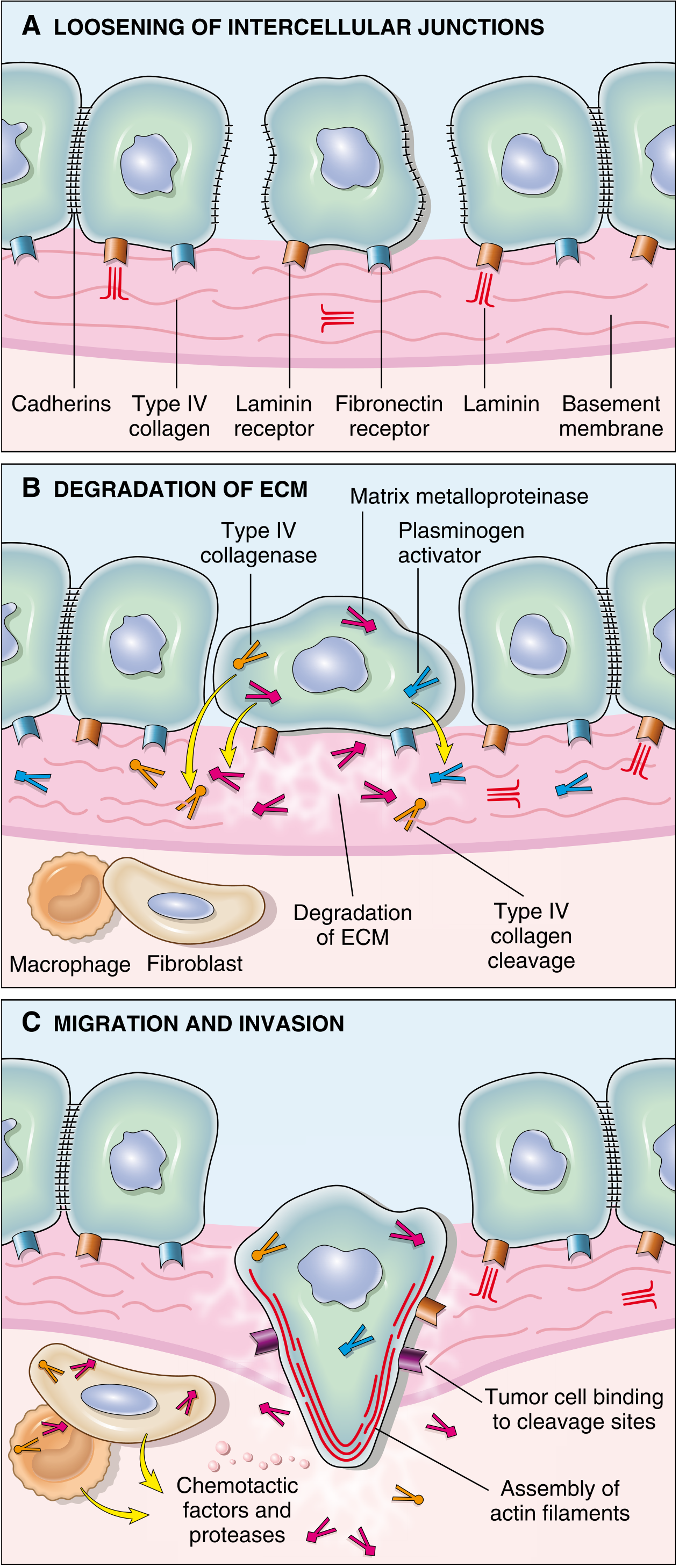
STEP 1: Loosening of Cell-Cell Contacts (Reduced Intercellular Adhesion)
- E-cadherin - the most important intercellular adhesion molecule for epithelial cells; homotypic transmembrane glycoprotein that holds cells together AND relays differentiation/growth-inhibitory signals
- Tight junctions (claudins, occludins)
- Gap junctions (connexins)
- E-cadherin function is lost by two mechanisms:
- Somatic mutation of CDH1 gene (e.g., hereditary diffuse gastric cancer - germline CDH1 mutation; some lobular breast cancers, diffuse gastric adenocarcinoma)
- Transcriptional silencing via Epithelial-Mesenchymal Transition (EMT)
The Epithelial-Mesenchymal Transition (EMT) - Central Event
- SNAIL (SNAI1, SNAI2/SLUG) - directly repress E-cadherin promoter; induce mesenchymal genes
- TWIST (TWIST1, TWIST2) - bHLH transcription factor; represses E-cadherin; upregulates mesenchymal markers
- ZEB1/ZEB2 (zinc finger E-box binding homeobox) - E-cadherin repressors
- Other inducers: TGF-β (from stroma), HGF/scatter factor (from fibroblasts), EGF, FGF, hypoxia (HIF-1α), Wnt, Notch signaling
| Lost (Epithelial Markers) | Gained (Mesenchymal Markers) |
|---|---|
| E-cadherin (CDH1) | N-cadherin (CDH2) - "cadherin switching" |
| Claudins, occludins (tight junctions) | Vimentin |
| Desmoplakin (desmosomes) | Fibronectin |
| Cytokeratins | Smooth muscle actin (α-SMA) |
| Epithelial polarity (apical-basolateral) | Actin stress fibers |
| Basement membrane attachment | Invasive/migratory filopodia |
- Loss of cell-cell adhesion → individual cells detach from tumor mass
- Loss of apical-basolateral polarity → cells become motile
- Resistance to anoikis (apoptosis triggered by loss of anchorage to ECM) - a critical survival advantage for circulating tumor cells
- Gain of stem cell-like properties (tumor initiating capacity)
- Upregulation of proteases (MMP production)
Key exam point: EMT is not binary but a spectrum/partial state - most invading cancer cells exhibit hybrid E/M phenotypes. Cells in full EMT may have superior invasive capacity but inferior proliferative capacity; colonization requires MET to restore proliferative potential.
STEP 2: Degradation of the Basement Membrane and ECM
| Protease Class | Key Members | ECM Targets | Notes |
|---|---|---|---|
| Matrix Metalloproteinases (MMPs) | MMP-2 (gelatinase A), MMP-9 (gelatinase B), MMP-1 (collagenase), MMP-3 (stromelysin), MT1-MMP (MMP-14) | Type IV collagen (BM), gelatin, laminin, fibronectin, proteoglycans | Most important class; zinc-dependent; membrane-anchored MT-MMPs activate soluble MMPs at cell surface |
| Serine proteases | uPA (urokinase plasminogen activator), tPA | Plasminogen → plasmin (activates MMPs) | uPA receptor (uPAR) concentrates proteolysis at leading edge |
| Cysteine proteases | Cathepsin D, cathepsin B, cathepsin L | Type IV collagen, fibronectin, laminin | Lysosomal; secreted by macrophages; active in acidic TME |
| Heparanase | HPSE1 | Heparan sulfate proteoglycans | Releases growth factors (FGF, VEGF) sequestered in ECM |
- MMP-9: cleaves type IV collagen in BM → releases VEGF stored in ECM → promotes angiogenesis; generates pro-invasive collagen/laminin fragments
- MMP-2: cleaves laminin-5 γ2 chain → generates promigratory laminin fragments; forms complex with αvβ3 integrin at invadopodia
- Generate cryptic binding sites in BM → novel integrin binding sites → support tumor cell migration
- Release ECM-sequestered growth factors (IGF, FGF, EGF-like) → feed tumor growth
- Cleavage products of ECM have chemotactic activity → direct tumor migration
STEP 3: Attachment to Novel ECM Components (Altered Integrin Signaling)
- Normal epithelial cells: integrins that bind laminin/collagen are restricted to basal surface → maintains polarity, quiescence
- In cancer: altered integrin expression profile:
- αvβ3 integrin - upregulated in many cancers; binds osteopontin, vitronectin, fibronectin → survival and migration signals
- α6β4 - laminin receptor, overexpressed in invasive carcinomas
- α5β1 - fibronectin receptor; promotes migration in desmoplastic stroma
- Resistance to anoikis: Tumor cells upregulate integrins and survival signals (PI3K/Akt) that override the normal apoptotic response to loss of ECM contact
- ECM cleavage products of collagen IV and laminin (by MMP-2/9) generate novel sequences that bind tumor cell receptors and stimulate migration - a positive feedback loop
STEP 4: Migration and Locomotion
- Cells extend lamellipodia and filopodia at leading edge (actin polymerization)
- Form new focal adhesions at leading edge (integrin-FAK-Rho GTPase signaling)
- Generate traction force via actomyosin contraction
- Release focal adhesions at trailing edge → net forward movement
- Invadopodia: specialized F-actin-rich protrusions containing concentrated MMPs → focal ECM degradation at leading edge
| Factor | Source | Receptor on Tumor Cell |
|---|---|---|
| HGF/Scatter factor | Cancer-associated fibroblasts (paracrine) | MET receptor tyrosine kinase |
| EGF | TAMs (in paracrine tumor-macrophage loop) | EGFR |
| IGF-1, IGF-2 | Liver, bone marrow | IGF-1R |
| CXCL12 (SDF-1) | Bone marrow, liver, lung endothelium | CXCR4 on tumor cells - key organotropism axis |
| Cleavage products of ECM | Released by MMPs | Various |
| Autocrine motility factors | Tumor cells themselves | Autocrine GF receptors |
- Most carcinomas invade as collective cell clusters (sheets, cords, nests) rather than single cells, with leader cells at the front
- Leading cells have more mesenchymal character (high SNAIL, low E-cadherin); trailing cells retain epithelial traits
- Collective migration preserves cell-cell contacts and may confer survival advantages
PHASE 2: VASCULAR DISSEMINATION, HOMING, AND COLONIZATION
STEP 5: Intravasation (Entry into Blood or Lymph Vessels)
- Lymphatics are preferred by many carcinomas (leakier, lack a well-formed BM compared to blood vessels)
- Blood vessel intravasation: requires same proteolytic machinery (MMPs) + directed migration toward tumor-associated blood vessels
- Tumor-associated macrophages (TAMs) at invasion fronts directly facilitate intravasation via paracrine EGF-CSF-1 signaling loops (TAMs secrete EGF → tumor cell migration; tumor cells secrete CSF-1 → TAM recruitment)
- Angiogenic vessels (leaky, incompletely formed BM) are preferred sites for intravasation
STEP 6: Survival in the Circulation (Circulating Tumor Cells - CTCs)
- Mechanical shear stress (particularly in arterial circulation)
- Anoikis (loss of ECM contact → apoptosis)
- Immune surveillance: NK cells, cytotoxic T cells, macrophages actively kill CTCs
- Complement system
| Mechanism | Details |
|---|---|
| Platelet aggregation around CTCs | Tumor cells activate platelets → platelet cloak → physical protection from NK cells and shear stress; platelet-derived TGF-β further suppresses NK cytotoxicity; fibrin deposition further stabilizes tumor emboli |
| Anoikis resistance | Upregulation of integrin αvβ3, EGFR, PI3K/Akt signaling; altered dependence on ECM signals |
| Aggregation into CTC clusters | Homotypic (tumor-tumor) + heterotypic (tumor-platelet) aggregates → tumor emboli → more likely to arrest en masse in capillary beds; collectively more likely to possess all properties needed for metastasis; contain cancer stem cells |
| EMT-conferred stemness | Cells in partial EMT state have stem-like properties → more fit to survive and colonize |
| Immune evasion | Downregulation of MHC-I; expression of PD-L1 on CTCs; platelet-derived TGF-β suppresses NK cells |
| Polyphosphate / coagulation activation | Tumor cells express anionic substances (polyphosphate) that activate factor XII (contact pathway) → fibrin deposition → stabilization of tumor emboli → enhance arrest in capillary beds |
Most CTCs die - the metastatic cascade is profoundly inefficient. Only rare CTCs with specific molecular features survive and colonize.
STEP 7: Arrest and Extravasation at Distant Sites
- Mechanical trapping: in capillary beds downstream of the primary tumor (anatomy-based - colon → portal vein → liver)
- Specific molecular adhesion: tumor cells express adhesion molecules and chemokine receptors whose ligands are preferentially expressed on the endothelium of target organs
| Mechanism | Example |
|---|---|
| Anatomical/vascular drainage | Colon → liver (portal vein); All organs → lung (systemic veins → right heart → pulmonary capillaries) |
| Chemokine receptor-ligand axis | Breast cancer cells express CXCR4; bone marrow, liver, lymph nodes, lung endothelium express CXCL12 (SDF-1) → breast cancer preferentially metastasizes to bone, liver, lung, lymph nodes |
| Adhesion molecule expression | CD44 on tumor cells binds hyaluronate on target organ high endothelial venules; α4β1 (VLA-4) on tumor cells binds VCAM-1 on bone marrow endothelium |
| Growth factor availability | Transferrin (high in lung, bone, brain) drives growth in those organs |
| Premetastatic niche (see below) | Primary tumor pre-conditions distant organs before cells arrive |
| Primary Tumor | Preferred Metastatic Sites |
|---|---|
| Carcinoma of colon | Liver (#1 - portal drainage), lung |
| Breast carcinoma | Bone, liver, lung, brain, adrenals |
| Prostate carcinoma | Bone (osteoblastic metastases) |
| Lung carcinoma | Brain, adrenals, liver, bone |
| Neuroblastoma | Liver, bone |
| Ocular malignant melanoma | Liver (remarkable tropism) |
| Cutaneous melanoma | Brain, liver, lung, bone |
| Renal cell carcinoma | Lung, bone, liver, brain ("brown tumor"-like) |
| Thyroid carcinoma | Lung, bone |
- Tumor cell transmigrates between endothelial cells → breaches vascular BM → enters organ parenchyma
- In fenestrated endothelium (liver sinusoids, bone marrow): relatively easy extravasation
- In tight junction endothelium (brain, blood-brain barrier): requires active disruption (metalloproteinases, inflammatory mediators)
- Same proteolytic machinery (MMPs, cathepsins) + integrin-mediated adhesion used in reverse
- Monocytes/neutrophils in the target tissue may also facilitate extravasation
STEP 8: The Premetastatic Niche
- Tumor-secreted factors (VEGF, TGF-β, osteopontin, PlGF, LOX) enter circulation → act on target organs
- Tumor-derived extracellular vesicles (exosomes): carry oncoproteins, miRNAs, integrins → activate resident stromal cells; exosome integrins determine which organ is "educated" (e.g., αvβ5 exosomes → liver; α6β4 exosomes → lung)
- In target organs: bone marrow-derived cells (BMDCs) are recruited → VEGFR1+ hematopoietic progenitors, TAM precursors → cluster at future metastatic sites before tumor cells arrive
- These BMDCs remodel ECM, increase vascular permeability → create a receptive niche
- Once the niche is formed, circulating tumor cells home to it preferentially
STEP 9: Colonization - Growth at the Distant Site
- Die immediately
- Enter a state of tumor dormancy (survive but do not proliferate) - can remain dormant for years to decades (clinically relevant in breast cancer, melanoma, prostate cancer)
- Lack of angiogenic signals at new site
- Active immune suppression by NK cells and CD8+ T cells
- Absence of required stromal signals
- Epigenetic reprogramming at new site
- MET (Mesenchymal-Epithelial Transition): reverts EMT → restores proliferative capacity
- Tumor cells secrete cytokines, growth factors, and ECM molecules that act on resident stromal cells → remodel the site to support cancer growth
- Example - bone metastasis (breast cancer): Breast cancer cells secrete PTHrP (parathyroid hormone-related protein) → stimulates osteoblasts to produce RANKL → activates osteoclasts → bone resorption → releases IGF, TGF-β embedded in bone matrix → feeds breast cancer cell growth (a vicious cycle)
- Cancer stem cells (within the metastatic deposit): essential for sustained proliferation and resistance to therapy; EMT process generates cells with CSC properties
4. EMT-MET Axis: The Full Arc
PRIMARY TUMOR
Epithelial cancer cells
↓ (EMT triggered by SNAIL, TWIST, TGF-β, hypoxia)
Partial/Full Mesenchymal Phenotype
• ↓ E-cadherin • ↑ N-cadherin, vimentin
• Motile, invasive, stem-like
• Anoikis-resistant
↓ (Intravasation)
CIRCULATION
• CTC (single or clustered)
• Protected by platelets, fibrin
↓ (Extravasation → colonization)
DISTANT SITE
↓ (MET triggered by microenvironment)
Re-epithelialization
• ↑ E-cadherin restored
• Proliferative capacity regained
• Histological resemblance to primary
METASTATIC COLONY
5. Metastasis Genes
| Class | Examples | Function |
|---|---|---|
| Metastasis-promoting genes (metastasis oncogenes) | SNAIL, TWIST, MET, CXCR4, CD44, Src | Drive EMT, intravasation, organotropism |
| Metastasis suppressor genes | NM23 (NDPK), KAI1/CD82, BRMS1, KISS1, RhoGDI2 | Suppress invasion, survival in circulation, colonization without affecting primary growth |
| Invasion signature genes | WNT, TGF-β, HGF/MET, RhoA/Rac/Cdc42 (cytoskeleton) | Drive active migration |
Key concept: metastasis suppressor genes suppress metastasis without affecting primary tumor growth - they act specifically at steps in the cascade downstream of primary tumor formation.
6. Molecular Summary by Step
STEP 1: Loosening of Cell-Cell Contacts
→ E-cadherin loss (mutation or EMT via SNAIL/TWIST)
→ Loss of tight/gap junctions
STEP 2: EMT
→ SNAIL/TWIST/ZEB1 transcription factors
→ Trigger: TGF-β, HGF, hypoxia (HIF-1α), Wnt, Notch
→ ↑ N-cadherin, vimentin, fibronectin, MMPs
STEP 3: ECM Degradation
→ MMPs (MMP-2, MMP-9, MT1-MMP)
→ uPA / plasmin cascade
→ Cathepsin D
→ Aided by CAFs and TAMs
STEP 4: Migration
→ Integrin-FAK-Rho GTPase signaling
→ Invadopodia (MMP-concentrated)
→ HGF/MET, CXCL12/CXCR4, EGF/EGFR
STEP 5: Intravasation
→ Proteolytic BM breach
→ TAM-facilitated (EGF-CSF-1 loop)
STEP 6: Survival in Circulation
→ Platelet cloak, fibrin stabilization
→ Anoikis resistance
→ CTC clusters (tumor emboli)
STEP 7: Extravasation
→ Organ-specific adhesion (CD44, CXCR4, integrins)
→ Proteolytic BM breach (reverse)
→ Facilitated by fenestrated endothelium or tight junction disruption
STEP 8: Premetastatic Niche
→ Primary tumor exosomes educate distant organs
→ BMDC recruitment → vascular permeability ↑
→ ECM remodeling → receptive soil
STEP 9: Colonization
→ Escape from dormancy
→ MET (proliferative re-epithelialization)
→ Stromal co-option (PTHrP-RANKL loop in bone)
→ Angiogenesis (VEGF release)
→ Formation of macrometastasis
7. Key Points for the Exam
- Metastasis causes ~90% of cancer deaths and is the feature that most clearly defines malignancy
- EMT (controlled by SNAIL, TWIST) is the central molecular program - ↓ E-cadherin + ↑ N-cadherin, vimentin; cells become invasive, migratory, anoikis-resistant, stem-like
- MET is the reverse - required for colonization (restores proliferative capacity at metastatic site)
- Four steps of local invasion: loosening of contacts → ECM degradation → attachment to novel ECM → migration/locomotion
- MMPs (especially MMP-2, MMP-9) are the key ECM-degrading enzymes; also release VEGF from ECM, generate pro-invasive matrix fragments
- Survival in circulation: platelet-fibrin cloak, CTC clusters (tumor emboli), anoikis resistance; MHC-I downregulation evades NK/T cells
- Organotropism depends on: anatomy/vascular drainage (colon→liver), chemokine receptor axes (CXCR4/CXCL12 in breast→bone/lung), adhesion molecules, growth factor availability, and the premetastatic niche
- Premetastatic niche: primary tumor-derived exosomes and soluble factors recruit BMDCs and remodel distant organs BEFORE tumor cell arrival
- Tumor dormancy = cells arrive but do not proliferate (clinically: late recurrence in breast cancer, melanoma); escape from dormancy = colonization
- Bone metastasis mechanism: PTHrP → osteoblasts → RANKL → osteoclasts → bone resorption → IGF/TGF-β release → feeds cancer cells (vicious cycle)
- Paget's seed and soil (1889): still valid conceptual framework - tumor cells grow only in permissive microenvironments
- ~1 in 10,000 cancer cells has the capacity to successfully colonize - metastasis is profoundly inefficient at each step
Liquid biopsy and clinical applications
"liquid biopsy" AND "circulating tumor DNA" AND "clinical"
PMID: 39617822
Giant-cell variants (giant-cell lesions) of bone
Giant-cell variants (giant-cell lesions) of bone
Molecular classification of urothelial carcinoma