
Short note on glaucoma
Reading File
Now let me search the medical library for glaucoma content in parallel.
Finding Sources
Searching PubMed
glaucoma
Good results. Now let me read the most authoritative sources in parallel - Kanski's Clinical Ophthalmology (the dedicated ophthalmology text) and Robbins Pathology for the pathological basis.
Reading File
Reading File
Reading File
Let me now get the main comprehensive glaucoma section from Kanski's and Goldman-Cecil for clinical details and management.
Finding Sources
Reading File
Loading Image
Short note on glaucoma physiology
Reading File
This is a broad + visual topic. Let me run parallel searches across the best sources.
Finding Sources
Finding Sources
Excellent. Now let me read the key physiology sections from Goodman & Gilman (aqueous humor dynamics), Ganong's (IOP physiology), and Robbins (pathophysiology) in parallel, plus fetch the diagram.
Reading File
Reading File
Reading File
Loading Image
I now have comprehensive physiology content from Goodman & Gilman, Robbins, and Ganong's. Let me compile the full short note.
Glaucoma - Physiology
Definition
Glaucoma is a collection of optic neuropathies characterized by progressive damage to retinal ganglion cells (RGCs) and their axons, most often driven by elevated intraocular pressure (IOP). The primary site of axonal injury is the lamina cribrosa, the sieve-like structure through which optic nerve fibers exit the eye. - Goldman-Cecil Medicine, p. 612
Normal Aqueous Humor Physiology
Understanding glaucoma requires understanding aqueous humor dynamics:
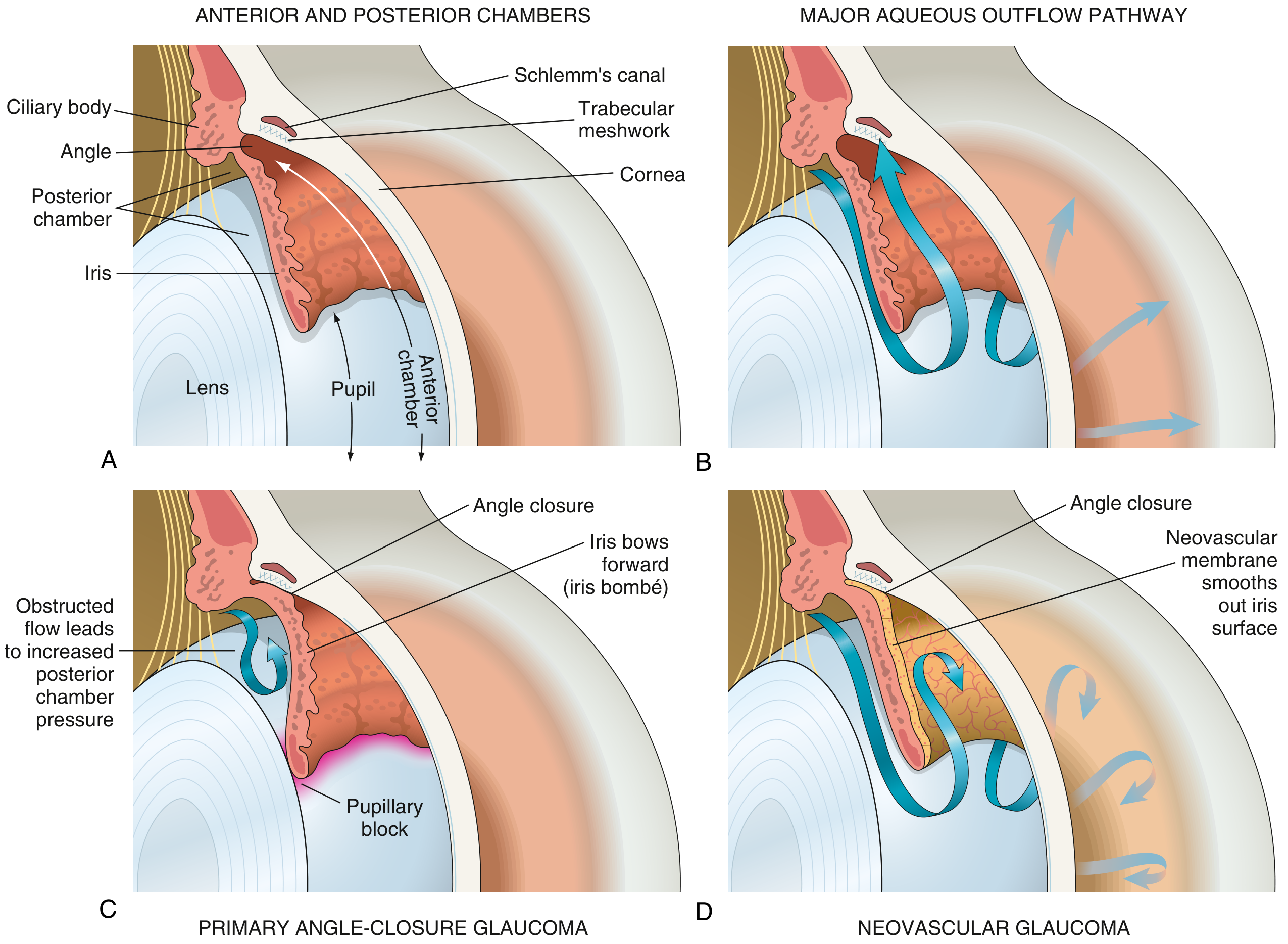
FIG: (A) Normal eye anatomy, (B) Normal aqueous outflow via trabecular meshwork → Schlemm's canal, (C) Angle-closure glaucoma with iris bombé, (D) Neovascular glaucoma with peripheral anterior synechiae. - Robbins & Kumar Basic Pathology
Production
- Aqueous humor is secreted by the non-pigmented ciliary epithelium of the ciliary body (pars plicata).
- It flows from the posterior chamber → through the pupil → into the anterior chamber.
Drainage Pathways
There are two outflow routes:
| Pathway | Route | Contribution |
|---|---|---|
| Conventional (trabecular) | Trabecular meshwork → Canal of Schlemm → episcleral venous plexus → systemic circulation | 80-95% of outflow |
| Uveoscleral | Ciliary muscles → suprachoroidal space | 5-20% of outflow |
The conventional trabecular pathway is the primary pharmacological target for most anti-glaucoma drugs. - Goodman & Gilman's, p. 636
Intraocular Pressure (IOP)
- Normal IOP: 10-20 mm Hg (population mean ~15 mm Hg)
- Ocular hypertension: IOP > 21 mm Hg (> 2 standard deviations above mean)
- IOP is determined by the balance between aqueous production and aqueous outflow
- IOP is the principal but not the only risk factor for glaucoma - in 20-50% of glaucoma patients, IOP is actually within the normal range ("normal tension glaucoma") - Ganong's Review of Medical Physiology, p. 1973
Formula concept: IOP ∝ Rate of aqueous production / Facility of outflow
Pathophysiology of Elevated IOP
1. Open-Angle Glaucoma (most common)
- The anterior chamber angle is anatomically open - there is no physical obstruction visible on gonioscopy
- Mechanism: Sclerosis and increased resistance of the trabecular meshwork tissue impairs aqueous filtration, raising IOP
- May also be caused by increased production of aqueous humor or deposition of particulate matter (red cells, pigment, necrotic tumor cells) blocking the meshwork in secondary forms
- Primary open-angle glaucoma (POAG) is the commonest type; it progresses silently and is often detected late - Robbins & Kumar Basic Pathology, p. 2392
2. Angle-Closure Glaucoma
- The anterior chamber angle is physically narrowed or occluded, preventing aqueous from reaching the trabecular meshwork
- Mechanism (pupillary block): In anatomically predisposed eyes (shallow anterior chambers, hypermetropic eyes), the iris apposes the lens at the pupillary margin, creating a ball-valve block
- Aqueous accumulates in the posterior chamber → pressure builds → iris bows forward (iris bombé) → iris root occludes the trabecular meshwork angle
- This can occur acutely (acute angle-closure attack with dramatic IOP spikes, pain, halos, corneal edema) or chronically - Goodman & Gilman's, p. 638
3. Optic Nerve Damage Mechanism
Elevated IOP damages the optic nerve at the lamina cribrosa by two main mechanisms:
- Direct mechanical compression of axons at the lamina cribrosa, disrupting axoplasmic flow
- Ischemia - reduced perfusion pressure to the optic nerve head (optic nerve perfusion pressure = mean arterial pressure - IOP)
This leads to cupping of the optic disc - loss of axons widens the optic cup, increasing the cup-to-disc (C:D) ratio (normal < 0.5). A high C:D ratio is a key clinical sign. - Goldman-Cecil Medicine, p. 614
Physiological Basis of Treatment
All anti-glaucoma therapies target one of two physiological levers:
| Mechanism | Drug Classes | Examples |
|---|---|---|
| Decrease aqueous production | β-blockers | Timolol |
| Carbonic anhydrase inhibitors | Dorzolamide, acetazolamide | |
| α2-agonists | Brimonidine | |
| Increase aqueous outflow | Prostaglandin analogues (uveoscleral ↑) | Latanoprost, travoprost |
| Cholinergic agonists (trabecular ↑ via ciliary muscle contraction) | Pilocarpine, carbachol | |
| Rho-kinase inhibitors (trabecular ↑) | Netarsudil |
- A common first-line combination is a β-blocker (reduces secretion) + prostaglandin analogue (increases outflow) - Ganong's Review of Medical Physiology, p. 1979
Summary Flow
Ciliary body secretes aqueous
         ↓
Posterior chamber → pupil → Anterior chamber
         ↓
Trabecular meshwork → Canal of Schlemm → episcleral veins (80-95%)
Uveoscleral route (5-20%)
         ↓
If OUTFLOW ↓ or PRODUCTION ↑ → IOP ↑
         ↓
Mechanical + ischemic injury at lamina cribrosa
         ↓
Retinal ganglion cell axon loss → optic disc cupping → visual field defects → blindness
Key Point: Glaucoma is fundamentally a pressure-flow imbalance disease of the eye. Aqueous humor physiology - specifically the balance between ciliary body secretion and trabecular/uveoscleral drainage - governs IOP, and disruption of this balance at any point (trabecular sclerosis, angle closure, neovascularization) leads to optic nerve damage through mechanical and ischemic mechanisms.
This is a shared conversation. Sign in to Orris to start your own chat.