
Details about CSOM
Reading File
Finding Sources
Finding Sources
Searching PubMed
"chronic suppurative otitis media"
Searching the Web
WHO chronic suppurative otitis media fact sheet management
Reading File
Reading File
Reading File
Finding Sources
Finding Sources
Finding Sources
Reading File
Chronic suppurative otitis media (CSOM)
CSOM is chronic inflammation/infection of the middle ear and mastoid associated with a persistent tympanic membrane perforation and recurrent or persistent ear discharge (otorrhoea). Clinically, persistent purulent discharge through a perforation is the key feature. A discharge lasting at least 2 weeks should raise suspicion, though definitions vary. The WHO review includes both active discharge and, in some classifications, a dry inactive perforation.
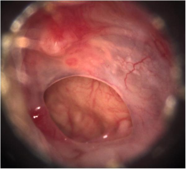
Example of a central tympanic-membrane perforation. This is a dry ear, not active suppurative disease.
Types
Modern classification describes chronic otitis media according to mucosal or squamous disease activity:
-
Mucosal CSOM
- Active mucosal disease: perforation with otorrhoea, inflamed middle-ear mucosa, and sometimes granulations/aural polyp.
- Inactive mucosal disease: dry, permanent pars tensa perforation without active inflammation.
- Usually corresponds to the older term tubotympanic or “safe” disease, but no chronic ear disease should be assumed entirely safe.
-
Squamous epithelial disease (cholesteatoma)
- Keratinizing squamous epithelium in the middle ear/mastoid, commonly arising from an attic or marginal retraction pocket.
- May erode ossicles, labyrinth, facial canal, or intracranial boundaries.
- Historically called atticoantral or “unsafe” CSOM.
Scott-Brown’s Otorhinolaryngology Head & Neck Surgery, Vol. 2, p. 1024.
Causes and risk factors
- Recurrent or inadequately resolved acute otitis media
- Persistent Eustachian-tube dysfunction
- Tympanic membrane perforation following infection or trauma
- Poor access to health care, overcrowding, malnutrition, and recurrent upper respiratory infections
- Cleft palate or craniofacial anomalies
- Water entering the ear through a perforation
- Cholesteatoma
Common pathogens include Pseudomonas aeruginosa, Staphylococcus aureus, Proteus species, Klebsiella, and mixed aerobic/anaerobic flora. Biofilm formation may contribute to persistence and treatment failure.
Clinical features
- Persistent or recurrent foul-smelling or mucoid/purulent ear discharge
- Conductive hearing loss, often fluctuating
- Tinnitus or a blocked-ear sensation
- Usually little pain in uncomplicated mucosal disease
- Otoscopy may show:
- Central, marginal, or attic perforation
- Mucopus in the canal/middle ear
- Granulation tissue or polyp
- Retraction pocket, keratin debris, or attic crust suggesting cholesteatoma
Chronic mucosal inflammation can involve the middle ear and mastoid, with oedema, fibrosis, hypervascularity, granulations, and possible ossicular-chain damage. Scott-Brown’s Otorhinolaryngology Head & Neck Surgery, Vol. 2, p. 1024.
Red flags requiring urgent ENT assessment
- Severe or increasing otalgia
- Vertigo, spontaneous nystagmus, or sudden worsening hearing
- Facial weakness
- Post-auricular swelling/tenderness or fever
- Severe headache, neck stiffness, altered consciousness, focal neurological signs
- Aural polyp, marginal/attic perforation, or keratin debris suspicious for cholesteatoma
Increasing pain, vertigo, or facial palsy may indicate a complication; CT is useful for bony erosion and MRI is used if intracranial involvement is suspected. Textbook of Family Medicine, 9e, p. 756.
Complications
Extracranial
- Mastoiditis
- Ossicular erosion and conductive hearing loss
- Labyrinthitis and sensorineural hearing loss
- Facial nerve palsy
- Petrositis
- Labyrinthine fistula
Intracranial
- Meningitis
- Extradural abscess
- Subdural empyema
- Brain abscess
- Lateral/sigmoid sinus thrombosis
Diagnosis
Diagnosis is primarily clinical:
- Otoscopy or preferably microscopic ear examination
- Suction clearance of debris/discharge, where available
- Hearing assessment: pure-tone audiometry
- Ear swab culture if disease is persistent, recurrent, severe, or unresponsive to initial therapy
- High-resolution CT temporal bone if cholesteatoma, bony erosion, complication, or surgical planning is suspected
- MRI when intracranial extension or labyrinthine/soft-tissue complications are suspected
Management principles
-
Aural toilet
- Careful dry mopping or microsuction to clear discharge and debris.
- Keep the ear dry. Avoid swimming and prevent water entry during bathing.
-
Topical antibiotic ear drops
- Topical quinolone drops are commonly preferred when there is a perforation because they cover common gram-negative organisms including Pseudomonas and avoid the ototoxicity concern of aminoglycoside-containing drops.
- Choice and duration should be individualized by a clinician, particularly for children, pregnancy, recurrent disease, or suspected fungal infection.
-
Treat contributing nasal/Eustachian-tube disease
- Address allergic rhinitis, chronic rhinosinusitis, adenoid disease, or recurrent upper respiratory infections when relevant.
-
Systemic antibiotics
- Not routinely required for uncomplicated CSOM.
- Considered for systemic illness, spreading infection, mastoiditis, or other complications, guided by clinical assessment and culture where possible.
-
Surgery
- Tympanoplasty/myringoplasty: closes a persistent perforation and may improve hearing after infection is controlled.
- Mastoidectomy with tympanoplasty: used selectively, especially for cholesteatoma, complications, or persistent/refractory disease.
- Cholesteatoma requires surgical assessment, since drops do not eradicate the keratinizing lesion.
Evidence for optimal antimicrobial strategies remains limited and uncertain in several comparisons. Recent Cochrane systematic reviews address systemic antibiotics and topical antiseptics for CSOM (2025).
Prevention and patient advice
- Complete prescribed therapy and attend follow-up until the ear is dry.
- Keep water out of the affected ear.
- Do not insert cotton buds, oils, or unprescribed drops into the ear.
- Obtain a hearing test in persistent or recurrent disease, especially in children because hearing loss may affect speech, learning, and development.
- Seek urgent care for the red-flag symptoms above.
This is a shared conversation. Sign in to Orris to start your own chat.