
Trachea histology
Reading File
Finding Sources
Searching Images
histology of trachea showing respiratory epithelium submucosal glands hyaline cartilage
Searching PubMed
trachea histology
Searching the Web
trachea histology respiratory epithelium cartilage submucosal glands authoritative
Reading File
Finding Sources
Reading File
Reading File
Trachea: histology
The trachea is lined by respiratory mucosa and supported by incomplete rings of hyaline cartilage.
Layers, from lumen outward
-
Mucosa
- Epithelium: pseudostratified ciliated columnar epithelium
- Major cells:
- Ciliated columnar cells: move mucus and trapped particles upward toward the pharynx.
- Goblet cells: secrete mucus.
- Basal cells: stem/progenitor cells that replace epithelial cells.
- Less conspicuous cells include brush cells and small granule neuroendocrine cells.
- Basement membrane: conspicuously thick.
- Lamina propria: loose, vascular connective tissue with elastic fibers and immune cells.
-
Submucosa
- Dense connective tissue containing numerous seromucous glands.
- Their secretion moistens the airway and contributes to mucociliary clearance.
-
Fibrocartilaginous layer
- Composed of C-shaped rings of hyaline cartilage that prevent collapse of the airway.
- The posterior gap in the rings is bridged by fibroelastic tissue and the trachealis muscle, a smooth muscle band adjacent to the esophagus.
-
Adventitia
- Outer connective tissue layer attaching the trachea to neighboring structures.
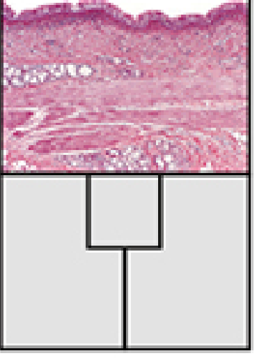
Key identification points on an H&E slide
- Wide lumen lined by pseudostratified ciliated epithelium
- Many pale goblet cells
- Thick basement membrane
- Submucosal seromucous glands
- Large C-shaped hyaline cartilage
- Posterior trachealis smooth muscle
One-line viva answer:
Trachea is lined by pseudostratified ciliated columnar epithelium with goblet cells; beneath it are lamina propria and seromucous submucosal glands, followed by C-shaped hyaline cartilage rings with posterior trachealis smooth muscle and an outer adventitia.
Trachea is lined by pseudostratified ciliated columnar epithelium with goblet cells; beneath it are lamina propria and seromucous submucosal glands, followed by C-shaped hyaline cartilage rings with posterior trachealis smooth muscle and an outer adventitia.
Source: Histology: A Text and Atlas with Correlated Cell and Molecular Biology, pp. 1834-1835.
Oesophagus histology
Reading File
Finding Sources
Searching Images
histology of esophagus H&E stratified squamous epithelium submucosal glands muscularis externa
Searching PubMed
esophagus histology
Searching the Web
esophagus histology stratified squamous nonkeratinized epithelium submucosal glands muscularis externa
Finding Sources
query_images_db
Reading File
Oesophagus: histology
The oesophagus is adapted to resist friction from swallowed food and to propel the bolus to the stomach.
Layers from lumen outward
-
Mucosa
- Epithelium: thick non-keratinized stratified squamous epithelium, protective against abrasion.
- Lamina propria: loose connective tissue; contains mucous oesophageal cardiac glands especially near the upper and lower ends.
- Muscularis mucosae: longitudinal smooth muscle, relatively thick in the lower oesophagus.
-
Submucosa
- Dense irregular connective tissue with blood vessels, lymphatics, and the submucosal (Meissner) plexus.
- Contains oesophageal glands proper, which are mucous/seromucous glands that lubricate and protect the lumen.
-
Muscularis externa
- Inner circular and outer longitudinal muscle layers, with the myenteric (Auerbach) plexus between them.
- Muscle type varies by level:
- Upper one-third: skeletal muscle
- Middle one-third: mixed skeletal and smooth muscle
- Lower one-third: smooth muscle
-
Outer covering
- Mostly adventitia, which anchors the oesophagus to surrounding mediastinal structures.
- Only the short abdominal part is covered by serosa.
Gastro-oesophageal junction
There is an abrupt transition from:
- Non-keratinized stratified squamous epithelium of oesophagus
to - Simple columnar mucous-secreting epithelium of stomach.
Identification points in an H&E slide
- Thick, multilayered squamous non-keratinized lining
- Submucosal mucous glands
- No villi or gastric pits
- Two muscle layers with changing muscle type along its length
- Predominantly adventitial outer coat
One-line viva answer:
The oesophagus is lined by non-keratinized stratified squamous epithelium, has submucosal oesophageal glands, and its muscularis externa changes from skeletal muscle above to smooth muscle below; it is mainly covered by adventitia.
The oesophagus is lined by non-keratinized stratified squamous epithelium, has submucosal oesophageal glands, and its muscularis externa changes from skeletal muscle above to smooth muscle below; it is mainly covered by adventitia.
Source: Histology: A Text and Atlas with Correlated Cell and Molecular Biology, p. 1610.
Duodenum histology
Reading File
Finding Sources
query_images_db
Searching PubMed
duodenum histology
Searching the Web
duodenum histology Brunner glands villi crypts authoritative
Finding Sources
Reading File
Duodenum: histology
The duodenum has the usual four layers of the gastrointestinal tract, but is identified by villi and Brunner glands in the submucosa.
1. Mucosa
- Lined by simple columnar epithelium with:
- Enterocytes with a brush border of microvilli for absorption
- Goblet cells that secrete mucus
- Has broad, leaf-shaped intestinal villi that increase absorptive surface area.
- Crypts of Lieberkühn lie between villi and contain stem cells, enteroendocrine cells, and Paneth cells.
- Lamina propria contains blood capillaries, immune cells, and a central lymphatic capillary called a lacteal.
- Muscularis mucosae forms the deepest part of mucosa.
2. Submucosa
- The distinguishing feature is Brunner glands.
- These are branched tubular mucous glands that secrete alkaline, bicarbonate-rich mucus.
- Their secretion neutralizes acidic chyme arriving from the stomach and protects the duodenal mucosa.
- Contains vessels, lymphatics, and the submucosal (Meissner) plexus.
3. Muscularis externa
- Inner circular smooth-muscle layer
- Outer longitudinal smooth-muscle layer
- Myenteric (Auerbach) plexus lies between them.
4. Outer layer
- First part of duodenum: partly covered by serosa
- Remaining retroperitoneal parts: covered mainly by adventitia
Identification points on H&E slide
- Mucosal villi and crypts
- Simple columnar epithelium with goblet cells
- Brunner glands in the submucosa: the key feature distinguishing duodenum from jejunum and ileum
- No Peyer patches as seen prominently in ileum
One-line viva answer:
The duodenum is lined by simple columnar epithelium with villi and crypts, and is distinguished by Brunner glands in the submucosa that secrete alkaline mucus to neutralize gastric acid.
The duodenum is lined by simple columnar epithelium with villi and crypts, and is distinguished by Brunner glands in the submucosa that secrete alkaline mucus to neutralize gastric acid.
Source: Histology: A Text and Atlas with Correlated Cell and Molecular Biology, section “Submucosa”; Junqueira’s Basic Histology - Text and Atlas, section “Other Layers.”
Hepatic lobule histology
Reading File
Finding Sources
query_images_db
Searching PubMed
hepatic lobule histology
Searching the Web
hepatic lobule histology central vein portal triad sinusoids authoritative
Reading File
Loading Image
Finding Sources
This is a shared conversation. Sign in to Orris to start your own chat.