

What's the treatment
acanthosis nigricans axilla hyperpigmented velvety plaque
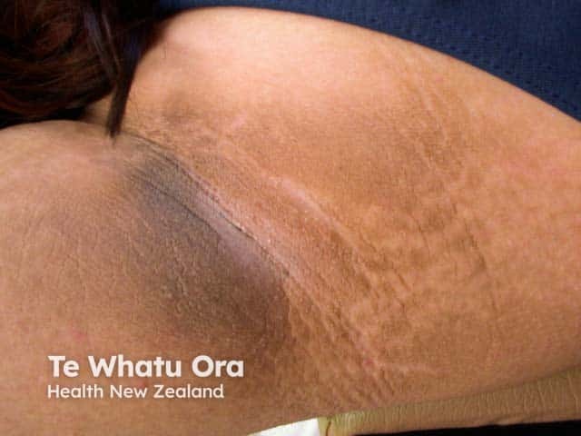
This dermatologic clinical photograph depicts classic axillary acanthosis nigricans: a velvety, hyperpigmented plaque within the skin folds of the axilla. The region shows diffuse brown-to-dark pigmented, lamellar texturing with parallel linear creases and fine wrinkling along skin folds. The texture appears velvety to the touch; there is mild skin thickening and uniform pigmentation without vesicles or erosions. The lesion is localized to the axillary area, typically associated with friction and perspiration, and commonly coexists with obesity, insulin resistance, or metabolic syndrome. On histology, epidermal hyperplasia with mild hyperkeratosis and papillomatosis would be expected, though not shown here. Clinically this finding signals possible systemic associations; in adults, screen for diabetes mellitus, impaired glucose tolerance, dyslipidemia, and obesity; in older patients or atypical presentations, consider paraneoplastic acanthosis nigricans associated with gastric or other malignancies. Differential diagnoses include frictional dermatitis, candidal intertrigo, and other pigmented dermatoses. The image supports educational usage in dermatology training, primary care screening, endocrinology workups, and radiology-dermatology correlation for cutaneous signs of systemic disease. The observation underscores the importance of comprehensive clinical assessment, lifestyle modification, and targeted laboratory evaluation to identify underlying etiologies and initiate appropriate management. Careful documentation of lesion characteristics improves reproducibility and supports longitudinal monitoring over time.
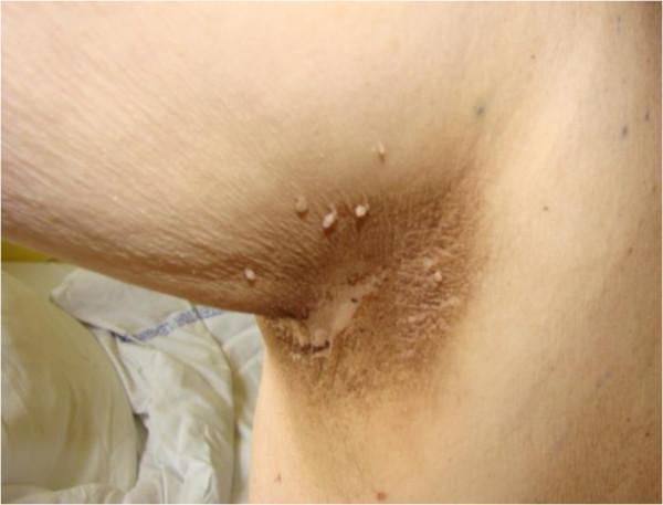
This clinical photograph displays the right axilla of a patient, demonstrating classic features of acanthosis nigricans. The primary finding is a large, focal area of brownish hyperpigmentation with a velvety or 'dirty' appearance. The skin texture within the affected region is notably altered, exhibiting lichenification and verrucous-like thickening with a non-uniform surface. Along the periphery and within the hyperpigmented plaque, several small, pale, pedunculated lesions resembling acrochordons (skin tags) are visible. The surrounding skin appears normal in color and texture. This presentation is characteristic of paraneoplastic acanthosis nigricans, often associated with underlying malignancies or metabolic conditions such as insulin resistance and type 2 diabetes. The educational focus of this image is the recognition of cutaneous markers for systemic disease and the distinctive morphological features of epidermal hyperproliferation in intertriginous areas.
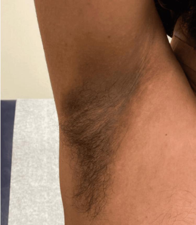
This clinical photograph displays a close-up view of the right axillary region of a patient, demonstrating classic features of acanthosis nigricans. The central axilla shows a poorly demarcated, symmetrical area of hyperpigmentation, characterized by a brownish to grayish-black discoloration that is markedly darker than the surrounding skin. The skin texture within the affected zone appears thickened with a velvety, rugose, or 'dirty' appearance, particularly visible along the natural skin folds. Terminal axillary hair is present, predominantly concentrated over the lower half of the hyperpigmented plaque. The image captures the transition between the medial upper arm and the lateral thoracic wall, providing anatomical context. In a clinical setting, this finding is often associated with insulin resistance, obesity, or certain medications, such as isoniazid as noted in this case, and serves as an important dermatological marker for underlying metabolic or systemic conditions.
Diagnosis: Acanthosis Nigricans (AN)
Treatment
1. Treat the Underlying Cause (Most Important)
| Underlying Cause | Action |
|---|---|
| Insulin resistance / Obesity / T2DM | Weight loss, dietary modification; metformin improves insulin sensitivity and may clear skin |
| PCOS | Insulin sensitizers (metformin); antiandrogen therapy is less reliable |
| Offending drug (steroids, nicotinic acid, estrogens, isoniazid) | Discontinue the causative medication - usually leads to clearance |
| Malignancy (rare - gastric or intra-abdominal adenocarcinoma) | Treat underlying tumor; skin improves with chemotherapy/remission |
2. Topical Treatments (Cosmetic improvement, partial benefit)
- Keratolytics: Salicylic acid, urea, glycolic acid peels
- Vitamin D analogue: Topical calcipotriol
- Retinoids: Topical or systemic tretinoin
- Ammonium lactate (moisturizing + mild keratolytic)
3. Procedural Options
- Long-pulsed alexandrite laser - reported effective in individual cases
4. Workup Before Treating
- Fasting glucose / HbA1c (screen for diabetes/pre-diabetes)
- Fasting insulin (insulin resistance)
- BMI / waist circumference
- Thyroid function (TSH)
- Androgens / LH:FSH in women (for PCOS)
- In older patients with rapid onset, extensive disease, or oral mucosal involvement - screen for internal malignancy (especially gastric)
Key point: As per Fitzpatrick's Dermatology (p. 2525), "treatment of acanthosis nigricans is generally ineffective" in isolation. Improvement or resolution with weight loss does occur in obese patients, and treating the root cause (insulin resistance, malignancy, or drug) is the most effective strategy. Referral to an endocrinologist is advisable if metabolic syndrome is suspected.
⚠️ Disclaimer: This is for educational reference only. A dermatologist or physician should examine the patient and recommend individualized treatment.

Treatment plans
Treatment Plan: Acanthosis Nigricans (Axillary) with Active Inflammation
Step 1 - Investigations First (Before treating)
| Test | Rationale |
|---|---|
| Fasting blood glucose + HbA1c | Rule out T2DM / pre-diabetes |
| Fasting insulin + HOMA-IR | Quantify insulin resistance |
| Lipid profile | Associated metabolic syndrome |
| TSH | Thyroid disease |
| LH, FSH, testosterone (if female) | Rule out PCOS |
| BMI + waist circumference | Obesity assessment |
| In older patients with rapid-onset/extensive AN | Consider malignancy workup (USG abdomen, endoscopy) |
Step 2 - Address the Root Cause (Primary Treatment)
- Lifestyle modification: Low-glycaemic index diet + regular aerobic exercise targeting 5-10% weight reduction - shown to improve or resolve AN
- Metformin 500-1000 mg BD (improves insulin sensitivity, has demonstrated benefit in AN associated with insulin resistance)
- Treat diabetes or pre-diabetes aggressively with glycaemic control
- Metformin + lifestyle modification
- Oral contraceptives if needed for cycle regulation
- Note: anti-androgen therapy alone is less reliable for skin clearance
- Identify and discontinue the offending drug (glucocorticoids, nicotinic acid, estrogens, isoniazid)
- Skin usually clears after stopping the medication
- Treat the underlying tumor; skin improves with chemotherapy and remits with recurrence
Step 3 - Topical Skin-Directed Therapy (Cosmetic / symptomatic)
| Agent | Use |
|---|---|
| Tretinoin cream (0.025-0.05%) | Nightly application to affected area; reduces thickening over weeks-months |
| Salicylic acid 2-6% (lotion/gel) | Keratolytic - reduces hyperkeratosis |
| Glycolic acid peel (20-30%) | In-clinic chemical peel every 4 weeks |
| Urea 10-20% cream | Daily moisturiser + keratolytic |
| Calcipotriol (topical vitamin D analogue) | Has shown improvement in AN; apply BD |
| Ammonium lactate 12% lotion | Daily use for softening |
Step 4 - Manage Active Inflammation (Visible in Image 2)
- Short-course mild-moderate topical corticosteroid (e.g., hydrocortisone 1% or mometasone furoate 0.1% cream) for 1-2 weeks to reduce inflammation
- Clotrimazole antifungal cream BD x 2 weeks if Candida intertrigo is suspected (moist axillary environment)
- Barrier zinc oxide paste to protect macerated skin folds
- Keep the axilla dry and clean - loose cotton clothing, avoid synthetic fabrics
- Avoid harsh deodorants/antiperspirants in the active phase
Step 5 - Procedural Options (If topical therapy fails)
- Long-pulsed alexandrite laser - reported effective in refractory cases
- Q-switched Nd:YAG laser - for pigmentation reduction
- Dermabrasion / Chemical peels (higher concentrations, in-clinic)
Summary Table
| Priority | Intervention |
|---|---|
| 1st | Investigate and treat underlying cause (insulin resistance, PCOS, drugs, malignancy) |
| 2nd | Weight loss + Metformin if insulin resistance confirmed |
| 3rd | Topical retinoids + keratolytics for skin texture |
| 4th | Short-term topical steroid ± antifungal for active inflammation |
| 5th | Laser/procedural therapy for persistent pigmentation |
⚠️ Note: Topical treatments alone provide cosmetic benefit but do not address the root cause. Long-term resolution requires managing the underlying systemic condition. A physician/dermatologist and endocrinologist review is recommended. - Fitzpatrick's Dermatology, Andrews' Diseases of the Skin