
What is bainbridge reflex
The Bainbridge reflex is an increase in heart rate triggered by increased venous return (right atrial filling). When blood volume/pressure rises in the right atrium and great veins, stretch receptors in the atrial walls send signals via the vagus nerve to the medulla, which then reduces vagal tone and/or increases sympathetic output - resulting in a faster heart rate. It acts as a compensatory mechanism to prevent blood from pooling in the venous system.
Searching sources...
Reading File
Finding Sources
Searching PubMed
Bainbridge reflex heart rate atrial stretch
Reading File
Reading File
Reading File
Also fetching the diagram for better illustration:
Reading File
Here is a detailed, textbook-based explanation of the Bainbridge reflex:
Bainbridge Reflex
Definition
The Bainbridge reflex is the tachycardia (increase in heart rate) produced by an increase in venous return and atrial pressure (volume loading). It was named after the British physiologist Francis Arthur Bainbridge who described it in 1915.
Stimulus
- Increased blood volume or venous return causes distension of the right atrium and great veins
- This stretches low-pressure B-type stretch receptors (mechanoreceptors) in the atrial walls
Reflex Arc
| Component | Details |
|---|---|
| Receptor | Low-pressure atrial stretch receptors (B fibers) in the atrial walls |
| Afferent limb | Signals travel via the vagus nerve to the nucleus tractus solitarius (NTS) in the medulla |
| Efferent limb | Response is carried back to the SA node via both vagal (parasympathetic) and sympathetic pathways |
| Effector | SA node - increases heart rate; minor effects on contractility |
Physiological Response
- Heart rate increases up to 75% (40-60% via the Bainbridge reflex + 10-15% by direct stretch of the SA node)
- Efferent signals also increase strength of cardiac contraction modestly
- The reflex saturates at high baseline heart rates - the increase is greatest when the resting heart rate is slow
Purpose / Significance
The Bainbridge reflex prevents blood from pooling in the veins, atria, and pulmonary circulation. When venous return is high, the heart must pump faster to "keep up" and clear the incoming blood. It acts as a complementary mechanism alongside:
- Frank-Starling law (more filling = more force)
- Baroreceptor reflex (arterial pressure regulation)
Bainbridge Reflex vs. Baroreceptor Reflex - Competition
This is a key physiological concept. Both reflexes respond to volume changes, but in opposite directions for the same stimulus:
| Situation | Bainbridge Reflex | Baroreceptor Reflex | Net Effect on Heart Rate |
|---|---|---|---|
| Volume loading (high blood volume) | Increases HR | Decreases HR (high BP activates baroreceptors) | Bainbridge dominates → HR rises |
| Volume depletion (low blood volume) | Minimal effect | Increases HR (low BP reduces baroreceptor firing) | Baroreceptor reflex dominates → HR rises |
| Normal volume | Neither strongly activated | Baseline | HR is at its minimum |
This explains why both volume loading and volume depletion cause tachycardia, but by different mechanisms. Heart rate is at its lowest when circulating volume is normal.
Key Points to Remember
- The Bainbridge reflex is a low-pressure receptor reflex (not arterial baroreceptors)
- The efferent limb involves both sympathetic and parasympathetic pathways to the SA node
- It primarily affects heart rate, not significantly contractility or stroke volume
- The reflex saturates - effective mainly when baseline HR is low
- Works in concert with the Frank-Starling mechanism and baroreceptor reflex to regulate cardiac output
Sources: Guyton and Hall Textbook of Medical Physiology; Medical Physiology (Boron & Boulpaep); Costanzo Physiology, 7th Edition
Chemiosmotic hypothesis
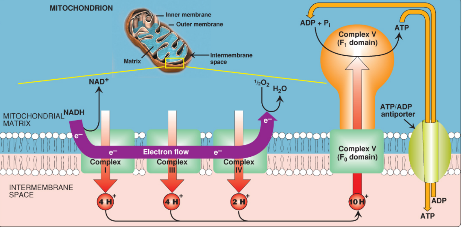
The chemiosmotic hypothesis, proposed by Peter Mitchell in 1961 (Nobel Prize 1978), explains how ATP is synthesized in mitochondria. Electron transport chain complexes pump protons (H+) from the mitochondrial matrix into the intermembrane space, creating an electrochemical gradient (proton motive force). Protons flow back down this gradient through ATP synthase (Complex V), and the energy released drives ATP synthesis from ADP + Pi.
Searching sources...
Finding Sources
Reading File
Reading File
Reading File
Reading File
Now let me fetch the key diagrams:
Loading Image
Loading Image
Chemiosmotic Hypothesis (Mitchell Hypothesis)
Proposed by Peter Mitchell in 1961 - Nobel Prize in Chemistry, 1978. It explains how the free energy generated by the electron transport chain (ETC) is used to produce ATP from ADP + Pi during oxidative phosphorylation.
Core Concept
The hypothesis states that the energy for ATP synthesis is provided by an electrochemical (proton) gradient across the inner mitochondrial membrane - NOT by a direct chemical intermediate as previously believed.
Step-by-Step Mechanism
Step 1 - Electron Transport and Proton Pumping
Electrons donated by NADH and FADH2 (from the citric acid cycle) pass through the ETC complexes embedded in the inner mitochondrial membrane:
| Complex | Name | Protons Pumped (per NADH) |
|---|---|---|
| Complex I | NADH dehydrogenase | 4 H+ |
| Complex II | Succinate dehydrogenase | 0 H+ (no pumping) |
| Complex III | Cytochrome bc1 complex | 4 H+ |
| Complex IV | Cytochrome c oxidase | 2 H+ |
| Total | Per NADH oxidized | 10 H+ pumped |
- At the end of the chain, O2 is the final electron acceptor, reduced to H2O
- Electrons from FADH2 enter at Complex II, yielding only 6 H+ (bypassing Complex I)
Step 2 - Formation of the Proton Motive Force (PMF)
The continuous pumping of H+ from the matrix → intermembrane space (IMS) creates two gradients:
- Chemical gradient (pH gradient, ΔpH) - IMS is more acidic (~0.75 pH units lower) than the matrix
- Electrical gradient (membrane potential, Δψ) - IMS is positively charged relative to the matrix
Together, these form the Proton Motive Force (PMF) - the combined electrochemical driving force that pushes protons back into the matrix.
The inner mitochondrial membrane is impermeable to protons, so they cannot simply diffuse back - they can only return through a specific channel (ATP synthase).
Step 3 - ATP Synthesis via ATP Synthase (Complex V / F0F1-ATPase)
H+ ions flow back down the electrochemical gradient through ATP synthase, and this flow drives ATP synthesis.
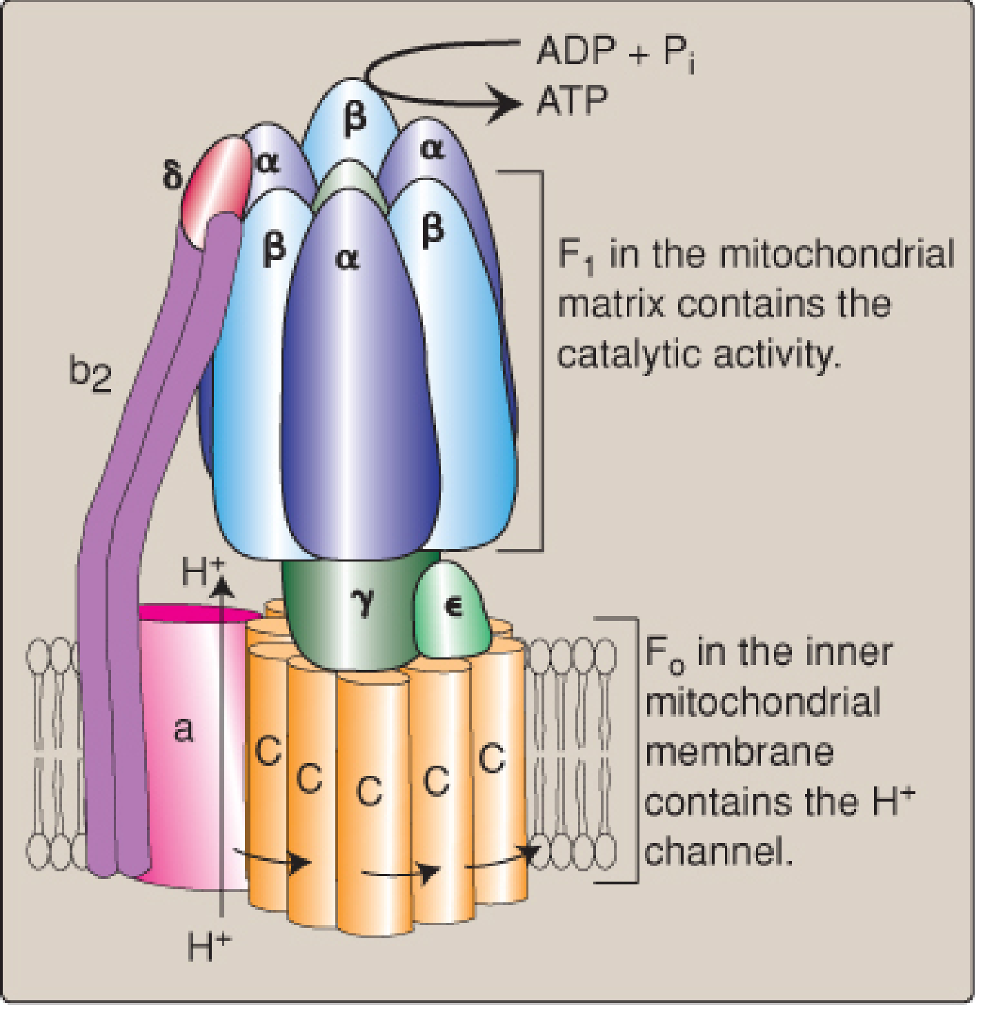
ATP synthase has two functional domains:
| Domain | Location | Function |
|---|---|---|
| F0 | Spans the inner mitochondrial membrane | Contains the H+ channel (c-ring subunits + subunit a) |
| F1 | Protrudes into the mitochondrial matrix | Contains the catalytic sites (3 αβ subunit pairs); synthesizes ATP |
Mechanism of rotation (Binding Change Mechanism):
- H+ enters from the IMS into a channel in the F0 domain
- Each H+ protonates a glutamyl carboxyl group on a c-subunit, causing the c-ring to rotate
- Rotation of the c-ring turns the γ-shaft (stalk), which is connected to the F1 domain
- As the γ-shaft rotates, it changes the conformation of the three β-subunits in F1 sequentially through three states: Open (O), Loose (L), Tight (T)
- In the Tight conformation, ADP + Pi are spontaneously phosphorylated to ATP
- Rotation to the Open conformation causes ATP release
- One complete rotation of the c-ring = 3 ATP synthesized
- In vertebrates, the c-ring has 8 subunits, so 8 H+ per full rotation = 3 ATP → ~2.7 H+ per ATP
Summary of Coupling
NADH/FADH2 → ETC → H+ pumped out → PMF created → H+ flows through F0F1 → ATP synthesized
(oxidation) (gradient) (phosphorylation)
This is why it is called oxidative phosphorylation - oxidation of NADH/FADH2 is directly coupled to phosphorylation of ADP via the H+ gradient as the common intermediate.
Inhibitors and Uncouplers
| Agent | Mechanism | Effect |
|---|---|---|
| Oligomycin | Blocks F0 H+ channel | Stops ATP synthesis AND electron transport (respiratory control) |
| Rotenone | Blocks Complex I | Inhibits NADH oxidation |
| Cyanide / CO | Blocks Complex IV | Stops O2 reduction; halts entire chain |
| 2,4-Dinitrophenol (DNP) | Lipophilic H+ carrier (ionophore) | Shuttles H+ across membrane, dissipates gradient; energy released as heat |
| UCP1 (Thermogenin) | Uncoupling protein in brown fat | Natural uncoupler - allows H+ back-flux, generating heat (non-shivering thermogenesis) |
| Aspirin (toxic doses) | Uncoupler | Dissipates gradient, causes fever |
Uncouplers allow electron transport to continue at a rapid rate (consuming O2 and fuel) but without generating ATP - energy is lost as heat.
Key Points
- The H+ gradient is the common intermediate that links oxidation to phosphorylation
- The inner mitochondrial membrane must be intact and impermeable for the hypothesis to work
- Coupling: electron transport and ATP synthesis are tightly linked through the H+ gradient; increasing ADP availability increases H+ flow, which accelerates electron transport
- The same principle applies in chloroplasts (light-driven proton pumping across the thylakoid membrane drives ATP synthesis in photosynthesis)
Sources: Biochemistry 8th ed, Lippincott Illustrated Reviews; Basic Medical Biochemistry - A Clinical Approach, 6th ed; Medical Physiology (Boron & Boulpaep)
Free radicals clear concept oriented
Free radicals are atoms or molecules with one or more unpaired electrons in their outer orbital, making them highly reactive. They steal electrons from nearby molecules (lipids, proteins, DNA), causing chain reactions of oxidative damage. The body has antioxidant defenses (SOD, catalase, glutathione, vitamins C and E) to neutralize them. When production exceeds defense capacity, oxidative stress results, contributing to aging, cancer, atherosclerosis, and many diseases.
Searching sources...
Reading File
Finding Sources
Good - I have several excellent sources. Let me pull the detailed content from multiple textbooks in parallel:
Reading File
Reading File
Reading File
Reading File
Reading File
Reading File
Reading File
Reading File
Reading File
Now let me fetch the key diagrams for visual clarity:
Loading Image
Loading Image
Excellent textbook diagrams obtained. Now here is the complete, concept-oriented explanation:
Free Radicals - A Clear Concept
1. What Is a Free Radical?
A free radical is any atom, molecule, or molecular fragment that has one or more unpaired electrons in its outer orbital.
- Unpaired electrons make the molecule highly unstable and extremely reactive
- To achieve stability, the radical steals an electron from the nearest molecule
- That molecule then becomes a radical itself - initiating a self-perpetuating chain reaction
- Free radicals persist for only 10⁻⁹ to 10⁻¹² seconds before colliding with another molecule
Think of it like a hot potato - the unpaired electron keeps getting passed from molecule to molecule, damaging each one in the process.
2. Reactive Oxygen Species (ROS) - The Main Players
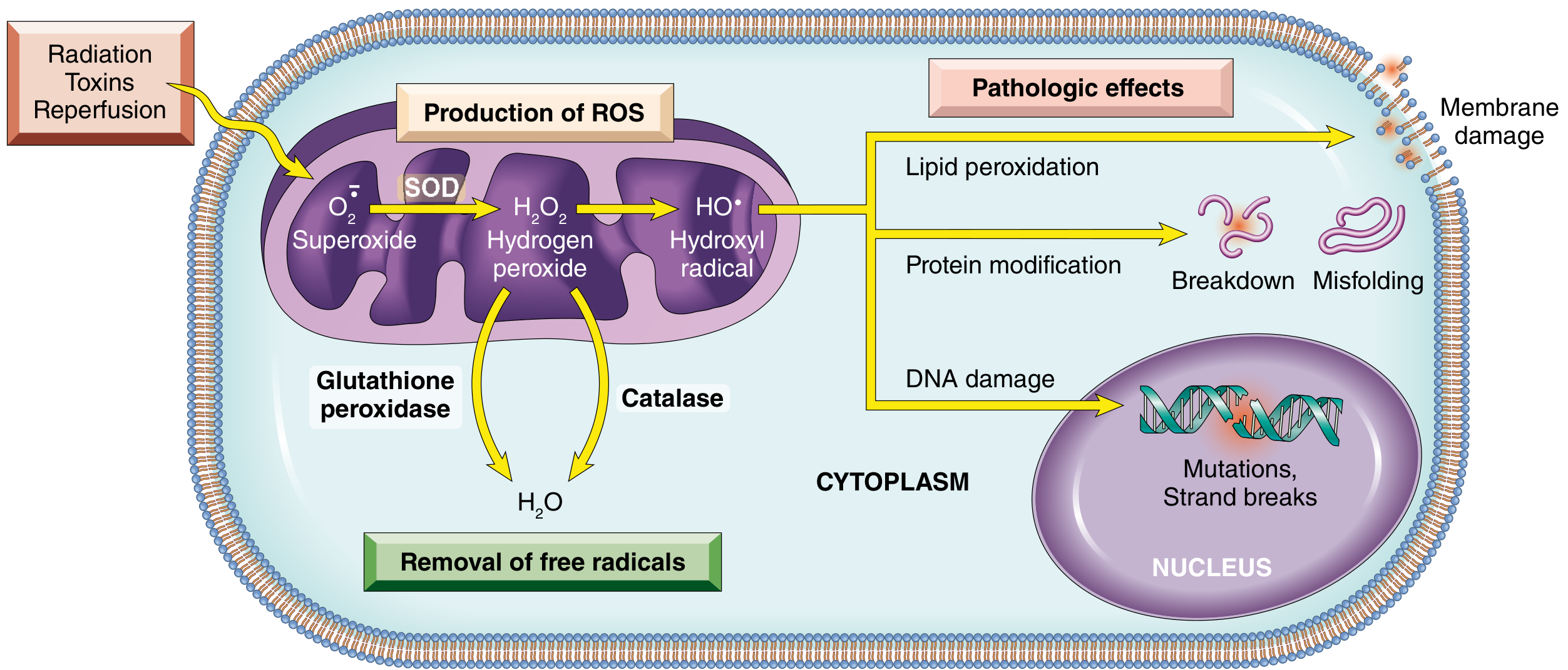
ROS are the most biologically important free radicals. They arise from sequential one-electron reductions of O₂:
O₂ → O₂•⁻ → H₂O₂ → •OH → H₂O
(+e⁻) (+e⁻+2H⁺) (+e⁻) (+e⁻+H⁺)
Superoxide Hydrogen Hydroxyl Water
anion peroxide radical
| ROS | Type | Key Property |
|---|---|---|
| Superoxide (O₂•⁻) | True radical | Cannot diffuse far; generates other ROS |
| Hydrogen peroxide (H₂O₂) | NOT a radical | Can diffuse through membranes; precursor to •OH |
| Hydroxyl radical (•OH) | True radical - most dangerous | Most reactive; damages lipids, proteins, DNA; no enzymatic removal |
| Peroxynitrite (ONOO⁻) | Not a radical | Formed from O₂•⁻ + NO; strong oxidant |
| Hypochlorous acid (HOCl) | Not a radical | Made by neutrophils during respiratory burst |
| Nitric oxide (NO•) | Radical | Signaling molecule; can combine with O₂•⁻ to form ONOO⁻ |
3. Sources / Generation of Free Radicals
Endogenous Sources (inside the body):
a) Mitochondrial electron transport chain (most important)
- Complexes I and III leak electrons onto O₂ → forms O₂•⁻ (superoxide)
- ~1% of all O₂ consumed in aerobic metabolism generates ROS - small but deadly if unchecked
b) Fenton Reaction (transition metals)
- Fe²⁺ + H₂O₂ → Fe³⁺ + •OH + OH⁻
- Cu⁺ can do the same
- Most intracellular free iron exists as Fe³⁺ but O₂•⁻ can reduce it back to Fe²⁺, making the reaction cyclical and self-amplifying
c) Haber-Weiss Reaction
- O₂•⁻ + H₂O₂ → O₂ + OH⁻ + •OH
- Generates the highly toxic hydroxyl radical from superoxide and hydrogen peroxide
d) NADPH Oxidase in Leukocytes (Respiratory Burst)
- During phagocytosis, activated neutrophils/macrophages deliberately produce large bursts of O₂•⁻ and HOCl to kill bacteria
- Defect in this → Chronic Granulomatous Disease (CGD)
e) Xanthine Oxidase (important in ischemia-reperfusion)
- Normally exists as xanthine dehydrogenase (uses NAD⁺)
- During ischemia, it converts to xanthine oxidase (uses O₂)
- On reperfusion when O₂ returns → floods the system with O₂•⁻ and H₂O₂
f) Cytochrome P450 enzymes - drug and xenobiotic metabolism
Exogenous Sources:
- Ionizing radiation - splits H₂O into •OH and H•
- UV light - generates singlet oxygen
- Cigarette smoke - contains preformed ROS and RNOS
- Chemicals and drugs (e.g., CCl₄ → •CCl₃ radical in the liver)
- Reperfusion after ischemia (as above)
4. Pathological Effects - How Free Radicals Damage the Cell
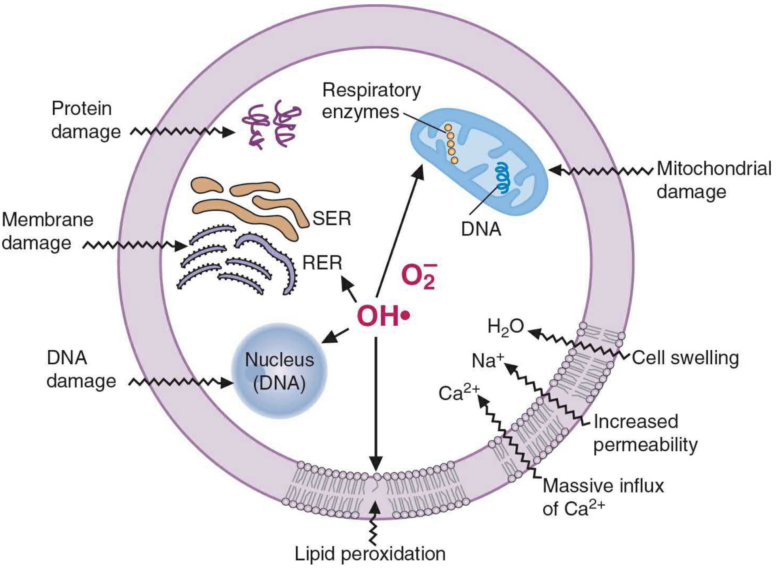
A. Lipid Peroxidation (most clinically significant)
A chain reaction in membrane phospholipids (especially those with polyunsaturated fatty acids):
- Initiation - •OH extracts a hydrogen atom from a PUFA → lipid radical (L•)
- Propagation - L• + O₂ → lipid peroxyl radical (LOO•) → attacks the next PUFA → forms lipid peroxide (LOOH) - self-perpetuating cycle
- Degradation - LOOH breaks down into malondialdehyde (MDA), ethane, pentane
- MDA is measured in blood/urine as a biomarker of oxidative stress
- Termination - two radicals meet to form a stable bond, or Vitamin E donates a hydrogen to LOO•, neutralizing the chain
Consequences: Membrane disruption → increased permeability → massive Ca²⁺ influx → further cell damage
B. Protein Modification
- Oxidation of amino acid side chains (especially cysteine -SH groups)
- Formation of covalent protein-protein cross-links (disulfide bonds)
- Damage to enzyme active sites → loss of enzyme function
- Enhanced proteasomal degradation of misfolded proteins
C. DNA Damage
- Single-strand and double-strand breaks
- Cross-linking of DNA strands
- Base modifications and adduct formation
- Leads to mutations, carcinogenesis, and cell aging
- Can trigger apoptosis in addition to necrosis
5. Defense Mechanisms - How the Body Fights Back
The body has a multi-layered antioxidant defense:
Enzymatic Defenses
| Enzyme | Location | Reaction | Notes |
|---|---|---|---|
| Superoxide Dismutase (SOD) | Mitochondria (Mn-SOD), Cytoplasm (Cu/Zn-SOD) | 2O₂•⁻ + 2H⁺ → H₂O₂ + O₂ | First line; converts superoxide to less reactive H₂O₂ |
| Catalase | Peroxisomes | 2H₂O₂ → O₂ + 2H₂O | Destroys H₂O₂ before it forms •OH |
| Glutathione Peroxidase (GPx) | Cytosol, Mitochondria | H₂O₂ + 2GSH → GSSG + 2H₂O | Also handles lipid peroxides; uses glutathione (GSH) |
The GSH:GSSG ratio is a key indicator of the cell's antioxidant capacity.
Non-Enzymatic (Antioxidant) Defenses
| Antioxidant | Where it works | Mechanism |
|---|---|---|
| Vitamin E (α-tocopherol) | Membranes (lipid-soluble) | Donates H to LOO•; terminates lipid peroxidation chain |
| Vitamin C (ascorbate) | Aqueous phase/cytosol | Scavenges O₂•⁻, •OH; also regenerates Vitamin E |
| Glutathione (GSH) | Cytosol | Directly reacts with •OH and HOCl; co-factor for GPx |
| β-carotene | Membranes | Quenches singlet oxygen |
| Ceruloplasmin, Transferrin, Ferritin | Blood/cells | Sequester free iron/copper - prevent Fenton reaction |
6. Oxidative Stress
Oxidative stress = a state where the rate of ROS production exceeds the cell's capacity to remove them.
This tips the balance toward cellular damage and underlies numerous diseases:
| Category | Diseases |
|---|---|
| Cardiovascular | Atherosclerosis, ischemia-reperfusion injury, heart failure |
| Neurological | Parkinson's disease, Alzheimer's disease, ALS |
| Cancer | DNA mutations, tumor promotion |
| Pulmonary | COPD, ARDS (oxygen toxicity) |
| Metabolic | Diabetes mellitus (advanced glycation end products) |
| Aging | Accumulation of oxidative macromolecular damage |
| Inflammatory | Inflammatory bowel disease, autoimmune conditions |
| Reperfusion injury | Post-MI reperfusion, organ transplantation |
7. Physiological Roles of Free Radicals (Not All Bad!)
Free radicals also serve important normal functions at controlled levels:
- Bacterial killing by neutrophils/macrophages (respiratory burst)
- Thyroid hormone synthesis - thyroid peroxidase uses H₂O₂ to oxidize iodide
- Signal transduction - ROS regulate kinase/phosphatase cascades, gene expression
- Nitric oxide (NO) - vasodilation, neurotransmission, immune modulation
Quick Concept Summary
STIMULUS (radiation, ischemia, toxin, inflammation)
↓
FREE RADICAL GENERATED (O₂•⁻, •OH, NO•)
↓
CHAIN REACTION - steals electrons from nearby molecules
↓
CELLULAR TARGETS:
Membrane lipids → lipid peroxidation → membrane disruption
Proteins → oxidation → enzyme inactivation
DNA → strand breaks → mutations / apoptosis
↓
IF DEFENSES OVERWHELMED → OXIDATIVE STRESS → DISEASE
↑
DEFENSES: SOD, Catalase, GPx, Vit E, Vit C, GSH
Sources: Robbins & Cotran Pathologic Basis of Disease; Basic Medical Biochemistry - A Clinical Approach, 6th ed; Medical Physiology (Boron & Boulpaep)
This is a shared conversation. Sign in to Orris to start your own chat.