
Pitting edema
"edema"[MeSH Terms] AND "physical examination"
pitting edema clinical assessment causes Merck Manual
- Ankles and lower legs when walking or sitting
- Sacrum/lower back in a bedbound person
- Chronic venous insufficiency or prolonged standing
- Heart failure
- Kidney disease, nephrotic syndrome, or excess salt/water retention
- Liver cirrhosis and low blood albumin
- Pregnancy
- Medicines such as calcium-channel blockers (for example, amlodipine), NSAIDs, estrogens, and corticosteroids
- Early lymphedema can pit, though longstanding lymphedema often becomes firm and non-pitting
How is pitting edema graded and documented?
"edema"[MeSH Terms] AND "physical examination"
pitting edema grading 1+ 2+ 3+ 4+ documentation clinical guidelines
Grading pitting edema
| Grade | Typical pit depth | Usual description |
|---|---|---|
| 1+ | ~2 mm | Slight pit; rebounds immediately or very quickly |
| 2+ | ~4 mm | Deeper pit; resolves within about 10-15 seconds |
| 3+ | ~6 mm | Noticeably deep pit with visible limb swelling; resolves in up to about 30 seconds |
| 4+ | ~8 mm | Very deep, persistent pit with marked distortion; lasts more than 30 seconds |
How to document it
- Grade and/or measured depth: 2+ or 4-mm pit
- Location: ankles, pretibial region, feet, sacrum
- Laterality/distribution: right, left, bilateral, symmetric/asymmetric
- Extent: for example, “to mid-calf”
- Skin findings: warmth, redness, tenderness, ulceration, weeping
- Change over time: improved, unchanged, worsening, response to elevation/diuretics
- Associated signs when relevant: weight, dyspnea, jugular venous distention, lung crackles
- “Bilateral 2+ pitting edema at ankles extending to lower calves; symmetric, nontender, no erythema.”
- “Right pretibial 3+ pitting edema to knee, left 1+ at ankle. Right calf tender and warm. Urgent medical evaluation requested.”
- “Sacral 2+ pitting edema in bedbound patient; skin intact, no erythema.”
Nephrotic syndrome
"nephrotic syndrome"[MeSH Terms]
NIDDK nephrotic syndrome symptoms diagnosis treatment
Core features
- Heavy proteinuria: usually >3.5 g/day
- Hypoalbuminemia: low serum albumin, often <2.5 g/dL
- Edema: commonly bilateral, dependent pitting edema of feet/legs; may progress to generalized edema (anasarca), ascites, or pleural effusions
- Hyperlipidemia and sometimes lipiduria
Why pitting edema occurs
Symptoms/signs
- Puffy eyelids, particularly in the morning
- Leg, ankle, or sacral swelling
- Foamy urine
- Weight gain and fatigue
- Possible shortness of breath if pleural effusions or significant fluid overload occur
Causes
- Minimal change disease
- Focal segmental glomerulosclerosis (FSGS)
- Membranous nephropathy
- Diabetes mellitus
- Systemic lupus erythematosus
- Amyloidosis
- Infections, medications, and some malignancies
Important complications
- Thrombosis, including renal-vein thrombosis and deep-vein thrombosis, because anticoagulant proteins are lost in urine.
- Infection, due in part to urinary loss of immunoglobulins and impaired immune function.
- Acute kidney injury, severe fluid overload, and cardiovascular risk from dyslipidemia.
Evaluation and treatment
What are the main causes of nephrotic syndrome?
"nephrotic syndrome"[MeSH Terms] AND etiology
NIDDK nephrotic syndrome adults causes primary secondary diabetes lupus amyloidosis
1. Primary glomerular diseases
- Minimal change disease: most common cause in children
- Focal segmental glomerulosclerosis (FSGS): a major cause in adults and more common in Black adults
- Membranous nephropathy: common primary cause in White adults
- Membranoproliferative glomerulonephritis and, less often, IgA nephropathy
2. Secondary causes
- Diabetes mellitus: a leading overall cause in adults
- Systemic lupus erythematosus and other autoimmune diseases
- Amyloidosis
- Infections: hepatitis B, hepatitis C, HIV, and sometimes malaria
- Drugs: notably NSAIDs; other medication exposures can also cause particular glomerular lesions
- Malignancy: especially associated with membranous nephropathy in some adults
- Genetic or congenital disorders, especially in infants and young children
- Children: minimal change disease predominates.
- Adults: FSGS and membranous nephropathy are major primary causes; diabetes, lupus, and amyloidosis are important secondary causes.
Pleural effusion
"Pleural Effusion"[MeSH Terms]
Merck Manual pleural effusion causes diagnosis treatment Light criteria
chest x-ray pleural effusion blunting costophrenic angle
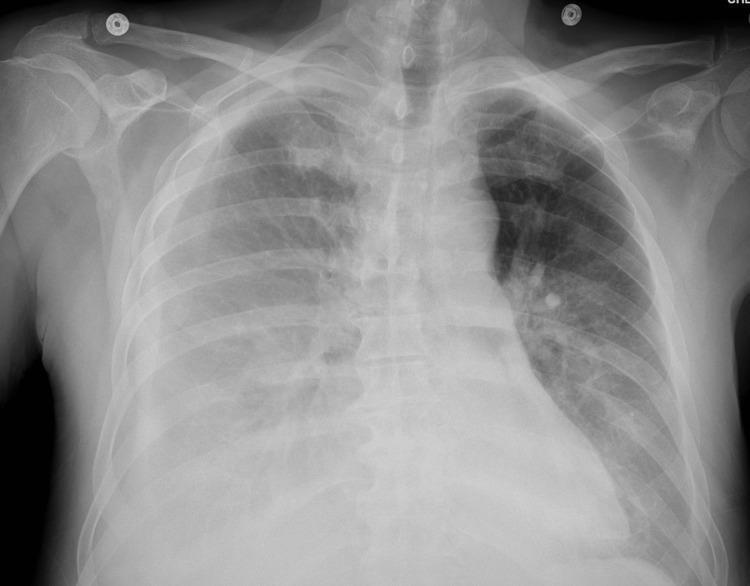
This anteroposterior (AP) chest X-ray demonstrates bilateral pleural effusions in a patient with a clinical history of renal cell carcinoma and recent pericardiocentesis. The right hemithorax shows a moderate-sized pleural effusion, evidenced by a significant homogenous opacity in the lower lung field with blunting of the costophrenic angle. Associated compressive atelectasis is visible in the adjacent right lung tissue. The left hemithorax exhibits a smaller, less pronounced pleural effusion with minimal blunting of the left costophrenic angle. The cardiac silhouette appears enlarged (cardiomegaly), consistent with the patient's recent history of large pericardial effusion. ECG leads are visible on the superior aspect of the chest, indicating active monitoring. The image illustrates typical radiographic findings of fluid accumulation within the pleural space and secondary parenchymal changes, serving as an educational example of postoperative or systemic disease complications in a critical care setting.
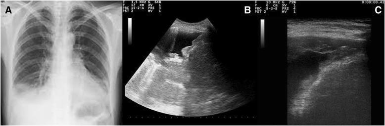
This composite educational graphic compares radiographic and ultrasonographic findings of a pleural effusion. Panel A is a posteroanterior chest X-ray showing a right-sided pleural effusion, characterized by a homogeneous opacity in the lower lung zone with blunting of the costophrenic angle and a visible meniscus sign. Panels B and C are bedside chest ultrasound images (sonograms) of the same patient. These point-of-care ultrasound (POCUS) views provide a more detailed assessment of the pleural space, showing an anechoic (fluid-filled) space consistent with effusion. Notably, the ultrasound images reveal irregular, hyperechoic nodular variations and thickening along the parietal pleura, diaphragm, and costophrenic sinus. These morphological irregularities, which are not visible on the conventional X-ray, are highly suggestive of neoplastic pleural involvement or malignant pleural disease. This comparison illustrates the increased diagnostic sensitivity of ultrasound for evaluating pleural surface morphology in the context of persistent effusion.
Symptoms and signs
- Shortness of breath
- Pleuritic chest pain, worse with a deep breath or cough
- Dry cough
- On examination: reduced breath sounds, reduced chest expansion, and dullness to percussion over the fluid
Main types and causes
| Type | Mechanism | Typical causes |
|---|---|---|
| Transudate | Systemic imbalance in hydrostatic or oncotic pressure | Heart failure, cirrhosis, nephrotic syndrome, kidney failure |
| Exudate | Local pleural inflammation, infection, malignancy, or injury | Pneumonia/parapneumonic effusion, tuberculosis, cancer, pulmonary embolism, autoimmune disease |
Diagnosis
- Chest X-ray and, especially, chest ultrasound to detect and characterize fluid
- Diagnostic thoracentesis for a new or unexplained effusion, unless a clear small effusion from heart failure responds to treatment
- Pleural fluid protein and LDH, with matching serum samples, classify fluid using Light's criteria. An effusion is exudative if at least one is true:
- Pleural-fluid protein/serum-protein ratio >0.5
- Pleural-fluid LDH/serum-LDH ratio >0.6
- Pleural-fluid LDH > two-thirds the upper normal limit for serum LDH
Treatment
- Diuretics and treatment of heart, kidney, or liver disease for a transudate
- Antibiotics and sometimes chest-tube drainage for infected pleural fluid or empyema
- Therapeutic thoracentesis for a large, symptomatic effusion
- Cancer-directed therapy and recurrent-effusion procedures when malignant