
TRALI
TRALI "transfusion-related acute lung injury"
TRALI transfusion related acute lung injury pathophysiology
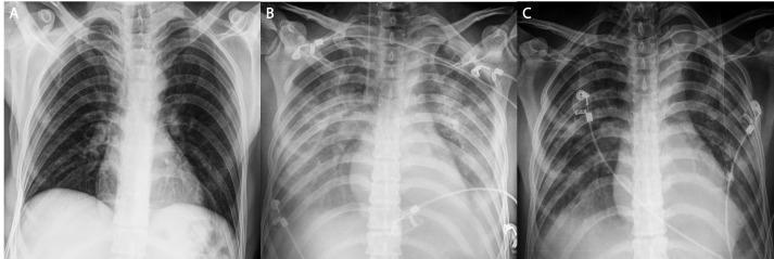
This composite of three anteroposterior (AP) chest x-rays illustrates the progression and resolution of Transfusion-Related Acute Lung Injury (TRALI). Image A represents a preoperative baseline showing clear lung fields, normal bronchovascular markings, and no medical devices. Image B demonstrates the acute phase of TRALI following blood transfusion, characterized by diffuse, bilateral pulmonary infiltrates and 'white-out' appearance consistent with non-cardiogenic pulmonary edema. Visible interventions in Image B include an endotracheal tube for mechanical ventilation, a right-sided central venous catheter, and ECG leads. Image C shows significant interval resolution of the bilateral infiltrates after several days of intensive care support. While the lungs have largely cleared, suggesting improved pulmonary compliance and gas exchange, the endotracheal tube has been removed (extubation), though central lines and monitoring electrodes remain. This sequence serves as a classic educational example of the rapid onset and potential for resolution of TRALI under appropriate ventilatory management.
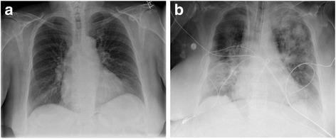
This diagnostic image set consists of two side-by-side anteroposterior (AP) chest X-rays labeled 'a' and 'b,' demonstrating a case of Transfusion-Related Acute Lung Injury (TRALI). Image (a) represents the pre-transfusion baseline, showing relatively clear lung fields with normal lung volumes, distinct vascular markings, and a normal cardiac silhouette. Image (b) depicts the post-transfusion state, showing significant pathological changes. Key findings include diffuse, bilateral, and heterogeneous airspace opacities (patchy and confluent densities) throughout both lung fields, consistent with acute pulmonary edema. There is a visible reduction in lung volumes compared to the baseline. Multiple medical support lines and monitoring leads are now present across the thoracic cavity, indicating an escalation in clinical care. The mediastinal and cardiac contours remain within normal limits, helping to distinguish this non-cardiogenic pulmonary edema from heart failure. This comparison is an educational resource for identifying the radiological hallmarks of TRALI following blood product administration, emphasizing the rapid onset of bilateral pulmonary infiltrates.
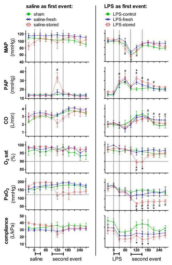
A multi-panel line graph illustrating hemodynamic and respiratory changes in a sheep model of Transfusion-Related Acute Lung Injury (TRALI). The figure is divided into two columns: the left represents groups receiving saline as a first event (sham, saline-fresh, saline-stored), and the right represents groups receiving lipopolysaccharide (LPS) as a first event (LPS-control, LPS-fresh, LPS-stored). Six parameters are plotted over a 240-minute period: Mean Arterial Pressure (MAP), Pulmonary Artery Pressure (PAP), Cardiac Output (CO), Oxygen Saturation (O2 sat), arterial partial pressure of oxygen (PaO2), and static pulmonary compliance. Key events are marked: first event (0–30 min) and second event (90–150 min), involving infusions of saline, fresh packed red blood cells (PRBC), or stored PRBC. Horizontal dashed lines indicate clinical hypoxemia thresholds (O2 sat = 90%; PaO2 = 125 mmHg). Notably, the LPS-stored group shows significant pathological trends, including decreased MAP, CO, PaO2, and O2 sat, alongside increased PAP, compared to controls, characteristic of a two-hit model of acute lung injury.
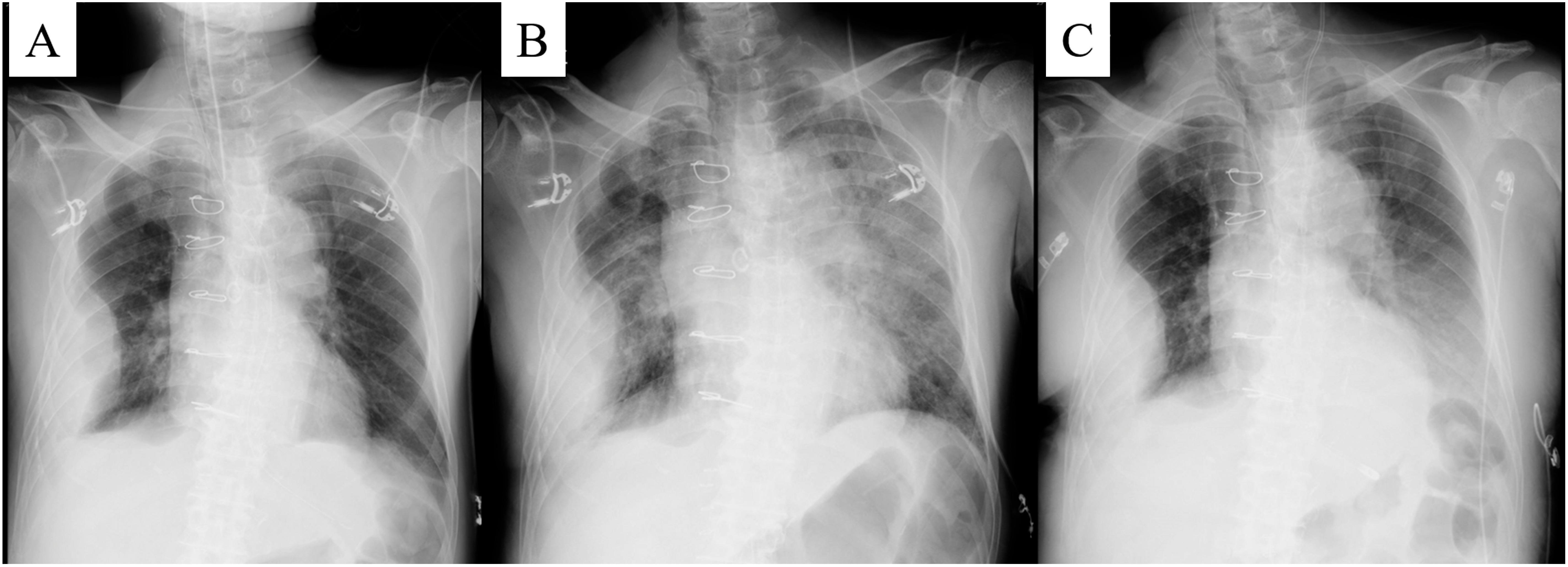
This sequence of three anteroposterior (AP) chest X-rays (labeled A, B, and C) demonstrates the longitudinal progression and resolution of acute pulmonary edema, consistent with Transfusion-Related Acute Lung Injury (TRALI). All images show sternotomy wires and surgical clips, indicating a post-thoracic surgery status. Image A (immediate postoperative) shows relatively clear lung fields with a stable nodular mass in the right middle zone. Image B (onset of respiratory distress) reveals a dramatic bilateral increase in diffuse, confluent alveolar and interstitial opacities, characteristic of pulmonary edema, with a stable cardiomediastinal silhouette. Image C (recovery phase) demonstrates significant clearing of the bilateral infiltrates and improved aeration of the lung parenchyma following treatment. The trachea remains midline and there is no evidence of cardiomegaly throughout the sequence, supporting a non-cardiogenic etiology for the pulmonary findings. These images serve as an educational example of rapid-onset pulmonary edema and its radiographic evolution in a critical care setting.
TRALI - Transfusion-Related Acute Lung Injury
Definition
Epidemiology
- Incidence: 1 in 5000 transfusions (some estimates range 1:1,000 to 1:100,000 depending on the product and population)
- Mortality: 5-10% overall; causes ~15% of fatal transfusion reactions
- Risk varies by product type - plasma-containing products (FFP, platelets, pooled products) carry the highest risk, though PRBCs have become increasingly implicated due to volume of use
- Underreported due to difficulty distinguishing from other causes of ARDS
- Goldman-Cecil Medicine, p. 975
Pathophysiology - The "Two-Hit" Model
Hit 1 (Priming)
Hit 2 (Activation) - Two mechanisms:
- Donor plasma contains anti-HLA class I or II antibodies or anti-neutrophil (antigranulocyte) antibodies
- These attach to recipient leukocytes, triggering release of injurious oxidative and non-oxidative products
- HLA antibodies develop in multiparous women (sensitized during pregnancy) - hence multiparity in female donors is the major donor risk factor
- Bioactive lipids and cytokines accumulate in stored blood products over time
- These activate primed neutrophils in the pulmonary circulation
- Level of these mediators increases with prolonged blood storage
Risk Factors
| Category | Factors |
|---|---|
| Donor | Multiparity, HLA antibodies, anti-neutrophil antibodies |
| Blood product | Plasma-containing components (platelets > FFP > pRBCs), pooled products, stored products |
| Recipient | Positive fluid balance, shock, sepsis, surgery, cardiac bypass, chemotherapy for hematologic malignancies, pre-existing lung injury |
Clinical Presentation
- Acute dyspnea, tachypnea, hypoxemia
- Fever (distinguishes from cardiogenic pulmonary edema)
- Cyanosis
- Bilateral crackles and decreased breath sounds in dependent zones
- Copious pink, frothy edema fluid
- Hypotension (or hypertension in milder cases)
- Rigors, headache, nausea - Goldman-Cecil Medicine, p. 975
Diagnostic Criteria (NHSN Hemovigilance Protocol)
- No evidence of acute lung injury BEFORE transfusion
- Acute lung injury onset during or within 6 hours of transfusion cessation
- Hypoxemia: PaO₂/FiO₂ ≤ 300 mmHg OR SpO₂ < 90% on room air
- Bilateral infiltrates on CXR
- No evidence of left atrial hypertension (no circulatory overload)
- Rosen's Emergency Medicine, p. 2432
TRALI Type I vs. Type II (updated classification)
| Type I (Classic TRALI) | Type II (formerly "Possible TRALI") | |
|---|---|---|
| Pre-existing ALI? | No | Mild pre-existing ALI; stable for ≥12 h before transfusion |
| Other ALI risk factors? | None | May be present |
| Temporal onset | Within 6 h of transfusion | Within 6 h of transfusion |
Laboratory & Imaging Findings
- CXR: Diffuse bilateral patchy alveolar infiltrates (no cardiomegaly, no effusions typically)
- ABG: Reduced PaO₂, low PaO₂/FiO₂
- Transient thrombocytopenia or leukopenia
- Confirmatory (not required for diagnosis): anti-HLA class I/II or antigranulocyte antibodies in donor plasma
- No readily available bedside confirmatory test
Differential Diagnosis
| Condition | Distinguishing Feature |
|---|---|
| TACO (Transfusion-associated circulatory overload) | Elevated BNP, elevated PCWP, responds to diuresis, hypertension |
| Anaphylaxis | Wheezing, urticaria, lacks bilateral infiltrates, responds to epinephrine |
| Transfusion-related sepsis | Culture positive, hemodynamic features predominate |
| Hemolytic transfusion reaction | Positive DAT, hemolysis, hemoglobinuria |
| ARDS (other causes) | No temporal relationship to transfusion |
Treatment
- Stop the transfusion immediately
- Supportive care is the cornerstone - most cases resolve within 48-96 hours
- Supplemental oxygen and respiratory support as needed
- Mechanical ventilation if required - use low tidal volume strategy (as in ARDS management)
- Volume resuscitation ± vasopressors for hypotension (intravascular pressures are often LOW - use diuretics cautiously or not at all)
- Corticosteroids: not routinely recommended
- Survivors recover within 72 hours typically
Prognosis
- Mortality ~5%
- Most cases self-limited, resolving within hours to days
- Patients are NOT at increased risk for future TRALI episodes from other (non-implicated) donors
- If an implicated donor is identified, the recipient should not receive further products from that donor
Prevention
-
Exclude implicated donors from future blood donation
-
Screen female multiparous donors for HLA antibodies; exclude plasma from HLA-antibody-positive donors
-
Use male-predominant or never-pregnant female plasma for transfusion (policy now adopted by many blood banks)
-
Use leukoreduced blood products (reduces cytokine accumulation)
-
Limit unnecessary transfusions (restrictive transfusion strategy)
-
Quick Compendium of Clinical Pathology 5th ed., p. 93 | Goldman-Cecil Medicine, p. 975 | Rosen's Emergency Medicine, p. 2432
TRALI vs. TACO - Quick Comparison
| Feature | TRALI | TACO |
|---|---|---|
| Onset | During or within 6 h | 6-12 h |
| Mechanism | Non-cardiogenic (immune/inflammatory) | Cardiogenic (volume overload) |
| BP | Usually hypotension | Hypertension |
| BNP | Normal | Elevated |
| PCWP | Low/normal | Elevated |
| CXR | Bilateral infiltrates, no cardiomegaly | Cardiomegaly + infiltrates |
| Response to diuretics | May worsen | Improves rapidly |
| Fever | Common | Less common |