
Management of diabetes and it's complications
"diabetes mellitus"[MeSH Terms] AND "management"[MeSH Terms]
"type 2 diabetes" AND "treatment"
diabetic retinopathy fundus photograph
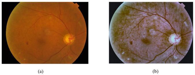
Diagnostic Image: Fundus Photographs. This figure shows two side-by-side retinal fundus images illustrating Diabetic Retinopathy (DR) manifestations and the effect of digital preprocessing. Image (a) is the original raw fundus photograph with a standard reddish-orange hue, showing the optic disc and retinal vasculature. Pathological findings include red lesions (microaneurysms and hemorrhages) appearing as dark spots, and bright lesions representing hard exudates (yellowish spots) and soft exudates/cotton wool spots (fluffy white areas). Image (b) shows the same retina after preprocessing to enhance contrast and sharpen clinical features. In the processed version, the overall tone shifts to a muted brownish-purple, making the vascular network more distinct and the lesions more defined. The preprocessing helps differentiate between red lesions and bright exudative lesions, which are critical for DR staging. This comparison demonstrates the role of image processing in improving the detectability of subtle retinal pathology for diagnostic AI models or clinical review.
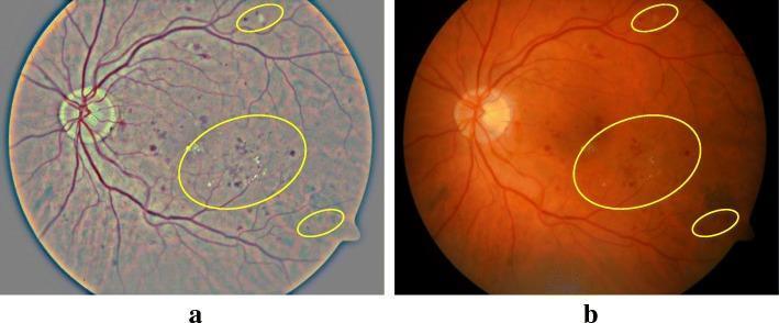
This clinical comparison shows side-by-side retinal fundus photographs demonstrating the effect of contrast enhancement (CE) on the visualization of diabetic retinopathy (DR). Image (a) is the original fundus photograph, presenting a somewhat washed-out, grayish-blue tone with low contrast between retinal features. Image (b) shows the result of the image enhancement technique, featuring a more natural orange-red retinal hue with significantly improved contrast and sharpness. Both images utilize yellow elliptical annotations to highlight specific pathological signs of DR, including hard exudates (bright, yellowish spots) and microaneurysms or small hemorrhages (dark red spots). The optic disc is visible on the left side of each image, with retinal vessels radiating outward. The enhancement in image (b) allows for better singularization and identification of lesions, particularly small microaneurysms and subtle exudates that are less distinct in the raw image. This demonstrates the clinical utility of computational image processing in improving the diagnostic sensitivity of fundus imaging for automated or manual screening.
diabetic nephropathy kidney pathology
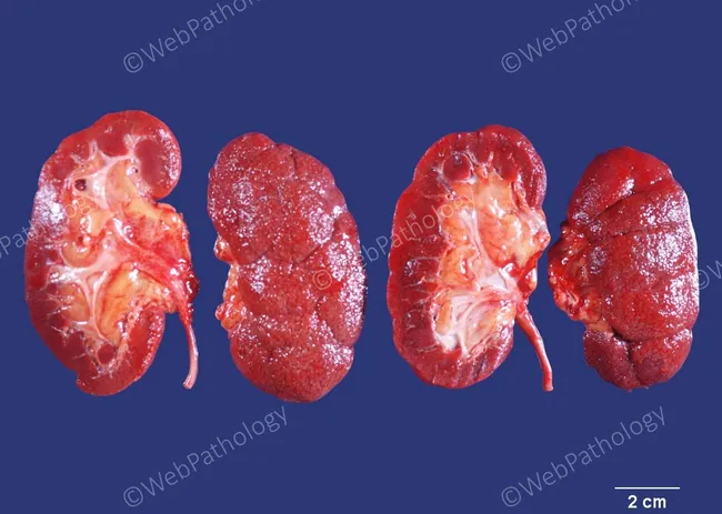
Autopsy gross photograph of the kidneys (human, bilateral) illustrating advanced diabetic nephropathy with nephrosclerosis. Modality: macroscopic pathology imaging; view: anterior/sagittal kidney sections arranged for direct comparison. Anatomical location: renal cortex with outer medulla; both kidneys show marked cortical thinning and a finely granular, leathery subcapsular surface consistent with chronic scarring. External contour is reduced in size, indicating symmetrically shrunken kidneys typical of end-stage nephropathy. Internal features reveal shallow cortical scars with sclerotic glomeruli, tubulointerstitial atrophy, and hyalinized arterioles with narrowed lumens (arteriolosclerosis), consistent with diabetic microangiopathy. Secondary findings include diffuse interstitial fibrosis and vascular remodeling. Pathophysiology: long-standing type 2 diabetes mellitus has driven glomerular and arteriolar hyalinosis, ischemic tubular loss, and progressive loss of cortical nephrons, culminating in renal insufficiency. Clinically significant: this image demonstrates the morphologic endpoint of diabetic nephropathy contributing to ESRD risk; supports diagnosis and educates on end-organ damage. Diagnostic considerations include hypertensive nephrosclerosis and age-related nephrosclerosis; differential guarded by clinical history. Clinical correlation: in life, patient had type 2 DM for 27 years; death due to myocardial infarction; underscores DM-related microvascular complications and their contribution to morbidity and mortality in diabetics. This image serves as a teaching aid for renal pathology and DM complications.
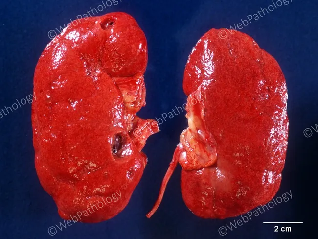
Autopsy gross pathology photograph documenting bilateral renal nephrosclerosis in an elderly patient with long-standing diabetes mellitus and poor glycemic control. The kidneys are slightly reduced in size with a finely granular, subcapsular surface characteristic of nephrosclerosis. External cortex shows coarse granularity and reduced corticomedullary contrast, with a reddish-brown coloration suggestive of chronic ischemic injury. A few simple cortical cysts are present. The image provides macroscopic corroboration of diabetic nephropathy as a microvascular complication, where glomerulosclerosis and arteriolar hyalinosis produce progressive renal scarring, cortical atrophy, and diminished renal function. While histology is not shown, gross findings align with late-stage diabetic kidney disease and hypertension-related vascular changes. The scale bar indicates 2 cm for reference, enabling size assessment and morphologic comparison with other cases. Clinically, this morphology corresponds to microalbuminuria evolving to proteinuria, reduced GFR, and potential progression to end-stage renal disease if the disease persists. This specimen demonstrates typical end-organ damage in diabetes mellitus, with nephrosclerosis accompanied by incidental cortical cysts—a finding frequently observed in elderly individuals and in long-standing metabolic disease. Educationally, it serves as a reference image for medical students, pathologists, and researchers studying diabetic nephropathy and autopsy pathology.
Management of Diabetes Mellitus and Its Complications
Overview
I. Glycemic Targets
| Parameter | Target |
|---|---|
| HbA1c (general) | < 7.0% (ADA) / < 6.5% (ACE) |
| HbA1c (elderly/CVD/hypoglycemia-prone) | 7.5-8.0% |
| Blood pressure | < 130/80 mmHg |
| LDL cholesterol | As low as possible (statin for all) |
- 21% reduction in any diabetes-related endpoint
- 14% reduction in MI
- 12% reduction in stroke
- 37% reduction in microvascular disease risk
II. Non-Pharmacological Management
- Medical nutrition therapy: Reduced caloric intake, low glycaemic-index foods, reduced saturated fats
- Physical activity: At least 150 minutes/week of moderate aerobic exercise
- Weight loss: Even 5-10% weight reduction improves insulin sensitivity significantly
- Smoking cessation: Mandatory for all patients
- Self-monitoring of blood glucose (SMBG)
- Diabetes education: Patient empowerment in self-care
III. Pharmacological Management
First-line: Metformin (Biguanide)
- Mechanism: Decreases hepatic glucose production; improves insulin sensitivity
- Benefits: No hypoglycemia risk; weight-neutral to modest weight loss; reduces acute cardiovascular events by 38% (UKPDS)
- Caution: Avoid if eGFR < 30 mL/min; monitor renal function and vitamin B12 levels
- Preferred first-line agent for type 2 DM unless contraindicated
Second-line Agents (Summary Table)
| Drug Class | Example | Mechanism | Hypoglycemia Risk | Key Notes |
|---|---|---|---|---|
| Sulfonylureas | Glimepiride, Glipizide | Stimulate insulin secretion | Yes | Weight gain; avoid glyburide in renal impairment |
| Meglitinides | Repaglinide, Nateglinide | Short-acting insulin secretagogues | Yes (rarely) | Taken with meals; postprandial effect |
| Thiazolidinediones (TZDs) | Pioglitazone | PPAR-γ agonist; reduces insulin resistance | No | Avoid in heart failure, liver disease |
| DPP-4 Inhibitors | Sitagliptin, Linagliptin | Increase glucose-dependent insulin; decrease glucagon | No | Once daily; risk of pancreatitis; renal adjustment (except linagliptin) |
| SGLT2 Inhibitors | Empagliflozin, Dapagliflozin | Inhibit renal glucose reabsorption | No | Cardiovascular and renal protective; stop if eGFR < 30; risk of UTI and DKA (T1DM) |
| GLP-1 Receptor Agonists | Semaglutide, Liraglutide, Dulaglutide | Glucose-dependent insulin release; suppress glucagon; delay gastric emptying | No | Weight loss; CV and renal protection; injectable (oral semaglutide available) |
| Alpha-glucosidase inhibitors | Acarbose | Delay intestinal carbohydrate absorption | No | GI side effects limit use |
| Insulin | Various formulations | Replaces/supplements endogenous insulin | Yes | Required for T1DM; added in progressive T2DM |
Modern Cardio-Renal Priority Framework
- SGLT2 inhibitors: Preferred in diabetic kidney disease (reduce intraglomerular pressure via tubuloglomerular feedback, lower BP, reduce CV events). Discontinue when eGFR < 30 mL/min.
- GLP-1 agonists: Alternative when SGLT2 inhibitors cannot be used; can continue until eGFR falls to 15 mL/min. Semaglutide (weekly), liraglutide (daily), dulaglutide (weekly) are options.
IV. Insulin Therapy
- Basal insulin (e.g., glargine, detemir): Once-daily; controls fasting glucose
- Bolus (prandial) insulin (e.g., lispro, aspart): Given before meals; controls postprandial glucose
- Premixed insulin: Convenient but less flexible
- Dose titration based on fasting glucose readings
- Risk of hypoglycemia - patient education mandatory
V. Complications and Their Management
1. Diabetic Retinopathy
- Nonproliferative DR (NPDR): Microaneurysms, retinal hemorrhages, cotton wool spots, venous beading
- Proliferative DR (PDR): Neovascularization, vitreous hemorrhage, retinal detachment
- Diabetic macular edema (DME): Now the leading cause of vision loss in diabetics; increased vascular permeability causes macular swelling
- Type 1 DM: First eye exam within 5 years of diagnosis, then annually
- Type 2 DM: Eye exam at diagnosis, then annually (every 2 years if well controlled with no retinopathy)
- Tight glycemic control: 76% reduced risk of developing DR in type 1 DM; 16-25% reduction in type 2 DM
- Blood pressure control slows progression
- Laser photocoagulation: Indicated for PDR and selected severe NPDR
- Anti-VEGF injections (intravitreal): Improves vision in diabetic macular edema
- Fenofibrate: Evidence supports slowing DR progression (recommended in Australia and Canada for secondary prevention)
2. Diabetic Neuropathy
- Distal symmetric polyneuropathy (DSPN): "Stocking-glove" distribution; burning pain, paresthesias
- Focal mononeuropathies: CN III or VI (cranial); median nerve most common limb site
- Diabetic amyotrophy: Severe asymmetric muscle weakness/wasting of iliopsoas and quadriceps
- Autonomic neuropathy: Cardiovascular (orthostatic hypotension, fixed HR, resting tachycardia), GI (gastroparesis, diabetic diarrhea, constipation), genitourinary (bladder dysfunction, erectile dysfunction)
- Monofilament testing (5.07/10g Semmes-Weinstein): LR+ = 10.2 for abnormal perception
- Tuning fork (128 Hz) vibration testing: LR+ = 18.5 for timed vibration
- Annual foot exam for all diabetics
| Condition | Management |
|---|---|
| Glycemic control | Reduces neuropathy risk 60% in type 1 DM |
| Peripheral neuropathic pain | Pregabalin, duloxetine (FDA-approved); gabapentin, TCAs (amitriptyline), venlafaxine |
| Refractory pain | Tramadol; opioids only with pain specialist |
| Orthostatic hypotension | Head of bed elevation, compression garments, midodrine (FDA-approved) |
| Gastroparesis | Prokinetics (metoclopramide, domperidone) |
| Diabetic diarrhea | Tetracycline, clonidine |
| Bladder dysfunction | Bethanecol; intermittent self-catheterization |
| Erectile dysfunction | Sildenafil and PDE5 inhibitors |
3. Diabetic Nephropathy (Diabetic Kidney Disease)
- Microalbuminuria appears 10-15 years after onset
- 80% progress to macroalbuminuria over the next 10-15 years
- 50% develop ESRD within 10 years of macroalbuminuria; 75% by 20 years
- Normal: < 30 mcg/mg
- Microalbuminuria: 30-299 mcg/mg
- Macroalbuminuria: ≥ 300 mcg/mg
- Glycemic control (HbA1c < 7%)
- BP control (target < 130/80 mmHg)
- RAAS blockade: ACE inhibitors or ARBs - preferred antihypertensives; reduce proteinuria and slow progression
- SGLT2 inhibitors: First-line add-on; reduce intraglomerular pressure, promote natriuresis, lower BP - stop at eGFR < 30
- GLP-1 agonists: Alternative when SGLT2 inhibitors not tolerated; use until eGFR falls to 15 mL/min
- Dietary protein restriction: 0.8 g/kg/day
- Preparation for RRT: Erythropoietin for anemia; manage calcium-phosphate metabolism
4. Macrovascular Disease (CVD, Stroke, PAD)
- Advanced glycation end-products (AGEs) damage the vascular endothelium
- Pro-oxidative and pro-inflammatory state
- Hypercoagulability: Increased coagulation factors, platelet reactivity, reduced tPA, increased PAI-1
- Atherogenic dyslipidemia: Low HDL, elevated triglycerides, small dense LDL
| Risk Factor | Target/Intervention |
|---|---|
| Glycemia | HbA1c < 7% |
| Blood pressure | < 130/80 mmHg (RAAS preferred) |
| LDL | Statin therapy for all diabetics (CARDS trial, 4S trial) |
| Antiplatelet therapy | Aspirin prophylactically (unless contraindicated) |
| Smoking | Complete cessation |
| Obesity | Weight loss; GLP-1 agonists aid weight reduction |
| CVD-high risk | SGLT2 inhibitors and/or GLP-1 agonists |
5. Diabetic Foot
- Foot ulcers affect up to 25% of diabetics over their lifetime
- Leading cause of non-traumatic lower limb amputation
- Annual comprehensive foot exam
- Monofilament and Doppler ABI (ankle-brachial index) screening
- Patient education: proper footwear, daily inspection
- Multidisciplinary wound care for active ulcers
- Revascularization for significant PAD
VI. Monitoring Summary
| Parameter | Frequency |
|---|---|
| HbA1c | Every 3 months (uncontrolled); every 6 months (stable) |
| Fasting lipid panel | Annually |
| Urine albumin/creatinine ratio | Annually |
| eGFR/serum creatinine | Annually |
| Dilated eye exam | Annually (see above) |
| Foot exam | At every visit |
| BP | Every visit |
| Dental exam | Annually |
| Vaccinations | Influenza, pneumococcal, Hep B per schedule |
Recent Evidence Update
-
GLP-1 RAs + SGLT2 inhibitors combo - Circulation 2024 (PMID 39210781): Combined therapy provides additive cardiovascular and kidney protection beyond either drug class alone.
-
GLP-1 RAs in kidney/CV disease - Lancet Diabetes Endocrinol 2025 (PMID 39608381): GLP-1 receptor agonists significantly reduce progression of diabetic kidney disease and major cardiovascular events in RCTs.
Management of hyperthyroidism and hypothyroidism
"hyperthyroidism"[MeSH Terms] AND "treatment"[MeSH Terms]
"Graves disease" OR "hyperthyroidism" AND "antithyroid drugs"
Graves disease exophthalmos thyroid goiter clinical photo
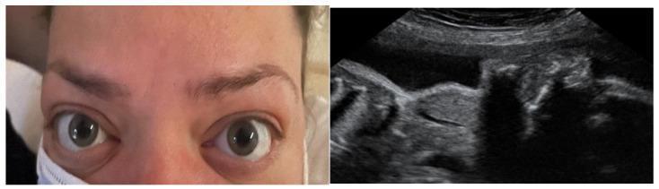
This dual-panel image demonstrates maternal and fetal manifestations of Graves' disease. The left panel is a clinical photograph of a pregnant woman's face, showing classic signs of thyroid-associated orbitopathy (TAO). Key features include bilateral exophthalmos (proptosis), lid retraction with visible superior scleral show, and periorbital edema, indicative of active Graves' ophthalmopathy. The right panel displays an obstetric ultrasound image focusing on the fetal neck. The ultrasound reveals a significant hyperechoic mass in the anterior cervical region, consistent with a fetal goiter. This finding in the context of maternal Graves' disease suggests fetal thyroid dysfunction caused by the transplacental passage of maternal thyroid-stimulating hormone receptor antibodies (TRAb). The image serves as a clinical illustration of the correlation between maternal autoimmune thyroid activity and potential neonatal hyperthyroidism or goitrous development during pregnancy.
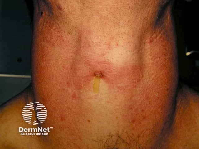
Clinical photograph of the anterior neck illustrating diffuse enlargement of the thyroid region consistent with a goiter. The image is a frontal view capturing the patient from the chin to the upper chest, suitable for documentation and educational purposes in endocrinology and dermatology. The thyroid gland appears expanded with smooth contour and midline projection; there is mild erythema and skin textural change over the lower neck, with a small vertical scar and minimal hair growth noted on the chest. Visual assessment alone cannot differentiate diffuse goiter from nodular disease; no discrete nodules are clearly identifiable in this image. The photograph emphasizes external neck anatomy, including the sternocleidomastoid borders and thyroid isthmus region just below the laryngeal prominence. This image is most applicable to clinical evaluation of neck swelling, patient education, and baseline documentation prior to ultrasound or other thyroid imaging. Potential clinical uses include tracking goiter progression, educating patients about thyroid enlargement, and serving as a visual aid in differential diagnosis discussions with Graves disease, Hashimoto thyroiditis, endemic iodine deficiency, and multinodular goiter. When combined with laboratory tests (TSH, free T4), and neck ultrasonography, this photo supports diagnosis and management planning, including consideration of fine-needle aspiration if focal nodularity is detected or suspicion of malignancy arises.
Management of Hyperthyroidism and Hypothyroidism
PART 1: HYPERTHYROIDISM
Definition and Diagnosis
- Suppressed TSH (< 0.1 mIU/L; often < 0.01 mIU/L in Graves disease)
- Elevated free T4 (FT4) and/or free T3 (FT3)
Common Causes
| Cause | Key Feature |
|---|---|
| Graves disease | Most common; TSH receptor-stimulating antibodies (TRAb); often with goiter and ophthalmopathy |
| Toxic multinodular goiter (Plummer disease) | Multiple autonomously functioning nodules |
| Toxic (autonomous) solitary nodule | Single "hot" nodule |
| Subacute thyroiditis | Transient; destructive release of stored hormone |
| Factitious (exogenous) thyrotoxicosis | Exogenous T4/T3 ingestion |
| TSH-secreting pituitary adenoma | Rare; elevated TSH with elevated thyroid hormones |
Clinical Features (Hypermetabolic State)
- Tachycardia, wide pulse pressure, systolic hypertension
- Fever, fine tremor, warm moist skin
- Anxiety, hyperactivity, insomnia
- Diarrhea, weight loss despite increased appetite
- Atrial fibrillation (particularly in elderly and subclinical hyperthyroidism)
- Osteoporosis with prolonged hyperthyroidism
- Exophthalmos, lid lag, lid retraction (Graves ophthalmopathy)
Management of Hyperthyroidism
1. Antithyroid Drugs (Thionamides)
| Drug | Dose | Notes |
|---|---|---|
| Methimazole (MMI) | 10-40 mg/day (single or divided) | Preferred in most situations; once-daily dosing possible |
| Propylthiouracil (PTU) | 100-200 mg every 6-8h | Preferred in: thyroid storm, 1st trimester pregnancy; also blocks T4→T3 conversion |
| Carbimazole | 20-60 mg/day | Prodrug of MMI; used widely outside USA |
- Titration regimen: Start high, reduce dose as euthyroidism is achieved (target TSH within normal range)
- Block-and-replace regimen: Full-dose thionamide + low-dose levothyroxine added once euthyroid
- Agranulocytosis (0.2-0.5%) - warn patients to report fever/sore throat; stop drug immediately if suspected
- Rash, urticaria (5-10%)
- Hepatotoxicity (PTU > MMI) - monitor LFTs
- Teratogenicity: avoid MMI in 1st trimester (methimazole embryopathy); switch to PTU; after 1st trimester, switch back to MMI
2. Radioactive Iodine (¹³¹I)
- Most commonly used definitive therapy in the USA
- Destroys thyroid tissue over weeks to months
- Results in hypothyroidism in the majority (then treated with levothyroxine)
- Contraindicated in: pregnancy, breastfeeding, active/severe Graves ophthalmopathy
- May worsen ophthalmopathy (risk mitigated by concurrent glucocorticoids)
- Give antithyroid drugs first to render patient euthyroid before ¹³¹I if severely thyrotoxic; stop thionamide 5-7 days before treatment
3. Surgery (Thyroidectomy)
- Near-total or total thyroidectomy preferred
- Results in permanent hypothyroidism (requires lifelong levothyroxine)
- Indications: Large goiter causing compressive symptoms; suspected malignancy; failed medical therapy; patient preference; severe or active Graves ophthalmopathy; pregnancy in 2nd trimester
- Render patient euthyroid with antithyroid drugs pre-operatively
- Complications: Hypoparathyroidism, recurrent laryngeal nerve injury
Symptomatic Control: Beta-Blockers
- Propranolol 40-80 mg every 6-8h (preferred - also inhibits T4→T3 conversion; improves tremor, palpitations, anxiety, heat intolerance)
- Atenolol or metoprolol for patients with asthma or COPD (beta-1 selective)
- Do NOT use propranolol in decompensated heart failure
Special Populations
| Population | Management |
|---|---|
| Pregnancy (1st trimester) | PTU (avoid MMI - embryopathy risk) |
| Pregnancy (2nd/3rd trimester) | Switch to MMI; PTU causes hepatotoxicity in mother |
| Neonatal hyperthyroidism | Maternal TRAb crosses placenta; treat neonate with PTU/MMI + propranolol |
| Postpartum | High risk of Graves relapse; restart antithyroid drugs |
| Elderly | Subclinical hyperthyroidism carries major AF and osteoporosis risk; treat nodular disease |
| Children | Methimazole preferred (avoid PTU); prolonged course (≥3 years) before considering definitive therapy |
Subclinical Hyperthyroidism
- Defined as TSH < 0.1 mIU/L with normal FT4 and FT3
- Associated with 2-3x increased risk of atrial fibrillation (Framingham data)
- Associated with reduced bone mineral density and fracture risk
- AACE recommends treatment for subclinical hyperthyroidism caused by nodular thyroid disease
- ¹³¹I preferred for toxic nodule/TMNG in this setting
Graves Ophthalmopathy
- Mild: Artificial tears, dark glasses, tape eyelids at night, smoking cessation, selenium 100 mcg bid
- Moderate-to-severe: IV methylprednisolone (500 mg weekly x 6 weeks, then 250 mg weekly x 6 weeks) - preferred over oral steroids
- Refractory: Orbital external beam radiotherapy; orbital decompression surgery
- Teprotumumab (IGF-1 receptor monoclonal antibody): Improves proptosis and diplopia in active moderate-to-severe disease
THYROID STORM (Thyrotoxic Crisis)
| Step | Drug | Dose |
|---|---|---|
| 1. Beta-blocker | Propranolol | 60-80 mg PO every 4h; or 2 mg IV every 4h. Use esmolol if HF present |
| 2. Thionamide | PTU (preferred) | 500-1000 mg loading dose; then 250 mg every 4h (NG or PR if unconscious) |
| or | Methimazole | 20 mg every 6h (if PTU unavailable) |
| 3. Iodine (≥1h after thionamide) | SSKI | 5 drops every 6h - blocks hormone release (Wolff-Chaikoff effect) |
| or | Lugol's solution | 8 drops every 6h |
| 4. Glucocorticoids | Hydrocortisone | 300 mg IV bolus, then 100 mg every 8h (inhibits T4→T3 conversion; treats relative adrenal insufficiency) |
| or | Dexamethasone | 2-4 mg IV every 6h |
| 5. Cholestyramine | 1-4 g twice daily | Blocks enterohepatic recirculation of thyroid hormone; for severe/refractory cases |
- Cooling (fans, ice packs, cooling blankets) + acetaminophen (avoid aspirin - displaces T4/T3 from binding proteins)
- IV fluids with dextrose (glycogen stores depleted)
- Benzodiazepines for agitation
- Treat precipitating cause (antibiotics empirically if infection suspected)
PART 2: HYPOTHYROIDISM
Definition and Diagnosis
- Elevated TSH (> 4.5 mIU/L; often > 10 mIU/L in overt hypothyroidism; > 25 mIU/L in severe/prolonged cases)
- Low or low-normal FT4
Causes
| Cause | Notes |
|---|---|
| Hashimoto thyroiditis (Chronic autoimmune thyroiditis) | Most common cause in developed world; anti-TPO antibodies destroy thyroid |
| Post-radioiodine or post-thyroidectomy | Intentional or incidental |
| Iodine deficiency | Leading cause worldwide |
| Subacute thyroiditis (post-inflammatory) | Transient; usually resolves in 3-6 months |
| Central/secondary hypothyroidism | Pituitary or hypothalamic failure; TSH low or inappropriately normal |
| Drug-induced | Amiodarone, lithium, interferon-alpha |
| Head and neck radiation | External beam treatment for cancer |
Clinical Features (Hypometabolic State)
- Fatigue, cold intolerance, weight gain
- Dry skin, coarse hair, hair loss, brittle nails
- Constipation, bradycardia
- Depression, cognitive slowing, memory impairment
- Periorbital puffiness, non-pitting edema (myxedema)
- Delayed relaxation of deep tendon reflexes
- Menstrual irregularities, infertility
- Dyslipidemia (elevated LDL and total cholesterol)
- Hyponatremia (in severe cases)
Management of Hypothyroidism
Standard Treatment: Levothyroxine (L-T4)
- Drug of choice for primary hypothyroidism
- Mechanism: Exogenous T4 is peripherally converted to active T3 in tissues
- Standard replacement dose: 1.6 mcg/kg/day (average adult: 75-150 mcg/day)
- Half-life: 7 days - permits once-daily dosing; steady state achieved at 6-8 weeks
- Take on an empty stomach, 30-60 minutes before breakfast (avoid calcium, iron, antacids which impair absorption)
| Patient | Starting Dose |
|---|---|
| Young, healthy adult | Full replacement (1.6 mcg/kg/day) immediately |
| Age > 50, no cardiac disease | 50 mcg/day; titrate up |
| Elderly with cardiac disease | 12.5-25 mcg/day, increase by 12.5-25 mcg every 2 weeks |
| Severe long-standing hypothyroidism | Start low, titrate slowly - cardiac sensitivity |
T4 + T3 Combination Therapy
- Considered when patient has normal TSH on T4 alone but persistent hypothyroid symptoms
- Add low-dose liothyronine (L-T3) to T4
- Monitor carefully - T3 is more cardiotoxic and harder to titrate
- Maintain TSH above 1.0 mIU/L
- Not standard first-line; reserve for symptomatic patients who fail T4 monotherapy
Subclinical Hypothyroidism
- TSH 4.5-10 mIU/L with normal FT4; patient asymptomatic or mildly symptomatic
- 3-5% progress to overt hypothyroidism annually (most represent early Hashimoto disease)
- Check TPO antibodies - positive TPO Ab predicts faster progression
- Treatment considerations (ATA/AACE 2012 guidelines):
- Treat if: TPO Ab positive, symptoms of hypothyroidism, history of atherosclerotic vascular disease, TSH > 10 mIU/L
- Observe if: TSH 4.5-10 mIU/L, no antibodies, truly asymptomatic
- Associated with increased risk of CHD events, heart failure, and adverse pregnancy outcomes
Hypothyroidism in Pregnancy
- Target TSH: 0.1-2.5 mIU/L in 1st trimester; 0.2-3.0 mIU/L in 2nd/3rd trimester
- Levothyroxine dose typically increases 25-30% in first weeks of pregnancy
- Maternal hypothyroidism impairs fetal neurodevelopment - early and aggressive treatment is essential
- Check TSH every 4 weeks in 1st trimester; every 6-8 weeks thereafter
Hypothyroidism with Coronary Artery Disease
- If coronary revascularization is indicated: perform revascularization BEFORE correcting hypothyroidism
- Use very low starting doses (12.5-25 mcg/day); titrate slowly over months
- Monitor for angina with dose changes
MYXEDEMA COMA
- Hypothermia (often severe, < 35°C)
- Respiratory depression (CO2 retention)
- Decreased or absent consciousness
- Hyponatremia, hypoglycemia, bradycardia, hypotension
| Intervention | Details |
|---|---|
| Airway/ventilation | Intubate and mechanically ventilate if needed |
| Levothyroxine (T4) IV | Loading dose 300-400 mcg IV initially, then 50-100 mcg IV daily |
| Liothyronine (T3) IV | Optional add-on: 5-20 mcg initially, then 2.5-10 mcg every 8h. More cardiotoxic - use caution in elderly/cardiac patients |
| Hydrocortisone | 100 mg IV every 8h (empiric, until adrenal/pituitary insufficiency excluded - may coexist with Schmidt's syndrome) |
| IV fluids | Cautious - risk of fluid overload and worsening hyponatremia. Use dextrose-containing solutions for hypoglycemia |
| Warming | Passive external warming (avoid aggressive active rewarming - can cause peripheral vasodilation and shock) |
| Treat precipitant | Antibiotics for infection; treat any underlying cause |
Summary Comparison Table
| Feature | Hyperthyroidism | Hypothyroidism |
|---|---|---|
| TSH | Low (< 0.1 mIU/L) | High (> 4.5-10 mIU/L) |
| FT4 | High | Low |
| Metabolic state | Hypermetabolic | Hypometabolic |
| Most common cause | Graves disease | Hashimoto thyroiditis |
| Primary drug treatment | Methimazole / PTU | Levothyroxine (T4) |
| Definitive options | ¹³¹I, surgery | None (lifelong replacement) |
| Life-threatening emergency | Thyroid storm | Myxedema coma |
| Emergency drug sequence | Propranolol → PTU → Iodine → Hydrocortisone | IV T4 loading + T3 + Hydrocortisone |
| Monitoring | TSH, FT4, FT3 | TSH (target 0.5-2.5 mIU/L) |