
High yield info about strokes everything as localization types diagnose mangmant
Stroke: high-yield study map
-
Stroke basics
- Definition, ischemic vs hemorrhagic stroke, TIA
- Time-critical approach and mimics
-
Vascular anatomy and localization
- ACA, MCA, PCA territories
- Lacunar syndromes
- Vertebrobasilar and brainstem stroke syndromes
- Cortical versus subcortical findings
-
Clinical presentation
- Motor, sensory, language, visual, cerebellar, and cranial-nerve deficits
- Dominant versus non-dominant hemisphere findings
-
Diagnosis
- ABCs, glucose, NIHSS, focused neurologic exam
- Non-contrast CT, CT angiography, CT perfusion, MRI-DWI
- ECG, vascular imaging, echocardiography, stroke-cause workup
-
Acute ischemic stroke management
- Immediate stabilization and eligibility for IV thrombolysis
- Mechanical thrombectomy selection
- Antiplatelets, blood-pressure targets, glucose, temperature, swallow screening
-
Intracerebral hemorrhage and subarachnoid hemorrhage
- Recognition, CT diagnosis, reversal of anticoagulation, BP management, neurosurgical indications
- SAH red flags and aneurysm workup
-
Secondary prevention
- Antiplatelet versus anticoagulation
- Carotid disease, statins, hypertension, diabetes, smoking cessation, rehabilitation
-
Exam tables and algorithms
- Territory localization table
- “Sudden focal deficit” diagnostic pathway
- Thrombolysis and thrombectomy contraindications
- High-yield differentiators and common traps
One-minute overview
Create a stroke localization comparison table
stroke localization
stroke syndrome localization MCA ACA PCA lacunar posterior circulation clinical features
Stroke Localization Comparison Table
Major cerebral arterial territories
| Territory / artery | Main area affected | Contralateral motor/sensory pattern | Cortical signs | Visual / eye signs | Key high-yield clue |
|---|---|---|---|---|---|
| MCA - Middle cerebral artery | Lateral frontal, parietal, temporal cortex; insula. Deep MCA branches supply basal ganglia and internal capsule. | Face and arm > leg weakness and sensory loss | Dominant hemisphere: aphasia. Non-dominant hemisphere: hemispatial neglect, anosognosia, impaired visuospatial function. Apraxia may occur. | Contralateral homonymous hemianopia; gaze preference toward lesion in large hemispheric stroke | Face/arm-predominant deficit + aphasia or neglect |
| MCA superior division | Lateral frontal lobe, motor cortex for face/arm | Face and arm weakness > leg; may have dysarthria | Broca aphasia if dominant side: non-fluent, effortful speech with relatively preserved comprehension | Usually no major visual field defect | Non-fluent aphasia + right face/arm weakness = left superior MCA |
| MCA inferior division | Lateral temporal and inferior parietal cortex | Weakness often minimal or absent | Wernicke aphasia if dominant side: fluent but nonsensical speech, impaired comprehension; neglect if non-dominant side | Contralateral superior quadrantanopia, “pie in the sky” | Fluent aphasia with poor comprehension, little weakness |
| ACA - Anterior cerebral artery | Medial frontal and parietal lobes, supplementary motor area, leg motor/sensory cortex, cingulate gyrus | Leg > arm/face weakness and sensory loss | Abulia, apathy, reduced spontaneous speech, impaired initiation; grasp reflex; urinary incontinence. Dominant side may cause transcortical motor aphasia. | Usually no primary visual-field deficit | Contralateral leg-predominant weakness + abulia/incontinence |
| PCA - Posterior cerebral artery | Occipital lobe, inferomedial temporal lobe; may involve thalamus or midbrain | Usually little motor weakness. Thalamic involvement can cause contralateral sensory loss. | Dominant occipital + splenium lesion: alexia without agraphia. Memory impairment may occur with medial temporal involvement. | Contralateral homonymous hemianopia, often with macular sparing | Sudden isolated visual-field loss suggests PCA stroke |
| ICA - Internal carotid artery | May affect ipsilateral retina plus MCA and/or ACA territories | Contralateral weakness/sensory loss, often severe if MCA territory is involved | Aphasia or neglect depending on hemisphere | Ipsilateral monocular vision loss (amaurosis fugax) may precede stroke; may also cause contralateral homonymous hemianopia | Monocular blindness plus contralateral hemispheric deficit |
| Anterior choroidal artery | Posterior limb of internal capsule, optic tract/radiations, parts of thalamus and basal ganglia | Contralateral hemiparesis, often dense | Usually no aphasia or neglect unless larger/adjacent infarct | Contralateral homonymous hemianopia | Classic triad: hemiplegia + hemisensory loss + homonymous hemianopia |
| Watershed / border-zone infarct | Junctions of ACA-MCA or MCA-PCA perfusion zones, often due to hypoperfusion | Proximal arm and leg weakness may predominate | Can cause language or cognitive deficits depending on side | Variable | “Man-in-the-barrel” syndrome: bilateral proximal arm weakness after severe hypotension |
Lacunar stroke syndromes
| Lacunar syndrome | Typical location | Findings | Exam clue |
|---|---|---|---|
| Pure motor hemiparesis | Posterior limb internal capsule, corona radiata, basis pontis | Contralateral weakness of face, arm, and leg without sensory or cortical deficits | Dense motor deficit but no aphasia/neglect |
| Pure sensory stroke | Thalamus | Contralateral numbness/paresthesia involving face, arm, leg, or hemibody | Sensory-only hemisyndrome |
| Sensorimotor stroke | Thalamocapsular region | Contralateral weakness plus sensory loss | Combined motor and sensory deficit, no cortical sign |
| Ataxic hemiparesis | Pons, internal capsule, corona radiata | Ipsilateral-to-weakness limb ataxia with contralateral weakness, often leg-predominant | Weakness plus marked incoordination |
| Dysarthria-clumsy hand syndrome | Basis pontis or genu of internal capsule | Dysarthria, facial weakness, clumsy hand, mild hemiparesis | Slurred speech plus hand clumsiness |
Posterior circulation and brainstem localization
| Vessel / syndrome | Main findings | Localization clue |
|---|---|---|
| PICA - lateral medullary (Wallenberg) syndrome | Ipsilateral facial pain/temperature loss; contralateral body pain/temperature loss; dysphagia, hoarseness, reduced gag; vertigo, nystagmus, ipsilateral ataxia; ipsilateral Horner syndrome | Crossed sensory findings + dysphagia/hoarseness. Motor weakness is usually absent. |
| AICA - lateral pontine syndrome | Ipsilateral facial weakness, reduced lacrimation/salivation, loss of taste anterior 2/3 tongue; vertigo, nystagmus, hearing loss; ipsilateral facial pain/temperature loss; contralateral body pain/temperature loss; ataxia | Lateral pontine symptoms plus facial paralysis and hearing loss |
| SCA - superior cerebellar artery | Ipsilateral limb and gait ataxia, dysarthria, nystagmus; contralateral pain/temperature loss may occur | Prominent cerebellar syndrome, generally no facial paralysis or hearing loss |
| Basilar artery occlusion | Quadriplegia, dysarthria/anarthria, impaired consciousness; may cause locked-in syndrome | Patient may be awake with preserved vertical eye movements but unable to speak or move limbs |
| Paramedian pontine infarct | Contralateral weakness and sensory loss; ipsilateral abducens palsy or horizontal gaze palsy may occur | “Crossed” cranial-nerve and long-tract signs |
| Midbrain infarct, Weber syndrome | Ipsilateral CN III palsy with contralateral hemiparesis | Ptosis, dilated pupil, eye “down and out” plus opposite-sided weakness |
| Thalamic infarct | Contralateral hemisensory loss; may later develop central post-stroke pain | Prominent sensory deficit, often with unpleasant burning pain later |
Fast pattern-recognition rules
| If you see... | Think... |
|---|---|
| Face/arm weakness more than leg weakness | MCA |
| Leg weakness more than face/arm weakness | ACA |
| Isolated homonymous hemianopia | PCA |
| Aphasia | Usually dominant hemisphere, commonly left MCA |
| Neglect | Usually non-dominant hemisphere, commonly right MCA |
| Pure motor or pure sensory deficit without cortical signs | Lacunar stroke |
| Vertigo, diplopia, dysarthria, dysphagia, ataxia, or crossed deficits | Posterior circulation / brainstem stroke |
| Ipsilateral cranial-nerve palsy with contralateral body weakness/sensory loss | Brainstem lesion |
| Sudden monocular visual loss | Ipsilateral retinal/optic ischemia, often ICA disease |
| Coma or rapid decline with quadriplegia | Consider basilar artery occlusion or large hemorrhage |
Memory anchors
- MCA = Mouth and Manual function: face, arm, speech, neglect.
- ACA = Ankle and Attitude: leg weakness, abulia.
- PCA = Picture: visual-field loss.
- Lacune = Local deep lesion: no cortical findings.
- Brainstem = Crossed signs: ipsilateral cranial nerve, contralateral body.
Add brainstem crossed-syndrome diagrams
brainstem crossed syndrome diagram lateral medullary Wallenberg stroke anatomy clinical findings
brainstem stroke crossed cranial nerve long tract deficits diagram pons midbrain medulla
brainstem stroke syndromes
brainstem crossed stroke syndromes diagrams Wallenberg Weber Millard Gubler authoritative
Educational labeled diagram showing three crossed brainstem stroke syndromes side by side: left lateral medullary Wallenberg syndrome, central ventral pontine Millard-Gubler syndrome, and ventral midbrain Weber syndrome. For each, show a simplified transverse brainstem section with the lesion on the left, an ipsilateral cranial nerve deficit on the left side of a face icon, and a contralateral long-tract body deficit on the right side of a body icon. Clearly label: Wallenberg - ipsilateral facial pain and temperature loss, dysphagia and hoarseness, Horner syndrome, ataxia; contralateral body pain and temperature loss. Millard-Gubler - ipsilateral facial weakness with possible abducens palsy; contralateral hemiparesis. Weber - ipsilateral oculomotor palsy, ptosis and dilated pupil; contralateral hemiparesis. Include a concise central rule: cranial nerve signs are ipsilateral, body long-tract signs are contralateral. Medical exam revision diagram.
Brainstem crossed-syndrome diagram
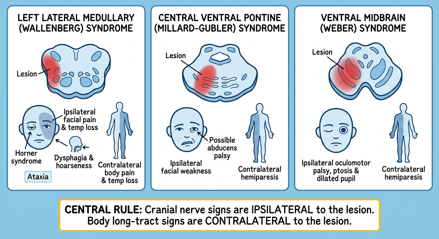
Core rule
- Ipsilateral cranial-nerve signs: face and eye findings on the same side as the lesion.
- Contralateral long-tract signs: body weakness or sensory loss on the opposite side.
| Level | Syndrome / artery | Ipsilateral findings | Contralateral findings | Fast recognition |
|---|---|---|---|---|
| Midbrain | Weber syndrome | CN III palsy: ptosis, dilated pupil, eye “down and out” | Hemiparesis, sometimes lower facial weakness | III + opposite weakness |
| Pons | Millard-Gubler syndrome | LMN facial palsy, sometimes CN VI palsy | Hemiparesis | VII + opposite weakness |
| Lateral medulla | Wallenberg syndrome, usually PICA/vertebral artery | Facial pain-temperature loss, dysphagia, hoarseness, Horner syndrome, ataxia | Body pain-temperature loss | Hoarseness/dysphagia + crossed pain-temperature loss |
| Medial medulla | Dejerine syndrome, anterior spinal artery | Tongue weakness with deviation toward lesion | Hemiparesis and impaired vibration/proprioception | XII + opposite weakness |
Exam tip
- Crossed face-body sensory loss strongly suggests a lateral brainstem lesion.
- Crossed cranial-nerve palsy plus hemiparesis suggests a medial or ventral brainstem lesion.
- Vertigo, nystagmus, diplopia, dysphagia, dysarthria, ataxia, skew deviation, or an internuclear ophthalmoplegia should raise concern for posterior-circulation stroke.
Give me like brain stim strok like this as location
brainstem infarction syndromes
brainstem stroke localization medial lateral midbrain pons medulla syndrome signs
Brainstem stroke localization: learn by level + side
The key rule: “Crossed signs”
- Same-side face/cranial nerve signs
- Opposite-side body weakness or sensory loss
1. Midbrain stroke
| Location / syndrome | Vessel | Ipsilateral signs | Contralateral signs | Localization shortcut |
|---|---|---|---|---|
| Ventral midbrain: Weber syndrome | PCA perforators | CN III palsy: ptosis, dilated pupil, eye down-and-out | Hemiparesis, often lower facial weakness | CN III + opposite hemiparesis |
| Midbrain tegmentum: Benedikt syndrome | PCA perforators | CN III palsy | Tremor, choreoathetosis, ataxia, sometimes weakness | CN III + opposite tremor/ataxia |
| Dorsal midbrain | Variable | Vertical gaze palsy, convergence-retraction nystagmus, light-near dissociation | Usually no classic crossed motor syndrome | Cannot look up |
Think: III and IV cranial nerves.
2. Pontine stroke
| Location / syndrome | Vessel | Ipsilateral signs | Contralateral signs | Localization shortcut |
|---|---|---|---|---|
| Ventral caudal pons: Millard-Gubler syndrome | Basilar paramedian branches | LMN CN VII facial palsy. Sometimes CN VI palsy | Hemiparesis | Facial paralysis + opposite body weakness |
| Dorsal/tegmental pons: Foville syndrome | Paramedian pontine branches | Facial weakness plus horizontal gaze palsy toward lesion | Hemiparesis | Cannot look toward lesion + facial weakness + opposite weakness |
| Medial pons | Basilar paramedian branches | CN VI palsy or horizontal gaze palsy; may have internuclear ophthalmoplegia | Hemiparesis, sensory loss | Abduction/gaze problem + opposite weakness |
| Lateral pons: AICA syndrome | AICA | Facial weakness, reduced lacrimation/salivation, taste loss anterior 2/3 tongue, vertigo/nystagmus, hearing loss, ipsilateral facial pain-temperature loss, ipsilateral ataxia | Body pain-temperature loss | Facial palsy + hearing loss = AICA/lateral pons |
| Large ventral pons: locked-in syndrome | Basilar artery occlusion | Quadriplegia and anarthria, but consciousness and vertical eye movements may be preserved | Bilateral motor deficit | Awake, cannot speak/move, can blink/look vertically |
Think: VI, VII, VIII.
3. Medullary stroke
| Location / syndrome | Vessel | Ipsilateral signs | Contralateral signs | Localization shortcut |
|---|---|---|---|---|
| Lateral medulla: Wallenberg syndrome | Usually PICA or vertebral artery | Facial pain-temperature loss; dysphagia, hoarseness, reduced gag due to nucleus ambiguus; vertigo/nystagmus; Horner syndrome; ataxia | Body pain-temperature loss | Dysphagia/hoarseness + crossed pain-temperature loss |
| Medial medulla: Dejerine syndrome | Anterior spinal artery or vertebral paramedian branches | CN XII palsy, tongue deviates toward lesion | Hemiparesis plus loss of vibration/proprioception | Tongue deviation + opposite weakness |
| Hemimedullary syndrome | Vertebral artery proximal to PICA | Features of both medial and lateral medullary lesions | Mixed crossed motor and sensory signs | Severe combination of Wallenberg + Dejerine features |
Think: IX, X, XII.
One-look comparison
| If the patient has... | Most likely location |
|---|---|
| Ptosis, dilated pupil, eye down-and-out + opposite hemiparesis | Midbrain, Weber syndrome |
| Facial paralysis + opposite hemiparesis | Ventral pons, Millard-Gubler syndrome |
| Facial paralysis + hearing loss + vertigo | Lateral pons, AICA stroke |
| Dysphagia, hoarseness, Horner syndrome + crossed pain/temperature loss | Lateral medulla, Wallenberg syndrome |
| Tongue deviates to lesion + opposite weakness | Medial medulla, Dejerine syndrome |
| Vertigo, nystagmus, diplopia, dysarthria, ataxia | Posterior circulation, often brainstem or cerebellum |
| Awake patient with quadriplegia and no speech | Basilar artery occlusion with locked-in syndrome |
Sensory pathways shortcut
| Deficit pattern | Structure / location |
|---|---|
| Ipsilateral face pain-temperature loss | Spinal trigeminal nucleus/tract, lateral pons or medulla |
| Contralateral body pain-temperature loss | Spinothalamic tract, lateral brainstem |
| Contralateral loss of vibration/proprioception | Medial lemniscus, medial brainstem |
| Contralateral motor weakness | Corticospinal tract, medial/ventral brainstem |
| Ipsilateral facial weakness | Facial nucleus/fascicle, pons |
| Ipsilateral tongue weakness | Hypoglossal nucleus/fascicle, medial medulla |
Memory sequence
- Midbrain: III, IV -> eye findings
- Pons: V to VIII -> face, horizontal gaze, hearing
- Medulla: IX, X, XII -> swallowing, voice, tongue
What is the most Serious type of stroke
basilar artery occlusion prognosis
basilar artery occlusion life threatening stroke locked-in syndrome authoritative overview
Basilar artery occlusion
- Sudden vertigo, diplopia, dysarthria, dysphagia, ataxia
- Rapid decline in consciousness or coma
- Quadriplegia
- Locked-in syndrome: awake and aware but unable to speak or move, sometimes able to communicate only with vertical eye movements
| Stroke type | Why it is dangerous |
|---|---|
| Massive MCA infarct, “malignant MCA” | Severe swelling can shift the brain and cause herniation in the first days. May require decompressive hemicraniectomy. |
| Cerebellar infarct | Swelling in the confined posterior fossa can compress the brainstem or obstruct CSF flow, causing acute hydrocephalus and herniation. |
| Intracerebral hemorrhage | Bleeding expands or raises intracranial pressure; brainstem hemorrhage is particularly dangerous. |
| Aneurysmal subarachnoid hemorrhage | Sudden “worst headache,” possible rapid loss of consciousness and rebleeding. It has high early mortality. |
| Large hemispheric stroke with edema | Can produce coma and herniation, especially with large ICA or MCA territory involvement. |
Basilar artery occlusion is among the most devastating strokes because it can infarct the pons and cause coma, quadriplegia, or locked-in syndrome.