
Anaesthsia concern of lithotomy and head down position in Gynae patient
Reading File
Now let me search the medical library for relevant content on lithotomy position, Trendelenburg/head-down position, and anaesthesia concerns in gynaecological patients.
Finding Sources
Finding Sources
Finding Sources
Finding Sources
Finding Sources
Finding Sources
Good results. Let me now read the key sections from Miller's Anesthesia and Morgan & Mikhail on these topics.
Reading File
Reading File
Reading File
Reading File
Reading File
I now have rich textbook content. Let me compile the comprehensive answer.
Loading Image
Excellent - I have all the content needed for a comprehensive answer. Let me compile it now.
Anaesthesia Concerns: Lithotomy + Head-Down (Trendelenburg) Position in Gynaecological Patients
These two positions are routinely combined in gynaecological procedures (hysteroscopy, laparoscopy, robotic-assisted surgery, TURP in some lists, colporrhaphy). The physiological stresses are additive and sometimes multiplicative.
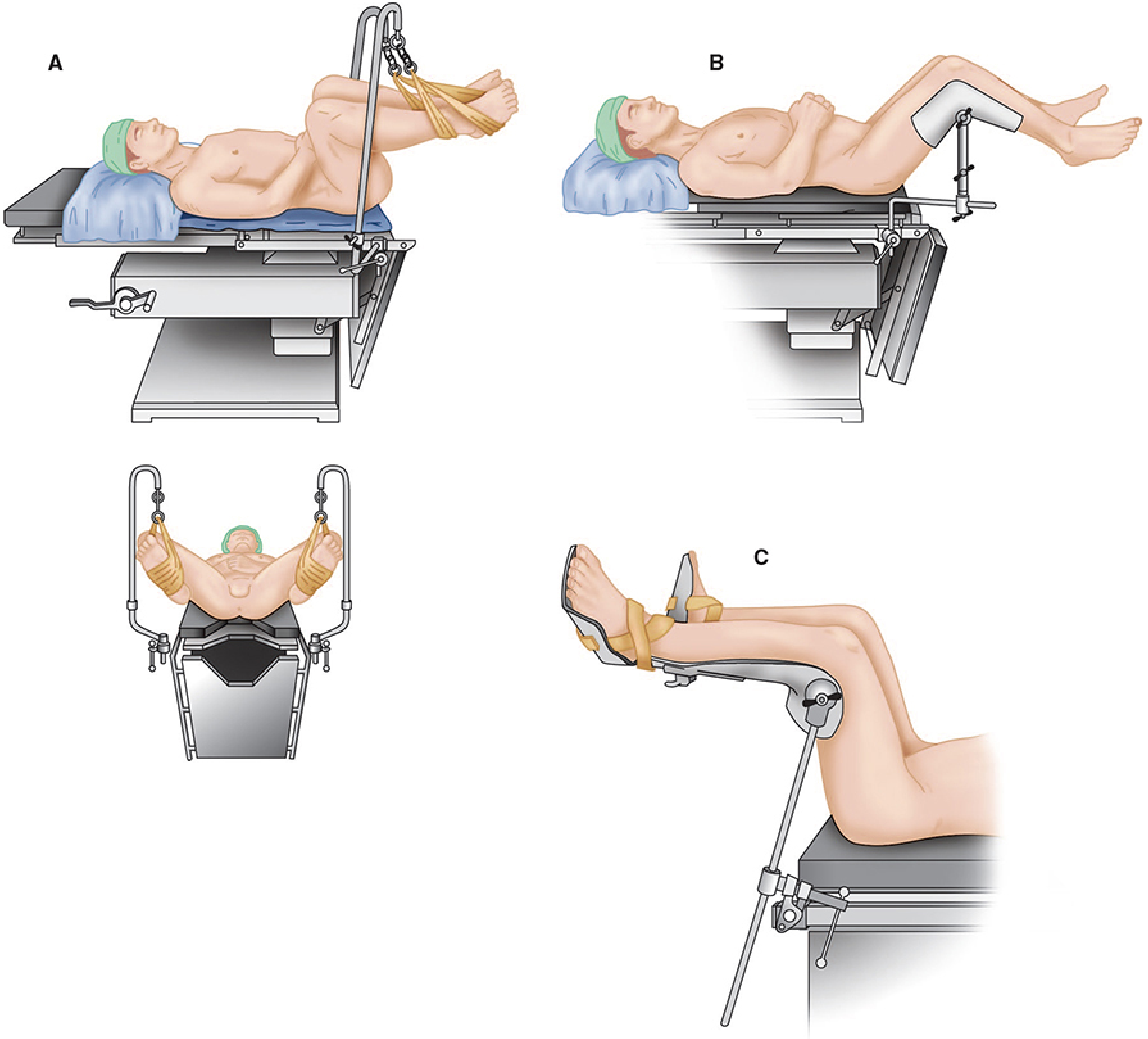
Lithotomy position: A - strap stirrups, B - Bier-Hoff stirrups, C - Allen stirrups (Morgan & Mikhail's Clinical Anesthesiology, 7e)
1. RESPIRATORY CONCERNS
| Problem | Mechanism |
|---|---|
| ↓ FRC | Abdominal contents pushed cephalad by both positions |
| ↓ Compliance | Diaphragm restricted, especially in obese patients |
| ↓ Vital capacity | Positional compressive effect |
| ↑ Peak airway pressures | Reduced thoracic volume |
| V/Q mismatch | Pulmonary congestion from cephalad shift |
| Atelectasis | Especially in prolonged procedures and obese patients |
| Hypercarbia + respiratory acidosis | CO2 absorption from pneumoperitoneum (laparoscopy) |
| ETT migration | Cephalad diaphragm shift can advance ETT into right main bronchus |
"Functional residual capacity decreases, predisposing patients to atelectasis and hypoxia. This effect is amplified by steep Trendelenburg positioning (30-45°)." - Morgan & Mikhail's Clinical Anesthesiology, 7e
Practical implications:
- Secure ETT carefully before positioning; check for bilateral air entry after final position
- Higher ventilation pressures required - may need increased tidal volumes or PEEP
- Steep Trendelenburg (>30°) is particularly risky in morbidly obese patients
2. CARDIOVASCULAR CONCERNS
On assuming the positions:
- Lithotomy: Leg elevation acutely drains blood centrally → ↑ venous return → transient ↑ MAP and cardiac output
- Trendelenburg: Adds further venous return and ↑ CVP
During pneumoperitoneum (if laparoscopy):
| Parameter | Effect |
|---|---|
| SVR | ↑ (up to 30%) |
| MAP | ↑ or maintained |
| Cardiac output | ↓ average 30% at induction of pneumoperitoneum |
| Myocardial oxygen consumption | ↑ |
| Renal, splanchnic, portal flow | ↓ |
On LOWERING the legs (reversing position):
- Acute hypotension - blood pools in previously elevated legs → ↓ venous return → ↓ CO
- Risk potentiated by vasodilation from general or regional anaesthesia
- "Blood pressure measurement should be taken IMMEDIATELY after the legs are lowered" - Morgan & Mikhail
Practical implications:
- Warn surgeons to lower legs slowly at the end of the procedure
- Have vasopressors ready; consider pre-loading with IV fluids
- Patients with poor cardiac reserve (LV dysfunction, poor EF) are at particular risk
3. CENTRAL NERVOUS SYSTEM CONCERNS
| Problem | Mechanism |
|---|---|
| ↑ Intracranial pressure (ICP) | Cephalad fluid shift, impaired venous drainage |
| ↑ Cerebral blood flow | ICP + hypercarbia from CO2 |
| ↑ Intraocular pressure (IOP) | Venous congestion; prolonged steep Trendelenburg |
| Catecholamine release | Neurohumoral response to pneumoperitoneum |
- Patients with intracranial tumors, raised ICP, or glaucoma need careful assessment
- Prolonged steep Trendelenburg (as in robotic surgery) has been associated with significant IOP elevation, with risk of postoperative visual loss
4. NERVE INJURY CONCERNS (Lithotomy-specific)
Nerve injuries are the second leading cause of anaesthesia liability (ASA Closed Claims Database).
| Nerve | Injury Mechanism | Deficit |
|---|---|---|
| Common peroneal (tibial) nerve | Lateral knee resting against strap | Loss of dorsiflexion ("foot drop") |
| Saphenous nerve | Compression against medially placed strap | Numbness along medial calf |
| Obturator nerve | Excessive hip flexion against groin | Adductor weakness |
| Femoral nerve | Excessive hip flexion; inguinal compression | Hip flexor/knee extensor weakness |
| Sciatic nerve | Extreme thigh flexion (stretching) | Combined sensorimotor loss |
| Lumbosacral plexus | Most common - prolonged compression | Mixed lower limb deficits |
| Brachial plexus | Arms improperly positioned (hyperextension) | Upper limb weakness/numbness |
"The most common nerve injuries directly associated with the lithotomy position involve the lumbosacral plexus." - Morgan & Mikhail's Clinical Anesthesiology, 7e
Key rules:
- Two people should move legs simultaneously into and out of lithotomy
- All leg support contact points must be padded
- Fingers must be protected from getting caught between table sections when arms are tucked
- Document pre-existing neuropathy pre-operatively
5. COMPARTMENT SYNDROME
- Elevated legs in lithotomy → reduced arterial perfusion pressure to calves
- Prolonged procedures (>4-5 hours) significantly increase risk
- Can occur in calves from prolonged pressure
- Associated with rhabdomyolysis, which itself causes AKI and hyperkalemia
- "Compartment syndrome of the lower extremities with rhabdomyolysis has been reported with prolonged time in the lithotomy position, after which lower extremity nerve damage is also more likely" - Morgan & Mikhail's Clinical Anesthesiology, 7e
6. AIRWAY AND ENDOTRACHEAL TUBE CONCERNS
- Cephalad shift of the diaphragm can displace a properly placed ETT distally → endobronchial intubation
- ETT position must be rechecked after final positioning
- Head-down + pneumoperitoneum increases risk of regurgitation and aspiration - secure airway (ETT preferred over LMA for laparoscopy) and consider rapid sequence induction
- Facial and airway edema can develop with prolonged procedures in steep Trendelenburg
7. SPECIAL CONCERNS FOR COMBINED LAPAROSCOPIC + LITHOTOMY + TRENDELENBURG (e.g., robotic hysterectomy)
When all three (pneumoperitoneum + lithotomy + steep Trendelenburg) are combined:
- All the above effects are additive
- Pneumoperitoneum insufflation pressure should be kept <12-15 mmHg where possible
- Adequate neuromuscular blockade helps keep insufflation pressures low
- Risk of venous gas embolism from CO2 entry into opened vessels
- Activation of renin-angiotensin system → fluid retention
8. SPECIAL POPULATIONS AT HIGHER RISK
| Population | Additional Concern |
|---|---|
| Obese patients | All respiratory effects magnified; higher compartment syndrome risk |
| Cardiac disease (poor EF) | Hypotension on lowering legs; cannot tolerate pneumoperitoneum |
| ICP raised / glaucoma | Trendelenburg worsens IOP and ICP |
| Prolonged procedures (>4 hr) | Nerve injury and compartment syndrome risk multiplied |
| Pre-existing peripheral neuropathy | Must document preoperatively; any new deficits medico-legally significant |
Summary Table
| System | Lithotomy Concern | Head-Down (Trendelenburg) Concern |
|---|---|---|
| Respiratory | ↓ FRC, atelectasis | Amplifies FRC reduction; ETT migration |
| Cardiovascular | ↑ venous return initially; ↓ CO on reversal | ↑ SVR, ↓ CO with pneumoperitoneum |
| CNS | - | ↑ ICP, ↑ IOP |
| Peripheral nerves | Lumbosacral plexus, sciatic, femoral, peroneal | Brachial plexus (neuropraxia) |
| Compartment syndrome | Calf compartments at risk | - |
| Airway | - | Regurgitation risk; ETT displacement |
Sources:
- Morgan & Mikhail's Clinical Anesthesiology, 7e - Chapter 32: Lithotomy Position and Urological/Gynaecological procedures
- Barash, Cullen, and Stoelting's Clinical Anesthesia, 9e - Table 50-5: Physiology of CO2 Pneumoperitoneum in Trendelenburg Position
- Campbell Walsh Wein Urology - Patient Positioning section
CVS Effect on lithomy position why bradycardia
When legs are elevated in lithotomy, blood drains centrally → increased venous return → increased preload → via the Bainbridge reflex and baroreceptor activation, heart rate can actually decrease (reflex bradycardia). This is the classic "increased venous return → stretch of right atrium → baroreceptor-mediated vagal slowing" mechanism.
Searching sources...
Finding Sources
Finding Sources
Finding Sources
Reading File
Finding Sources
This is the exact textbook content needed. Now let me synthesize a focused, clear answer.
CVS Effects of Lithotomy Position - Why Bradycardia?
This is a nuanced question because the lithotomy position produces opposing cardiovascular responses depending on the phase and the underlying mechanism.
Step 1: What Happens to Venous Return in Lithotomy?
When legs are elevated:
- Blood drains from the lower limbs into the central circulation
- Venous return acutely increases
- Right atrial pressure and preload rise
- MAP and cardiac output transiently increase
So far, this should produce tachycardia (more blood = harder pumping heart). But why does bradycardia occur instead?
Step 2: The Bainbridge Reflex - The Key Mechanism
The Bainbridge reflex is the central explanation:
"Baroreceptors in the right atrium and great veins produce an increase in HR when stretched by increased right atrial pressure. Reduced venous pressure decreases HR."
- Barash's Clinical Anesthesia, 9e
Wait - this says increased venous stretch → increases HR. So increased venous return in lithotomy should cause tachycardia. However, the Bainbridge reflex is rate-dependent:
"The magnitude and direction of the HR response are dependent on the prevailing HR at the time of stimulation."
- Barash's Clinical Anesthesia, 9e
| Baseline HR | Bainbridge Response to Volume Loading |
|---|---|
| Slow (bradycardic) | Causes tachycardia |
| Fast (tachycardic) | Causes bradycardia |
This means in an anaesthetized patient (who often has a relatively elevated resting HR), sudden volume loading from leg elevation can paradoxically slow the heart.
Step 3: The Dominant Mechanism for Bradycardia - Venous Baroreceptor Dominance
The deeper reason is that venous baroreceptors are dominant over arterial baroreceptors in the moment-to-moment regulation of cardiac output:
"In the unmedicated person, the venous baroreceptors are dominant over the arterial. A reduced venous pressure, therefore, slows HR."
- Barash's Clinical Anesthesia, 9e (quoting Greene)
This explains the paradox beautifully. The venous baroreceptors sample preload (right atrial stretch). When preload increases sharply (lithotomy position), the atrial stretch receptors signal the vagus nerve → vagal-mediated bradycardia.
The arterial baroreceptors would normally produce tachycardia when blood pressure drops, but venous baroreceptors override them.
Step 4: Vagal Mechanism
The Bainbridge reflex afferents travel via the vagus nerve, and Bainbridge himself showed the reflex "can be abolished by vagal resection."
So the pathway is:
Legs elevated → ↑ venous return → ↑ right atrial stretch
→ Atrial (venous) baroreceptors activated → Vagal afferents →
→ Medullary cardiovascular centre → ↑ Vagal efferent tone → BRADYCARDIA
Step 5: Regional Anaesthesia (Spinal/Epidural) Makes It Worse
In gynaecological patients receiving spinal anaesthesia:
- Spinal block at T1-T4 ablates the cardiac accelerator fibres (sympathetic)
- Vagus nerve is now unopposed
- Any additional venous pooling or hemodynamic perturbation triggers severe bradycardia
- This is the Bezold-Jarisch reflex variant - particularly dangerous with spinal anaesthesia in lithotomy position
"Blockade of the SNS levels of T1-T4 ablates the efferent limb of the cardiac accelerator nerves. This source of cardiac deceleration is obvious, as the vagus nerve is unopposed."
- Barash's Clinical Anesthesia, 9e
Step 6: The Reversal Problem - Bradycardia When Legs Are LOWERED
There is a second, different mechanism for bradycardia that occurs at the end of surgery when legs are suddenly lowered:
- Blood rapidly pools back into legs from the central circulation
- Venous return drops suddenly
- ↓ Venous pressure → ↓ right atrial stretch → Bainbridge reflex now produces bradycardia
- Combined with a drop in MAP → vasovagal-type bradycardia
This is why BP and HR must be measured immediately after lowering the legs.
Summary: Why Bradycardia in Lithotomy?
| Phase | Mechanism | Result |
|---|---|---|
| Legs elevated (position assumed) | Sudden ↑ venous return → atrial stretch → vagal dominance over sympathetic; Bainbridge reflex if baseline HR already elevated | Bradycardia |
| Spinal anaesthesia + lithotomy | T1-T4 block removes cardiac accelerators; vagus unopposed | Severe bradycardia |
| Legs lowered (end of procedure) | Sudden ↓ venous return → ↓ atrial stretch → Bainbridge slows HR further + hypotension | Bradycardia + hypotension |
| Vasovagal response | Distension of pelvic organs (e.g., during D&C, hysteroscopy) | Profound bradycardia, hypotension |
Clinical pearls:
- Have atropine (0.6-1.2 mg IV) immediately available in all gynaecological procedures in lithotomy
- In spinal anaesthesia, bradycardia is not primarily from block height - it is primarily from decreased venous return via Bainbridge-mediated slowing
- Monitor HR closely during position changes - both when raising and lowering legs
- Vasovagal episodes from uterine/cervical manipulation (cervical dilation) can cause dramatic bradycardia - the surgical stimulus activates pelvic parasympathetic fibres
Source: Barash, Cullen, and Stoelting's Clinical Anesthesia, 9e - Chapter 14: Baroreceptors and Cardiovascular Physiology
This is a shared conversation. Sign in to Orris to start your own chat.