
Inguinal hernia
Reading File
Now I'll search the medical library for comprehensive coverage of inguinal hernia - searching headings and paragraphs in parallel.
Finding Sources
Searching PubMed
"inguinal hernia"[MeSH Terms] AND repair
Excellent results. Now I'll read the most comprehensive sections - Schwartz's (very detailed), Bailey & Love's, and Gray's Anatomy in parallel.
Reading File
Reading File
Reading File
Now let me read the more detailed sections from Schwartz's and Current Surgical Therapy for anatomy, types, and management.
Reading File
Reading File
Now let me read Bailey & Love's more detailed content on anatomy, types, and management, plus search for adult hernia anatomy/classification.
Finding Sources
Reading File
Now let me get the key images and read the management section from Bailey & Love's.
Reading File
Loading Image
Loading Image
Now let me get the surgical repair details and complications from Bailey & Love's, plus the Nyhus/EHS classification.
Reading File
Finding Sources
Now let me read Schwartz's anatomy and Nyhus classification section, and the Bailey & Love repair section.
Reading File
Reading File
I now have comprehensive material. Let me read the laparoscopic section and complications from Bailey & Love's.
Reading File
I now have enough comprehensive information to write a thorough answer. Let me compile it.
Inguinal Hernia - Comprehensive Overview
Definition
An inguinal hernia is the protrusion of abdominal contents through a defect in the inguinal region of the abdominal wall. It is the most common hernia in both men and women, though it is approximately 10 times more common in men. Patients often refer to it as a "rupture."
Anatomy of the Inguinal Canal
The inguinal canal is an oblique passage (approximately 4 cm long in adults) running from the deep (internal) inguinal ring to the superficial (external) inguinal ring:
| Wall | Structure |
|---|---|
| Anterior | External oblique aponeurosis |
| Posterior | Transversalis fascia |
| Roof | Conjoint tendon (fused internal oblique + transversus abdominis) |
| Floor | Inguinal (Poupart's) ligament |
Contents:
- Males: spermatic cord (testicular artery, veins, vas deferens, lymphatics, cremasteric muscle), ilioinguinal nerve, iliohypogastric nerve, genital branch of genitofemoral nerve
- Females: round ligament, same nerves
Key landmark - Hesselbach's Triangle:
- Medial border: lateral edge of rectus abdominis
- Inferior border: inguinal ligament
- Lateral border: inferior epigastric vessels
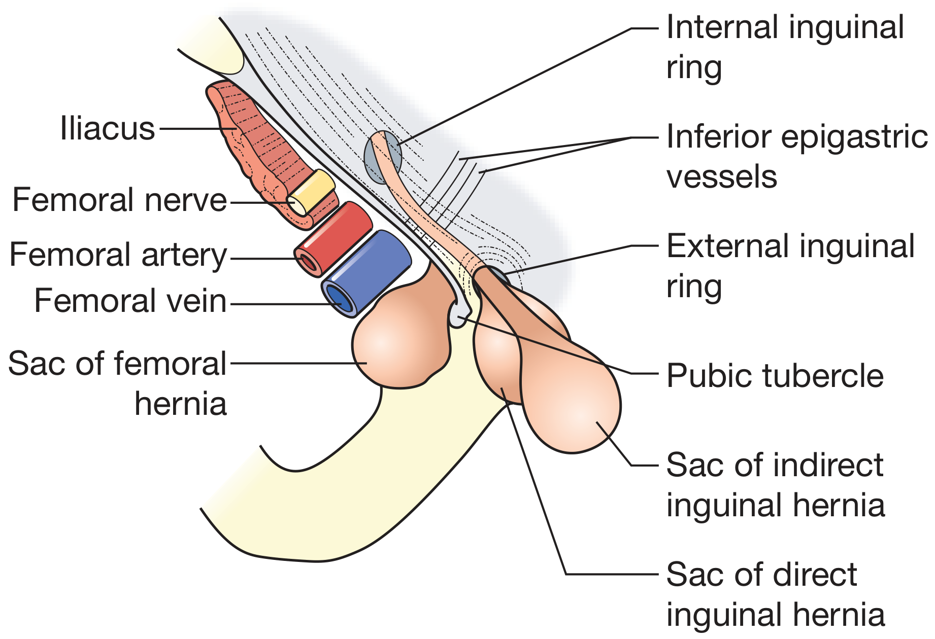
Types
1. Indirect (Lateral) Inguinal Hernia
- Most common type overall
- Protrudes lateral to the inferior epigastric vessels, through the deep inguinal ring
- Follows the path of the processus vaginalis into the scrotum
- Can be congenital (patent processus vaginalis) or acquired
- All congenital hernias in children are indirect by definition
- Higher risk of incarceration and strangulation due to narrow neck
2. Direct (Medial) Inguinal Hernia
- Always acquired; results from weakness/stretching of the posterior inguinal canal wall
- Protrudes medial to the inferior epigastric vessels, directly through Hesselbach's triangle
- Broad-based neck - unlikely to strangulate
- More common in elderly males
- The urinary bladder can be dragged into a direct hernia (sliding)
3. Sliding Hernia
- Retroperitoneal structures (sigmoid colon on left, caecum on right, bladder) form part of the sac wall
- Surgeons must take extra care not to damage bowel during repair
4. Pantaloon Hernia
- Both direct and indirect hernia coexist simultaneously, straddling the inferior epigastric vessels (resembles a pair of trousers)
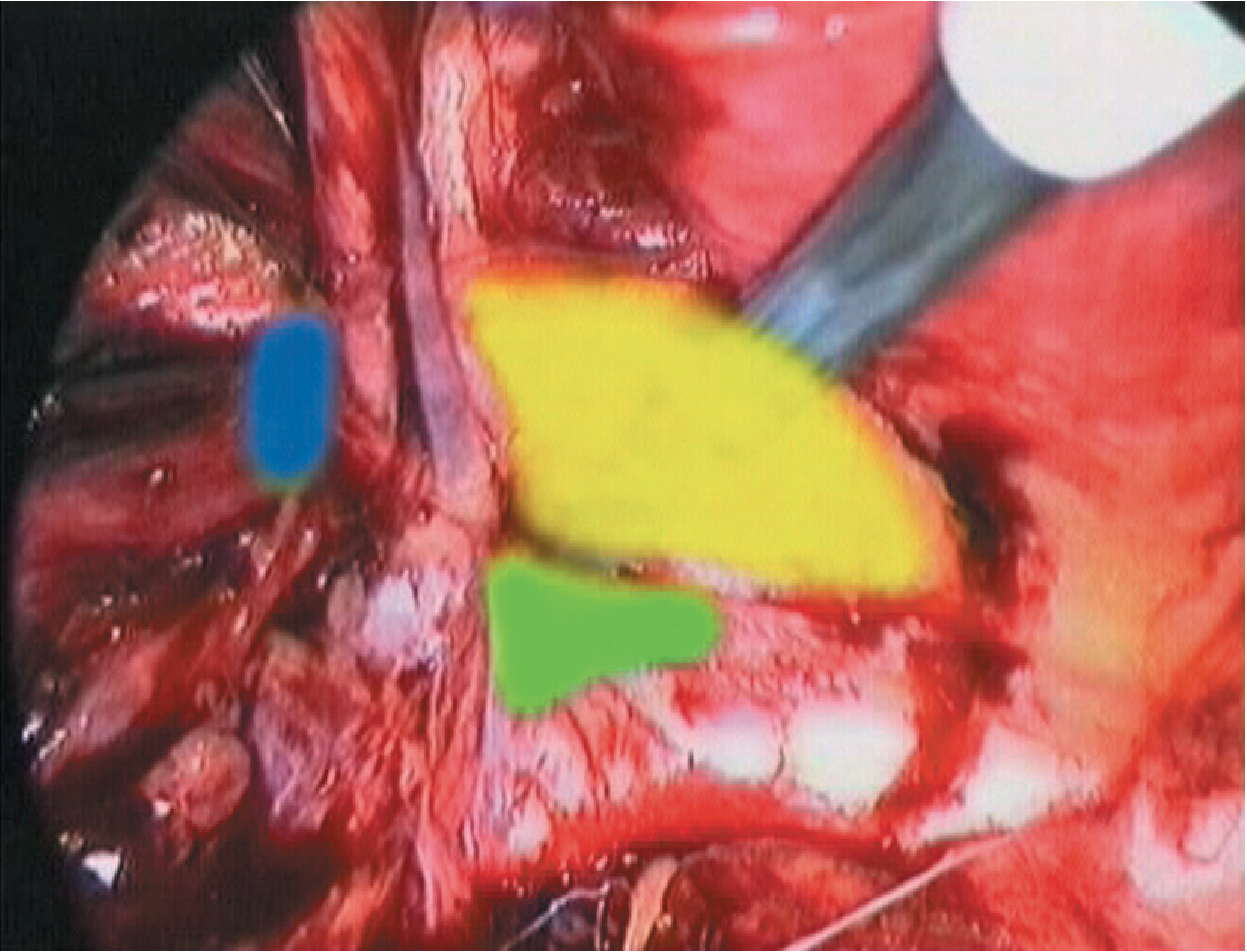
Classification
EHS (European Hernia Society) Classification
- P = Primary, R = Recurrent
- L = Lateral (indirect), M = Medial (direct), F = Femoral
- Defect size: 1 = ≤1 fingerbreadth (~1.5 cm), 2 = 1-3 fingerbreadths, 3 = ≥3 fingerbreadths
- Example: A primary indirect hernia with 3 cm defect = PL2
Nyhus Classification
Categorizes by location, size, and type (Schwartz's Principles of Surgery):
| Type | Description |
|---|---|
| I | Indirect, internal ring normal (pediatric) |
| II | Indirect, enlarged internal ring |
| IIIA | Direct |
| IIIB | Indirect, very enlarged/scrotal (including pantaloon) |
| IIIC | Femoral |
| IV | Recurrent (A=direct, B=indirect, C=femoral, D=combined) |
Epidemiology & Risk Factors
- Lifetime prevalence: 15% at age 25-34 rising to 47% at age 75+ in males (Schwartz's)
- Male:female ratio approximately 10:1
- Right > left side
- Higher incidence in premature infants (processus vaginalis not yet obliterated)
Risk factors:
- Increased intra-abdominal pressure (chronic cough, constipation, heavy lifting, BPH/straining)
- Collagen deficiency
- Prematurity
- Family history
- Prior ipsilateral hernia (recurrence)
Embryology
In males, as the testis descends into the scrotum, it carries a peritoneal sleeve called the processus vaginalis. This normally obliterates before birth (under probable hormonal control). Failure of obliteration leaves a patent communication between the peritoneal cavity and the scrotum - forming an indirect hernia. In premature infants, obliteration has not yet occurred, explaining the very high incidence in this group. - Schwartz's Principles of Surgery, p. 1771
Clinical Features
Symptoms
- Groin bulge, often described as a "lump in the groin"
- Ache or dragging discomfort, worse on standing/coughing/lifting
- Reducible: disappears on lying down, reappears on standing or Valsalva
- May present as scrotal swelling (indirect hernia reaching the scrotum)
Examination
- Examine the patient standing and supine
- Identify bony landmarks: ASIS and pubic tubercle
- Locate the deep inguinal ring (just above the midpoint of the inguinal ligament)
- Ask patient to cough - feel for an impulse
- Reduce the hernia and apply pressure at the deep ring:
- Controlled by pressure at deep ring → indirect
- Appears medial to pressure point → direct
Key distinction:
- Indirect hernia: sac lies superomedial to the pubic tubercle, within the spermatic cord
- Direct hernia: sac lies directly in front of the pubic tubercle
- Cannot "get above" a scrotal hernia (distinguishes from hydrocele; also transillumination negative unless omentum not present)
Complications
| Complication | Description |
|---|---|
| Irreducibility | Contents cannot be returned to abdomen; no cough impulse |
| Incarceration | Irreducible hernia with obstructed bowel; presents with colic, vomiting |
| Strangulation | Compromised blood supply → ischaemia; tender, erythematous swelling, systemic toxicity - surgical emergency |
| Obstruction | Small bowel obstruction if bowel is in the sac |
| Richter's hernia | Only part of the bowel wall is trapped (antimesenteric wall); can strangulate without obstruction |
Infants are at particularly high risk of incarceration due to the narrow inguinal ring. A strangulated hernia progresses to peritonitis, bowel necrosis, and systemic sepsis without emergency surgery.
Diagnosis
Primarily clinical. Investigations are reserved for atypical cases:
- Ultrasound: first-line imaging when clinical diagnosis is uncertain; identifies occult hernia, distinguishes from lymph node, lipoma, varicocele
- CT scan: for large/irreducible hernias, recurrent hernias, or suspected sliding hernia
- MRI (dynamic): for chronic groin pain, occult hernia, athletic pubalgia
Differential diagnosis of groin swelling:
- Femoral hernia (inferomedial to pubic tubercle)
- Hydrocele (transilluminates, can get above it)
- Lymphadenopathy
- Lipoma of the cord
- Varicocele ("bag of worms")
- Undescended testis
- Femoral artery aneurysm
- Psoas abscess
Management
Watchful Waiting
- Asymptomatic or minimally symptomatic hernias in high-risk patients may be observed
- Risk of acute incarceration/strangulation is low (~0.3-0.5% per year for reducible hernia)
- Symptomatic hernias and hernias in children always require repair
Surgical Repair - Principles
All inguinal hernia repairs in adults require either:
- Herniorrhaphy: tension repair with sutures (tissue-based)
- Hernioplasty: mesh-reinforced tension-free repair (preferred in adults)
In children, herniotomy (high ligation of the sac) alone is sufficient without floor reconstruction.
Open Anterior Repairs
1. Bassini Repair (1890)
- Opens external oblique aponeurosis, dissects the cord
- Sac excised/ligated; sutures placed between conjoint tendon and inguinal ligament
- Historical gold standard; over 150 modifications described
- Associated with tension - higher recurrence
2. Shouldice Repair
- Transversalis fascia is opened, then double-breasted (two-layer closure) to reconstruct the posterior wall
- External oblique similarly double-breasted
- Best tissue repair: expert centres report <2% lifetime failure
- Technically demanding
3. Lichtenstein Tension-Free Mesh Repair (most widely used today)
- Polypropylene mesh placed as a flat patch over the inguinal floor, sutured to the inguinal ligament below and the conjoint tendon above
- Introduced in the 1980s; dramatically reduced recurrence rates
- Recurrence <1-2% in most series
- Performed under local, regional, or general anaesthesia
- Recommended by 2018 EHS guidelines as the standard open repair
4. Plug and Patch (Millikan/Gilbert)
- Mesh plug placed into the defect + flat mesh overlay
- Simple but risk of meshoma, plug migration into bladder/bowel
- Not recommended by 2018 EHS guidelines
5. Open Preperitoneal (Stoppa/Rives)
- Large mesh placed in preperitoneal plane via midline or Pfannenstiel incision
- Useful for complex/recurrent hernias and bilateral repairs
- Largely superseded by laparoscopic TEP
Laparoscopic Repairs
Both laparoscopic techniques aim to place a large (≥10×15 cm) mesh in the preperitoneal plane, covering Hesselbach's triangle, the deep inguinal ring, and the femoral canal.
1. TEP (Totally Extraperitoneal)
- Balloons/blunt dissection develop the preperitoneal space without entering the peritoneal cavity
- No peritoneal breach; lower risk of intra-abdominal injury
- More technically challenging to learn
2. TAPP (Transabdominal Preperitoneal)
- Peritoneal cavity entered; peritoneal flap raised; mesh placed in preperitoneal space; peritoneum closed over mesh
- Better visualisation; easier learning curve
- Risk of intra-abdominal injury (bowel, bladder)
Advantages of laparoscopic repair:
- Less post-operative pain
- Faster return to work/activity
- Better for bilateral hernias (both sides repaired simultaneously)
- Better for recurrent hernias after open anterior repair (avoids previously dissected tissue)
- Lower incidence of chronic groin pain vs. open
Disadvantages:
- Requires general anaesthesia
- Longer operating time
- Risk of rare but serious vascular/visceral injury
- Recurrence rates similar to open Lichtenstein in experienced hands
Repair Approach - Decision Making
| Scenario | Preferred Approach |
|---|---|
| Primary unilateral hernia | Open Lichtenstein or TEP/TAPP (surgeon preference) |
| Bilateral hernia | Laparoscopic (TEP or TAPP) |
| Recurrent after open repair | Laparoscopic (TEP or TAPP) |
| Recurrent after laparoscopic | Open preperitoneal (Stoppa) or Lichtenstein |
| Paediatric | Open herniotomy (high ligation) |
| Incarcerated/strangulated | Emergency open surgery; may need bowel resection |
Complications of Surgery
| Complication | Details |
|---|---|
| Haematoma | Most common early complication |
| Wound infection | 1-2% |
| Urinary retention | Especially in elderly males |
| Vas deferens injury | Risk of infertility; higher in re-operations |
| Testicular ischaemia/atrophy | Damage to testicular artery or venous congestion |
| Chronic groin pain (inguinodynia) | Up to 10-20%; most important long-term complication; can be neuropathic (ilioinguinal, iliohypogastric, genitofemoral nerve) |
| Recurrence | <2% with mesh; 10-15% with tissue repair |
| Mesh complications | Infection, migration, seroma, meshoma (plug repair) |
Special Populations
Children
- All hernias are indirect by definition; repair as elective herniotomy
- Contralateral exploration debated (~30% have patent processus vaginalis on opposite side; rate decreases with age)
- Laparoscopic approach increasingly used under age 2
- Emergency repair if incarceration cannot be reduced
Females
- Hernias may contain the ovary and fallopian tube (often a sliding hernia)
- Risk of ovarian torsion if not repaired promptly
- Femoral hernias are relatively more common in women than men (though still less common overall)
Elderly
- Direct hernias more common
- Higher risk of recurrence with tissue repairs
- Mesh repair preferred
- Local anaesthesia is well tolerated
Key Anatomical Relationships to Remember
| Structure | Relation to Deep Inguinal Ring |
|---|---|
| Inferior epigastric vessels | Just medial |
| Indirect hernia sac | Lateral to inferior epigastric vessels |
| Direct hernia sac | Medial to inferior epigastric vessels |
| Femoral hernia | Below the inguinal ligament, medial to femoral vein |
Sources: Bailey and Love's Short Practice of Surgery 28th Edition, p. 1087-1090 | Schwartz's Principles of Surgery 11th Edition, p. 1628 | Schwartz's Principles of Surgery (Pediatrics), p. 1771 | Gray's Anatomy for Students | Tintinalli's Emergency Medicine
This is a shared conversation. Sign in to Orris to start your own chat.