
Whitish discharge per vagina with itching.. Dx?
Reading File
Whitish vaginal discharge + itching = most likely vulvovaginal candidiasis (VVC), though bacterial vaginosis and other causes must be considered.
Searching sources...
Here is a structured clinical answer:
Whitish Vaginal Discharge + Itching
Most Likely Diagnosis: Vulvovaginal Candidiasis (VVC)
The combination of white discharge + pruritus (itching) is the hallmark of VVC, caused most commonly by Candida albicans.
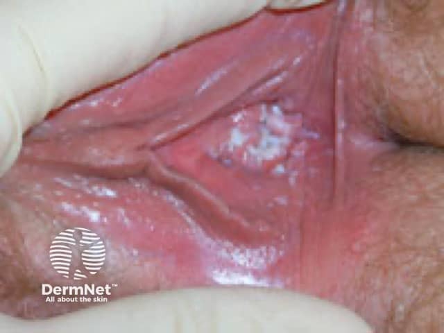
Differential Diagnosis of Vaginal Discharge with Itching
| Condition | Discharge | Itching | Odor | pH | Key Test |
|---|---|---|---|---|---|
| Candidiasis (VVC) | Thick, curd-like/cottage cheese, white | Prominent | None | 3.8-4.5 (normal) | KOH prep: pseudohyphae + budding yeast |
| Bacterial Vaginosis (BV) | Thin, gray, homogenous | Mild/absent | Fishy (amine) | >4.5 | Saline: clue cells; positive whiff test |
| Trichomoniasis | Frothy, yellow-green, profuse | Moderate | Fishy | 6-7 | Saline: motile trichomonads |
| Atrophic vaginitis | Thin, watery | Burning/dryness | None | >4.5 | History (postmenopausal) |
| Allergic/Contact vaginitis | Variable | Prominent | None | Any | History (irritant exposure) |
- Harrison's Principles of Internal Medicine 22E, Table 141-5
- Textbook of Family Medicine 9e, Table 25-7
Candidiasis: Clinical Features
- Discharge: thick, white, "cottage cheese" or curd-like; non-malodorous
- Itching/Pruritus: the dominant symptom - often intense
- Vulvar erythema, edema, fissures, dyspareunia, external dysuria
- pH: normal (3.8-4.5) - distinguishes from BV and Trichomonas
- KOH prep (10%): pseudohyphae and budding yeast cells
Causative organism: Candida albicans (most common), also C. glabrata, C. tropicalis
Risk Factors for VVC
- Recent antibiotic use (disrupts normal lactobacilli flora)
- Diabetes mellitus (uncontrolled)
- Pregnancy
- Immunosuppression (HIV, corticosteroids)
- Oral contraceptive use
- Tight synthetic clothing, poor hygiene
Treatment
| Setting | Drug | Dose |
|---|---|---|
| Uncomplicated (first-line) | Clotrimazole pessary/cream (topical) | 500 mg single dose vaginal pessary; or 1% cream for 7 days |
| Oral option | Fluconazole | 150 mg single oral dose |
| Recurrent VVC (≥4 episodes/year) | Fluconazole | 150 mg every 72h x3, then weekly for 6 months |
| Pregnancy | Topical azoles only (fluconazole avoided in 1st trimester) | 7-day course |
Quick Diagnostic Workup
- Vaginal pH - normal (≤4.5) favors candidiasis; >4.5 favors BV/Trichomonas
- KOH wet mount (10%) - pseudohyphae + yeast (confirms candidiasis)
- Saline wet mount - rules out Trichomonas (motile organisms) and BV (clue cells)
- Whiff/amine test - positive in BV, negative in VVC
- Culture if recurrent/resistant (to identify C. glabrata - azole-resistant)
Bottom line: Whitish discharge + itching without odor + normal pH = Vulvovaginal Candidiasis until proven otherwise. Confirm with KOH prep and treat with topical or oral azole.
Rx
Treatment for Vulvovaginal Candidiasis (VVC)
Searching sources...
Finding Sources
Reading File
Reading File
Treatment of Vulvovaginal Candidiasis
1. Uncomplicated VVC
(First episode / infrequent / mild-moderate / non-pregnant / immunocompetent)
Oral (Systemic) - Preferred by most patients:
| Drug | Dose | Duration |
|---|---|---|
| Fluconazole | 150 mg PO | Single dose |
Topical Intravaginal Azoles (equally effective, ~80-90% cure):
| Drug | Formulation | Regimen |
|---|---|---|
| Clotrimazole | 1% cream | 1 applicator intravaginally at bedtime x 7 days |
| Clotrimazole | 2% cream | 1 applicator intravaginally at bedtime x 3 days |
| Miconazole | 2% cream | 1 applicator intravaginally at bedtime x 7 days |
| Miconazole | 4% cream | 1 applicator intravaginally at bedtime x 3 days |
| Terconazole | 0.4% cream | 1 applicator intravaginally at bedtime x 7 days |
| Terconazole | 80 mg suppository | 1 suppository intravaginally at bedtime x 3 days |
| Tioconazole | 6.5% ointment | Single intravaginal dose |
| Nystatin | 100,000-unit vaginal tablet | 1 tablet at bedtime x 14 days (less effective) |
Topical azoles > nystatin in efficacy. All formulations (creams, suppositories, tablets) are equally effective.
- Tintinalli's Emergency Medicine, Table 102-5
- Harrison's 22E
2. Complicated VVC
(Recurrent ≥4 episodes/year, severe symptoms, C. glabrata, pregnant, or immunocompromised)
Recurrent VVC (RVVC) - Induction + Maintenance:
| Phase | Drug | Dose |
|---|---|---|
| Induction | Fluconazole 150 mg PO | Every 72 hours x 3 doses (Day 1, 4, 7) |
| Maintenance | Fluconazole 150 mg PO | Once weekly x 6 months |
Severe VVC:
- Fluconazole 150 mg PO x 2 doses, 72 hours apart
3. Pregnancy
- Topical azoles ONLY - 7-day course (extended duration)
- Fluconazole is CONTRAINDICATED in pregnancy (teratogenic risk in 1st trimester, Category C)
- Clotrimazole or miconazole topical preferred
4. C. glabrata / Azole-Resistant VVC
- Boric acid 600 mg vaginal capsule at bedtime x 14 days
- Nystatin vaginal suppositories
- Flucytosine vaginal cream (specialist use)
5. Partner Treatment
- Routine treatment of male partner is NOT indicated
- Treat only if male partner has symptomatic candidal balanitis
Key Points
- Topical azoles = Oral fluconazole in efficacy for uncomplicated cases
- Oral fluconazole is more convenient (single dose, patient preference)
- Treat underlying risk factors: control diabetes, limit unnecessary antibiotics, avoid tight synthetic clothing
- If no response to standard treatment - culture to identify C. glabrata or C. krusei (intrinsically azole-resistant)
This is a shared conversation. Sign in to Orris to start your own chat.