
Classification of antibiotics and all examples
Reading File
Antibiotics are broadly classified by chemical structure, mechanism of action, and spectrum. Here's a quick overview while I pull from the textbook library:
- Beta-lactams (penicillins, cephalosporins, carbapenems, monobactams) - inhibit cell wall synthesis
- Aminoglycosides (gentamicin, amikacin) - inhibit protein synthesis (30S)
- Macrolides (azithromycin, erythromycin) - inhibit protein synthesis (50S)
- Fluoroquinolones (ciprofloxacin, levofloxacin) - inhibit DNA gyrase
- Tetracyclines (doxycycline, tigecycline) - inhibit protein synthesis (30S)
Searching sources...
Finding Sources
Finding Sources
Goodman & Gilman's has the most relevant section. Let me pull that content along with another pharmacology source.
Reading File
Finding Sources
Now let me get the specific pharmacology chapters on each antibiotic class from Goodman & Gilman's and also look for a dedicated pharmacology textbook section:
Finding Sources
Finding Sources
Finding Sources
Finding Sources
Reading File
Reading File
Good, I have an excellent antibiotic sites diagram. Let me fetch that image and also read more of the Medical Microbiology antibiotic classification section:
Loading Image
Reading File
I have excellent content. Let me now read the rest of the antibiotic classes section from Medical Microbiology:
Reading File
I have enough rich content from the textbooks. Let me now compile the comprehensive classification. I have the key antibiotic diagram and detailed tables from Medical Microbiology 9e and Goodman & Gilman's. Let me now deliver the full answer.
Classification of Antibiotics
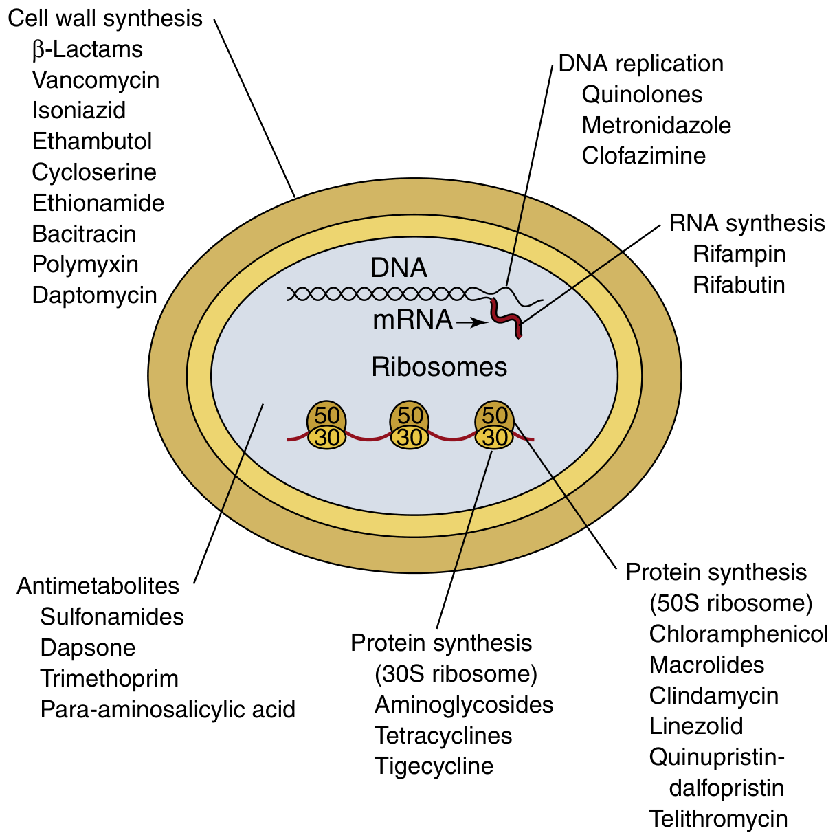
Antibiotics are classified along several dimensions: mechanism of action, chemical structure, and spectrum of activity. The primary classification by mechanism is the most clinically useful.
1. INHIBITORS OF CELL WALL SYNTHESIS
A. Beta-Lactams
All share a beta-lactam ring; bind penicillin-binding proteins (PBPs) and block peptidoglycan crosslinking.
i. Natural Penicillins
| Drug | Route | Notes |
|---|---|---|
| Penicillin G (benzylpenicillin) | IV/IM | Acid-labile; narrow spectrum |
| Penicillin V (phenoxymethylpenicillin) | Oral | Acid-stable oral form |
Spectrum: Beta-hemolytic streptococci, meningococci, most gram-positive anaerobes. Poor against gram-negatives.
ii. Penicillinase-Resistant Penicillins (Anti-staphylococcal)
| Drug |
|---|
| Methicillin |
| Nafcillin |
| Oxacillin |
| Cloxacillin / Dicloxacillin / Flucloxacillin |
Spectrum: Active against penicillinase-producing Staphylococcus aureus (MSSA). Useless against MRSA.
iii. Broad-Spectrum (Aminopenicillins)
| Drug |
|---|
| Ampicillin |
| Amoxicillin |
Spectrum: Gram-positive cocci + some gram-negatives (E. coli, Proteus, Haemophilus).
iv. Extended-Spectrum (Antipseudomonal) Penicillins
| Drug |
|---|
| Piperacillin |
| Ticarcillin |
| Carbenicillin |
Spectrum: Broad, including Pseudomonas aeruginosa.
v. Beta-Lactam + Beta-Lactamase Inhibitor Combinations
| Combination |
|---|
| Ampicillin-sulbactam (Unasyn) |
| Amoxicillin-clavulanate (Augmentin) |
| Ticarcillin-clavulanate (Timentin) |
| Piperacillin-tazobactam (Zosyn) |
| Ceftazidime-avibactam |
| Ceftolozane-tazobactam |
| Meropenem-vaborbactam |
Beta-lactamase inhibitors (clavulanic acid, sulbactam, tazobactam, avibactam) are relatively inactive alone but irreversibly inhibit beta-lactamases, restoring activity of the companion drug.
B. Cephalosporins
Derived from 7-aminocephalosporanic acid; same mechanism as penicillins but wider spectrum and more stable to many beta-lactamases. Classified by "generations":
| Generation | Key Drugs | Spectrum |
|---|---|---|
| 1st | Cephalexin, Cefazolin, Cephalothin, Cefadroxil | Gram-positive cocci (MSSA), limited gram-negatives |
| 2nd | Cefuroxime, Cefaclor, Cefprozil, Cefoxitin (cephamycin), Cefotetan | Broader gram-negative coverage (H. influenzae, Moraxella); cephamycins cover anaerobes |
| 3rd | Cefotaxime, Ceftriaxone, Ceftazidime, Cefixime, Cefdinir, Cefpodoxime | Extended gram-negative coverage including Enterobacteriaceae; ceftazidime covers Pseudomonas |
| 4th | Cefepime | Broad spectrum; Pseudomonas + gram-positives |
| 5th | Ceftaroline, Ceftobiprole | Active against MRSA (binds PBP2a) |
C. Carbapenems
Broadest spectrum beta-lactams; stable to most beta-lactamases including ESBLs. Not active against MRSA.
| Drug |
|---|
| Imipenem-cilastatin (cilastatin prevents renal inactivation) |
| Meropenem |
| Ertapenem (no Pseudomonas coverage) |
| Doripenem |
| Biapenem |
D. Monobactams
Monocyclic beta-lactam ring; active only against aerobic gram-negatives (including Pseudomonas). Safe in penicillin allergy (minimal cross-reactivity).
| Drug |
|---|
| Aztreonam |
E. Glycopeptides
Inhibit cell wall synthesis by binding D-Ala-D-Ala terminus of peptidoglycan precursors. Active against gram-positives only (including MRSA).
| Drug | Notes |
|---|---|
| Vancomycin | First-line for MRSA |
| Teicoplanin | Longer half-life |
| Dalbavancin | Long-acting (weekly dosing) |
| Oritavancin | Single-dose therapy |
| Televancin | Active against MRSA, VRE |
F. Other Cell Wall Inhibitors
| Drug | Class | Notes |
|---|---|---|
| Bacitracin | Polypeptide | Topical use; gram-positives |
| Fosfomycin | Phosphonic acid | Inhibits MurA; UTI treatment |
| Cycloserine | D-amino acid analog | Anti-TB (2nd line) |
| Isoniazid (INH) | Nicotinic acid analog | Anti-TB; inhibits mycolic acid synthesis |
| Ethambutol | Synthetic | Anti-TB; inhibits arabinogalactan synthesis |
| Ethionamide | Thioamide | Anti-TB (2nd line) |
2. INHIBITORS OF PROTEIN SYNTHESIS (30S Ribosome)
A. Aminoglycosides
Bind 30S ribosomal subunit; cause misreading of mRNA. Bactericidal. Concentration-dependent killing.
| Drug |
|---|
| Gentamicin |
| Amikacin |
| Tobramycin |
| Streptomycin (also anti-TB) |
| Neomycin (topical) |
| Kanamycin |
| Netilmicin |
| Spectinomycin |
Spectrum: Aerobic gram-negatives; synergistic with beta-lactams against enterococci.
B. Tetracyclines
Bind 30S subunit; reversibly block aminoacyl-tRNA binding. Bacteriostatic. Broad spectrum.
| Generation | Drugs |
|---|---|
| Classic | Tetracycline, Oxytetracycline, Chlortetracycline |
| Long-acting | Doxycycline, Minocycline |
| Glycylcycline | Tigecycline (overcomes tetracycline resistance) |
| New | Omadacycline, Eravacycline |
Uses: Atypicals (Chlamydia, Mycoplasma, Rickettsia), Brucella, acne, community-acquired pneumonia.
3. INHIBITORS OF PROTEIN SYNTHESIS (50S Ribosome)
A. Macrolides
Bind 50S subunit; block translocation. Bacteriostatic (bactericidal at high concentrations). Excellent intracellular penetration.
| Drug |
|---|
| Erythromycin (prototype) |
| Azithromycin (Z-pack) |
| Clarithromycin |
| Roxithromycin |
| Spiramycin |
| Fidaxomicin (GI-restricted; C. difficile) |
B. Ketolides
Structural derivatives of macrolides; overcome macrolide resistance.
| Drug |
|---|
| Telithromycin |
| Solithromycin |
C. Lincosamides
Bind 50S; similar spectrum to macrolides but excellent anaerobic coverage.
| Drug |
|---|
| Clindamycin |
| Lincomycin |
D. Chloramphenicol
Binds 50S; inhibits peptidyl transferase. Broad spectrum but reserved due to bone marrow toxicity (aplastic anemia).
| Drug |
|---|
| Chloramphenicol |
E. Oxazolidinones
Bind 50S; prevent formation of initiation complex. Active against MRSA, VRE, drug-resistant gram-positives.
| Drug |
|---|
| Linezolid |
| Tedizolid |
F. Streptogramins
Bind 50S in two sequential steps (synergistic combination). Active against MRSA and VRE (vanA-negative).
| Drug |
|---|
| Quinupristin-dalfopristin (Synercid) |
G. Pleuromutilins
Bind 50S (peptidyl transferase center).
| Drug |
|---|
| Lefamulin (systemic) |
| Retapamulin (topical) |
4. INHIBITORS OF DNA SYNTHESIS / REPLICATION
A. Fluoroquinolones (Quinolones)
Inhibit DNA gyrase (topoisomerase II) and topoisomerase IV; bactericidal.
| Generation | Drugs | Coverage |
|---|---|---|
| 1st (Quinolones) | Nalidixic acid, Cinoxacin | Gram-negative (urinary only) |
| 2nd | Ciprofloxacin, Ofloxacin, Norfloxacin, Enoxacin | Broad gram-negative + Pseudomonas |
| 3rd | Levofloxacin, Sparfloxacin | Adds improved gram-positive + atypicals |
| 4th | Moxifloxacin, Gatifloxacin, Gemifloxacin | Broadest including anaerobes; "respiratory fluoroquinolones" |
B. Nitroimidazoles
Reduced to toxic radicals inside anaerobic organisms; damage DNA. Active against strict anaerobes and certain protozoa.
| Drug |
|---|
| Metronidazole (Flagyl) |
| Tinidazole |
| Ornidazole |
| Secnidazole |
C. Nitrofurans
Reductive activation; multiple mechanisms including DNA damage. Used only for urinary tract infections.
| Drug |
|---|
| Nitrofurantoin |
| Furazolidone |
5. INHIBITORS OF RNA SYNTHESIS
Rifamycins
Inhibit DNA-dependent RNA polymerase (beta subunit). Bactericidal; excellent intracellular penetration.
| Drug |
|---|
| Rifampicin (Rifampin) - anti-TB |
| Rifabutin - anti-TB (MAC) |
| Rifaximin - non-absorbable (traveler's diarrhea, hepatic encephalopathy) |
| Rifapentine - anti-TB (once-weekly) |
6. INHIBITORS OF CELL MEMBRANE FUNCTION
A. Polymyxins
Disrupt gram-negative outer membrane by acting like detergents. Last-resort agents for MDR gram-negatives.
| Drug |
|---|
| Polymyxin B |
| Colistin (Polymyxin E) |
B. Lipopeptides
Depolarize gram-positive bacterial membrane; rapidly bactericidal.
| Drug |
|---|
| Daptomycin |
Spectrum: Gram-positives including MRSA, VRE. Inactivated by pulmonary surfactant (not for pneumonia).
7. ANTIMETABOLITES (Inhibitors of Folate Synthesis)
Sulfonamides and trimethoprim act at sequential steps in folate synthesis - powerful synergy when combined.
| Drug | Target |
|---|---|
| Sulfonamides: Sulfamethoxazole, Sulfadiazine, Sulfisoxazole, Sulfadoxine, Mafenide, Silver sulfadiazine | Inhibit dihydropteroate synthase (DHPS) - block PABA incorporation |
| Trimethoprim | Inhibits dihydrofolate reductase (DHFR) |
| Co-trimoxazole (TMP-SMX) | Sequential blockade - synergistic |
| Dapsone | Inhibits DHPS (anti-leprosy, PCP prophylaxis) |
| Para-aminosalicylic acid (PAS) | Anti-TB; inhibits folate/salicylate synthesis |
| Pyrimethamine | Inhibits DHFR (antiprotozoal) |
8. ANTI-MYCOBACTERIAL AGENTS (Special Group)
| Drug | Class | Primary Use |
|---|---|---|
| Isoniazid (INH) | Isonicotinic acid | TB (1st line) |
| Rifampicin | Rifamycin | TB (1st line) |
| Pyrazinamide | Nicotinic acid amide | TB (1st line) |
| Ethambutol | Synthetic | TB (1st line) |
| Streptomycin | Aminoglycoside | TB (2nd line) |
| Amikacin | Aminoglycoside | MDR-TB |
| Capreomycin | Cyclic peptide | MDR-TB |
| Cycloserine | D-amino acid | MDR-TB |
| Ethionamide / Prothionamide | Thioamide | MDR-TB |
| Bedaquiline | Diarylquinoline (ATP synthase inhibitor) | MDR-TB |
| Delamanid | Nitroimidazole | MDR-TB |
| Clofazimine | Phenazine | Leprosy, MDR-TB |
| Dapsone | Sulfone | Leprosy |
Summary Table by Mechanism
| Mechanism | Classes | Bactericidal / Static |
|---|---|---|
| Cell wall synthesis | Beta-lactams, Glycopeptides, Bacitracin, Fosfomycin | Cidal |
| 30S ribosome | Aminoglycosides, Tetracyclines | Cidal (AG), Static (Tet) |
| 50S ribosome | Macrolides, Clindamycin, Chloramphenicol, Linezolid, Quinupristin-dalfopristin | Static (mostly), Cidal (Linezolid variable) |
| DNA gyrase / Topoisomerase | Fluoroquinolones | Cidal |
| DNA damage | Metronidazole, Nitrofurantoin | Cidal |
| RNA polymerase | Rifamycins | Cidal |
| Cell membrane | Polymyxins, Daptomycin | Cidal |
| Folate synthesis | Sulfonamides, Trimethoprim | Static |
Sources: Medical Microbiology 9e (Murray et al.), Tables 17.2-17.3; Goodman & Gilman's Pharmacological Basis of Therapeutics, Ch. 56 - "Antimicrobial Chemotherapy: Classes and Actions"

IMG_0595.jpeg
Teach me and explain detailed
Reading File
analyse_media
This is a critically abnormal VBG. Let me search the textbook library for acid-base interpretation and lactic acidosis in parallel.
Finding Sources
Finding Sources
Finding Sources
Finding Sources
Reading File
Reading File
Reading File
Finding Sources
Reading File
Reading File
I now have everything needed for a thorough, teaching-focused explanation of this critically abnormal VBG. Let me compile the full analysis.
📋 Venous Blood Gas (VBG) - Full Teaching Analysis
Patient: aliakbar | NIMS Hospital | 11-07-2026 | Sample: Venous
Step 1: What Is a Venous Blood Gas (VBG)?
The sample is venous, not arterial. This is important because normal reference ranges differ:
| Parameter | Arterial (ABG) Normal | Venous (VBG) Normal |
|---|---|---|
| pH | 7.35-7.45 | 7.31-7.41 (slightly more acidic) |
| pCO2 | 35-45 mmHg | 41-51 mmHg (higher - CO2 returns from tissues) |
| pO2 | 80-100 mmHg | 30-40 mmHg (lower - O2 extracted by tissues) |
| HCO3- | 22-26 mmol/L | similar |
The machine here uses venous reference ranges (e.g., pCO2 normal = 41-51, pO2 normal = 30-40). This matters when interpreting the results below.
Step 2: Read Every Value - What Is Normal and What Is Not?
BLOOD GAS
| Parameter | Patient Value | Reference | Status | Meaning |
|---|---|---|---|---|
| pH | 6.88 | 7.31-7.41 | 🔴 CRITICALLY LOW (below reportable range) | Severe acidemia |
| pCO2 | 32.2 mmHg | 41.0-51.0 | 🔻 LOW | Respiratory compensation (hyperventilation blowing off CO2) |
| pO2 | 66 mmHg | 30-40 | 🔺 HIGH for venous | Unusual; discussed below |
pH 6.88 is a life-threatening value. Normal cellular enzyme function requires pH 7.35-7.45. Below 7.0, cardiac arrhythmias, vasodilation, and cellular death occur.
ELECTROLYTES / METABOLITES
| Parameter | Patient Value | Reference | Status | Clinical Meaning |
|---|---|---|---|---|
| Na⁺ (Sodium) | 133 mmol/L | 135-145 | 🔻 Low | Mild hyponatremia |
| K⁺ (Potassium) | 6.18 mmol/L | 3.50-4.50 | 🔺 HIGH | Dangerous hyperkalemia |
| Ca²⁺ (Ionized Calcium) | 0.97 mmol/L | 1.15-1.30 | 🔻 Low | Hypocalcemia (partly pH-related) |
| Cl⁻ (Chloride) | 99 mmol/L | 98-108 | ✅ Normal | |
| Lactate | 14.4 mmol/L | 0.5-1.6 | 🔺 CRITICALLY HIGH | Severe lactic acidosis (9x upper limit) |
DERIVED VALUES - Explained One by One
🔴 HCO₃⁻ (Bicarbonate) = 6.0 mmol/L (normal ~22-26)
- Bicarbonate is the body's main blood buffer.
- Normal reaction: H⁺ + HCO₃⁻ → H₂CO₃ → CO₂ + H₂O
- When massive acid accumulates (like lactate here), HCO₃⁻ is consumed to neutralize it → it falls dramatically.
- HCO₃⁻ of 6.0 means the buffer is nearly exhausted - catastrophic.
🔴 Standard Bicarbonate (cHCO₃⁻ P,st) = 7.2 mmol/L
- Standardized to normal pCO2 (40 mmHg) to isolate the metabolic component.
- Confirms this is a primary metabolic acidosis, not just a respiratory issue.
🔴 Base Excess = -27.4 mmol/L (normal: -2 to +2)
- Base excess (BE) tells you how much base (or acid) needs to be added to restore normal pH.
- Negative BE = base deficit = the patient needs base.
- -27.4 means the patient is 27.4 mmol/L short of base - this is incompatible with survival without urgent intervention.
- Normal BE = 0. Mild acidosis = -5. Moderate = -10. Severe = -15. This patient is at -27 - extreme.
🔴 Anion Gap = 27.5 mmol/L (normal <12)
- Formula: AG = Na⁺ - (Cl⁻ + HCO₃⁻)
- Patient: 133 - (99 + 6) = 28 (matches ~27.5 as calculated by machine)
- Normal AG = 6-12 mmol/L. This patient's AG = 27.5 → massively elevated.
- High AG means unknown negative ions (acid anions) are filling the gap - in this case, lactate (14.4 mmol/L) is a major contributor.
🔴 Anion Gap with K⁺ = 33.6 mmol/L
- Same calculation but includes potassium: (Na + K) - (Cl + HCO3)
- = (133 + 6.18) - (99 + 6) = 139.18 - 105 = 34.18 ≈ matches machine value.
- Even higher because of the elevated K⁺.
Ionized Ca²⁺ corrected to pH 7.40 = 0.73 mmol/L
- True calcium at normal pH.
- Very low - suggests significant hypocalcemia independent of pH effects (acidosis artificially raises ionized Ca²⁺, so correcting back to 7.40 reveals the true deficit is even worse: 0.73 vs normal 1.15-1.30).
Total CO₂ (ctCO₂) = 6.4 mmol/L (normal ~23-27)
- Includes dissolved CO₂ + HCO₃⁻ + carbamino compounds.
- Reflects the profoundly low bicarbonate. Critically low.
sO₂e (Estimated O₂ Saturation) = 77%
- For a venous sample, this is actually not unexpected (venous O2 sat is normally 60-80%).
- However, the pO2 of 66 mmHg is high for venous - this may indicate reduced oxygen extraction by tissues (cells cannot use O2 due to mitochondrial failure from severe acidosis/shock), OR the sample is partially arterial.
Step 3: Putting It Together - Diagnosis
🔴 PRIMARY DISORDER: Severe High-Anion Gap Metabolic Acidosis
Using the stepwise approach from Harrison's Principles of Internal Medicine:
Step 1 - Check pH: 6.88 → Acidemia (severely low)
Step 2 - Check primary component:
- HCO₃⁻ = 6 (very low) → Primary metabolic acidosis
- pCO2 = 32.2 (low) → Respiratory compensation (the lungs are trying to blow off CO2 to reduce acid - this is called Kussmaul breathing)
Step 3 - Check Anion Gap: AG = 27.5 → High AG metabolic acidosis
Step 4 - Is compensation adequate?
Using Winter's formula for expected respiratory compensation:
Expected pCO2 = 1.5 × HCO₃⁻ + 8 ± 2 = 1.5 × 6 + 8 = 17 ± 2 mmHg
Patient's actual pCO2 = 32.2 mmHg → Higher than expected (32 vs expected 17)
This means the respiratory compensation is INSUFFICIENT for the degree of acidosis - either:
- There is a concurrent respiratory acidosis (lungs cannot compensate fully, perhaps due to respiratory muscle fatigue or lung disease), OR
- The patient is tiring and losing their compensatory hyperventilation - a very ominous sign.
Step 5 - Identify cause of high AG:
The lactate = 14.4 mmol/L (normal <1.6) is massively elevated. This IS the anion filling the gap.
Diagnosis: Type A Lactic Acidosis (tissue hypoperfusion/shock state)According to Tintinalli's Emergency Medicine: "In critical illness, an oxygen debt develops when oxygen delivery is inadequate... This results in global tissue hypoxia, anaerobic metabolism, and lactate production."
Step 4: The Electrolyte Abnormalities - Explained
Hyperkalemia (K⁺ = 6.18)
Why does acidosis cause hyperkalemia?
- In acidosis, excess H⁺ ions enter cells to be buffered.
- To maintain electrical neutrality, K⁺ exits cells into the blood.
- For every 0.1 unit drop in pH, K⁺ rises approximately 0.5-0.7 mmol/L.
- pH dropped from 7.4 to 6.88 = 0.52 units → expected K⁺ rise of ~2.6-3.6 mmol/L.
- This explains most of the hyperkalemia here - it is redistributional (not total body excess in most cases), but still causes dangerous cardiac arrhythmias.
Hypocalcemia (Ca²⁺ = 0.97)
- May occur in septic shock, rhabdomyolysis, or pancreatitis.
- The corrected Ca²⁺ at pH 7.40 is only 0.73 (very low) - worsens cardiac function.
Hyponatremia (Na⁺ = 133)
- Mild; may reflect fluid shifts, or dilutional in resuscitation.
Step 5: Summary Table of All Abnormalities
| Value | Patient | Normal | Interpretation |
|---|---|---|---|
| pH | 6.88 | 7.31-7.41 | Severe acidemia - life-threatening |
| pCO2 | 32.2 | 41-51 | Low - respiratory compensation |
| HCO3⁻ | 6.0 | 22-26 | Nearly depleted buffer |
| Base Excess | -27.4 | -2 to +2 | Catastrophic base deficit |
| Lactate | 14.4 | 0.5-1.6 | Severe lactic acidosis - shock/hypoperfusion |
| Anion Gap | 27.5 | <12 | High AG - lactate driven |
| K⁺ | 6.18 | 3.5-4.5 | Hyperkalemia - acidosis-driven shift |
| Na⁺ | 133 | 135-145 | Mild hyponatremia |
| Ca²⁺ | 0.97 | 1.15-1.30 | Hypocalcemia |
| Hb | 13.2 g/dL | - | Normal - anemia not the cause |
Step 6: What Is Killing This Patient? - Clinical Correlation
The pattern here is:
- Profound metabolic acidosis (pH 6.88, HCO3 = 6, BE = -27)
- Massive lactic acidosis (lactate 14.4 - 9x normal)
- Hyperkalemia from acidosis
- Failed respiratory compensation (pCO2 higher than expected for the degree of acidosis)
This is a critically ill patient in severe shock - most likely:
- Septic shock (most common cause of lactic acidosis in ICU)
- Cardiogenic shock (low output → tissue hypoperfusion → anaerobic metabolism)
- Hypovolemic shock (hemorrhage, severe dehydration)
- Mesenteric ischemia (gut hypoperfusion gives massive lactate)
- Toxic/drug-induced (cyanide, metformin toxicity in renal failure, etc.)
"When lactic acidosis accompanies low-flow disease states such as sepsis, mortality increases by almost threefold." - Tintinalli's Emergency Medicine
Step 7: Memory Aid - How to Read Any VBG/ABG
1. pH < 7.35 = Acidosis  |  pH > 7.45 = Alkalosis
2. Primary cause: HCO3 ↓ or pCO2 ↑ → Acidosis
                  HCO3 ↑ or pCO2 ↓ → Alkalosis
3. Compensation: Metabolic acidosis → lungs blow off CO2 (pCO2 ↓)
                 Respiratory acidosis → kidneys retain HCO3 (HCO3 ↑)
4. Anion Gap = Na - (Cl + HCO3)   Normal < 12
   High AG causes: GOLD MARK
   G = Glycols (ethylene glycol)
   O = Oxoproline
   L = L-lactate (THIS PATIENT)
   D = D-lactate
   M = Methanol
   A = Aspirin (salicylates)
   R = Renal failure (uremia)
   K = Ketoacidosis (DKA)
5. Base Excess < -2 = base deficit = metabolic acidosis
6. Lactate > 2 mmol/L = hyperlactatemia; > 4 = severe lactic acidosis
Sources: Harrison's Principles of Internal Medicine 22E - "Metabolic Acidosis, Anion Gap"; Tintinalli's Emergency Medicine - "Blood Gas Analysis, Plasma Lactate"; Schwartz's Principles of Surgery 11E - "Respiratory and Metabolic Components of Acid-Base Disorders"
⚠️ Clinical urgency: A patient with pH 6.88, lactate 14.4, and base excess -27 requires immediate ICU-level resuscitation - airway management, identification and treatment of the underlying cause (sepsis workup, echo for cardiac function), IV sodium bicarbonate consideration, and urgent correction of hyperkalemia.
IMG_0595.jpeg
Explain
This is the same VBG report I already analyzed in detail above. Let me give you a focused, simplified explanation of what this report means clinically - in plain language.
🩸 This Patient's VBG - Plain Language Explanation
The Big Picture in One Sentence
This patient's blood is dangerously acidic because their tissues are not getting enough oxygen, causing massive lactic acid buildup - this is a medical emergency.
The 5 Most Critical Findings
🔴 1. pH = 6.88 (Normal: 7.31–7.41)
What is pH?
pH measures how acidic or alkaline the blood is. Like a swimming pool - too acidic or too alkaline and things stop working.
- pH 7.4 = normal blood
- pH 7.2 = mild-moderate acidosis
- pH 7.0 = severe, life-threatening
- pH 6.88 = extreme acidosis - below the machine's reportable range (the ‡ symbol means "too low to reliably measure")
At pH 6.88:
- Heart muscle stops contracting properly → cardiac arrest risk
- Blood vessels dilate → blood pressure collapses
- Enzymes in every cell stop working
- Brain function deteriorates → coma
🔴 2. Lactate = 14.4 mmol/L (Normal: 0.5–1.6)
What is lactate and why is it high?
Think of it like this: your cells normally burn oxygen to make energy (like a clean-burning engine). When oxygen is not available, they switch to a "dirty" backup mode called anaerobic metabolism - this produces lactic acid as a waste product.
Normal:   Glucose + O₂ → Energy + CO₂ + H₂O  (clean)
In shock: Glucose → Energy + LACTIC ACID      (dirty, acidic)
- Normal lactate < 1.6 mmol/L
- Mild elevation: 2-4 (concerning)
- Severe: > 4 (organ failure territory)
- This patient: 14.4 = nearly 9× the upper limit of normal
This tells us: massive, widespread tissue oxygen deprivation - the entire body is not getting enough blood/oxygen. This is called Type A Lactic Acidosis and it occurs in shock.
🔴 3. Bicarbonate (HCO₃⁻) = 6.0 mmol/L (Normal: 22–26)
What is bicarbonate and why does it matter?
Bicarbonate is the blood's main shock absorber for acid. Think of it as fire extinguisher foam - it neutralizes acid as fast as it's produced.
HCO₃⁻ + H⁺  →  H₂CO₃  →  CO₂ + H₂O
(buffer)   (acid)          (breathed out)
- Normal = 22-26 mmol/L (full extinguisher)
- This patient = 6.0 mmol/L (extinguisher nearly empty)
The body produced so much lactic acid so fast that the bicarbonate buffer was almost completely consumed. There is almost nothing left to fight the acid.
🔴 4. Base Excess = -27.4 mmol/L (Normal: -2 to +2)
What does base excess mean?
Base excess is a simple number that tells you: "How much base does this patient need to return to normal?"
- If base excess = 0 → perfect
- If base excess = -5 → mildly short of base
- If base excess = -10 → moderate deficit
- If base excess = -27.4 → catastrophically short of base
To put it in perspective: a base excess of -27.4 in a 70 kg person means roughly 190 mmol of bicarbonate would be needed just to correct the chemistry - an enormous amount.
🔴 5. Anion Gap = 27.5 mmol/L (Normal: < 12)
What is the anion gap?
The body always maintains electrical balance - positive charges must equal negative charges in blood. We can calculate:
Anion Gap = Sodium - (Chloride + Bicarbonate)
           = 133 - (99 + 6) = 28 mmol/L
When the AG is high, it means hidden acid anions are present that we haven't measured directly. Here, the culprit is lactate (14.4 mmol/L) - it's the "hidden anion" driving the gap up.
A mnemonic for causes of high anion gap (GOLD MARK):
| Letter | Cause |
|---|---|
| G | Glycols (ethylene glycol) |
| O | Oxoproline |
| L | L-Lactate ← THIS PATIENT |
| D | D-Lactate |
| M | Methanol |
| A | Aspirin |
| R | Renal failure |
| K | Ketoacidosis (DKA) |
The Compensation Attempt
The body is not passive - it fights back. With metabolic acidosis, the lungs immediately try to compensate by breathing faster and deeper (Kussmaul breathing) to blow off CO₂ (which is also an acid):
- Patient's pCO₂ = 32.2 mmHg (below normal 41-51)
- This confirms the lungs ARE trying to compensate
But using Winter's Formula to check if compensation is adequate:
Expected pCO₂ = (1.5 × HCO₃⁻) + 8 = (1.5 × 6) + 8 = 17 mmHg
The patient's actual pCO₂ (32.2) is much higher than expected (17) - meaning the lungs are NOT compensating enough. This suggests either:
- Concurrent lung disease limiting ventilation, OR
- The patient is tiring and losing their ability to hyperventilate - an ominous sign of impending respiratory failure
The Electrolyte Problems
K⁺ = 6.18 (High - Dangerous)
Why? In acidosis, excess H⁺ ions flood into cells. To maintain electrical balance, K⁺ is pushed out of cells into the blood. This is called a transcellular shift. The hyperkalemia here is largely pH-driven - but it is still dangerous and can cause fatal cardiac arrhythmias (ventricular fibrillation).
Ca²⁺ = 0.97 (Low)
The corrected calcium (at normal pH 7.40) is only 0.73 - even lower. Low calcium weakens cardiac muscle contraction and can cause tetany.
Na⁺ = 133 (Mildly Low)
Mild hyponatremia - likely from fluid shifts.
Overall Diagnosis
pH 6.88 + HCO3 6.0 + Lactate 14.4 + High Anion Gap
         ↓
SEVERE HIGH ANION GAP METABOLIC ACIDOSIS
CAUSED BY MASSIVE LACTIC ACIDOSIS
         ↓
Underlying cause = SHOCK (tissue not getting oxygen)
Most likely: Septic shock / Cardiogenic shock / Severe hemorrhage
Visual Summary
NORMAL PATIENT:
pH:       |--------7.40--------|  ✅
HCO3:     |------24------------|  ✅
Lactate:  |1.0|                    ✅
BE:       |0|                      ✅

THIS PATIENT:
pH:      6.88|----------------------7.40  🔴 Catastrophically acidic
HCO3:    6|---------------------------24  🔴 Buffer nearly gone
Lactate: |----14.4-----------------1.6|  🔴 9× normal
BE:      -27.4|---------------------0|   🔴 Massive base deficit
What Needs to Happen Immediately
- ICU admission - this patient cannot be on a ward
- Identify the cause of shock - blood cultures (sepsis?), echo (cardiac?), CT (source of bleeding or ischemia?)
- Restore tissue perfusion - IV fluids, vasopressors if needed
- Manage airway - if the patient tires from Kussmaul breathing, they need intubation
- Treat hyperkalemia - calcium gluconate (protects heart), insulin + glucose, Kayexalate
- Consider sodium bicarbonate - controversial, but when pH < 7.1 and patient is deteriorating, IV sodium bicarbonate may be given as a bridge
- Serial lactate measurements - lactate clearance (dropping by ≥25% in 6 hours) is a sign of recovery
Bottom line: This is one of the most critically abnormal blood gases you can see. The pH of 6.88 with lactate of 14.4 and base excess of -27 represents a patient in severe shock with end-organ failure. Without urgent diagnosis and treatment of the underlying cause, survival is very unlikely.
Sources: Harrison's Principles of Internal Medicine 22E; Tintinalli's Emergency Medicine; Schwartz's Principles of Surgery 11E
This is a shared conversation. Sign in to Orris to start your own chat.